

Abstract
Canine babesiosis is a worldwide tick borne disease. Dogs with fever, in appetence and enlarged lymph nodes were screened for the presence of haemoprotozoan parasites during the one year period of study at T.V.C.C., C.V.Sc., Proddatur. Based on the stained peripheral blood smears examination, six dogs were found to be affected with babesiosis. Clinical examination of the dogs revealed ticks over the body, congested conjunctival mucus membranes, dullness, fever, tachycardia, tachypnoea in all the dogs. Some of the dogs had icterus, lymphadenopathy, haemoglobinuria. Laboratory examination of the clinical samples revealed reduction in haemoglobin concentration, erythrocyte count, platelet count, serum total protein, serum albumin and glucose levels. Increased serum SGPT, creatinine, BUN levels were recorded. Dogs were treated with inj. Diminazene aceturate (Berenil RTU) @ 5 mg/kg body weight, single dose along with supportive and symptomatic therapy in individual cases.
Keywords: Babesiosis, Clinical signs, Dogs, Laboratory findings
Introduction
Canine babesiosis is a clinically significant and geographically widespread haemoprotozoan disease of domesticated dogs and wild canids (Irwin 2010). The large Babesia canis and the small Babesia gibsoni are two organisms commonly known to infect the dogs. The life cycle of Babesia includes two stages: inside the host RBCs, in which the sporozoites convert into piroplasms, and the other inside the tick vector (Uilenberg 2006). A typical intra erythrocytic piroplasma is pear-shaped and often occurs in pairs (Homer et al. 2000). Clinically, the illness is characterized by fever, pale mucous membrane, anorexia, anemia, icterus, lymphadenopathy and splenomegaly (Boozer and Macintire 2003). Clinical signs are exceedingly variable; the classical presentation is a febrile illness with perceptible anemia. The severity of babesiosis varies from sub clinical infection to extensive organ failure and death. The most common hematologic alterations associated with B. canis infections are anemia and thrombocytopenia (Schetters et al. 2009). The parasite has been reported to be transmitted by ixodid tick vectors from different geographical locations. Depending on the species of Babesia parasite present in an area, the common tick vectors reported to serve in transmission of canine babesiosis include Rhipicephalus species, Haemaphysalis species and Dermacentor species (Matjila et al. 2004). The parasite also transmitted by blood exchange and transplacental route can also possible (Jefferies et al. 2007). The objectives of this study were to describe the clinical signs and hemato-biochemical alterations in dogs affected with Babesia.
Materials and methods
The present study was performed on dogs presented and referred to the Teaching Veterinary Clinical Complex, College of Veterinary Science, Proddatur from November 2012 to November 2013. In this study, a total number of 300 dogs belongs to the different breeds were examined for the presence of Babesia in peripheral blood smears. Each suspected dog was subjected to physical and clinical examination. During the clinical examination, the presence of ticks on the coat of dogs were observed and collected for their identification. Peripheral blood was collected from the ear tips of the dogs for Giemsa-stained thin blood smear examination to detect Babesia species in RBC. The smears were fixed in methanol for 5 min and stained by 10 % Giemsa. Two blood smears were made from each sample and a minimum of 50 fields of each stained blood smear were examined under oil immersion to determine parasitaemia. Blood was collected in 10 % EDTA solution from all the dogs was used for estimation of packed cell volume (PCV), total leucocyte count (TLC), total erythrocyte count (TEC) and haemoglobin (Hb). The peripheral blood smears were stained by Leishman’s stain to study the differential count (DLC) by battlement method following which; the absolute counts were also calculated. Thrombocyte count was carried out only in the three seriously affected dogs with babesiosis. Serum was also collected to study the sero biochemical parameters including SGPT, BUN, total protein, serum albumin by using Span diagnostics Ltd. Kits. The procedures followed as per the Sivajothi et al. (2013). Haematological and biochemical findings of the infected dogs were compared with haemato-biochemical findings of apparently healthy dogs of the local area (Reddy et al. 2014). Urine samples were collected from the Babesia infected dogs for urine analysis by urine dipstic method and microscopic examination of urinary sediment. The data in respect of each parameter was tabulated and statistical analyses were carried out by using Statistical Package for Social Sciences (SPSS) using Student's t-test.
Results
Microscopic evaluation of the stained peripheral blood smears demonstrated the presence of typical large pyriform, paired parasites of Babesia in the erythrocytes (Fig. 4) of six dogs in this study. Dogs included in this study had 1–8 years age belongs to the both sex. Clinical examination of the six infected dogs revealed presence of ticks over the body (6/6), dullness (6/6), variations in the appetite (6/6), rise in rectal temperature (6/6), tachycardia (6/6), tachypnoea (5/6), dyspnoea (5/6), congested mucus membranes with sunken eye balls (5/6), lymphadenopathy (5/6), pallor (3/6), haemoglobinuria (3/6), tensed abdomen (2/6), yellowish mucus membranes (2/6), yellowish discoloration of abdomen (2/6), vomitions (2/6), diarrhoea (2/6), edema at the legs (1/6) and constipation (1/6) (Figs. 1, 2, 3). Based on the morphological characteristics, collected ticks were identified as Rhipicephalus spp.
Fig. 4.

Giemsa-stained peripheral blood smear of infected dog showing the pear-shaped large B. canis inside the RBCs (× 1,000)
Fig. 1.
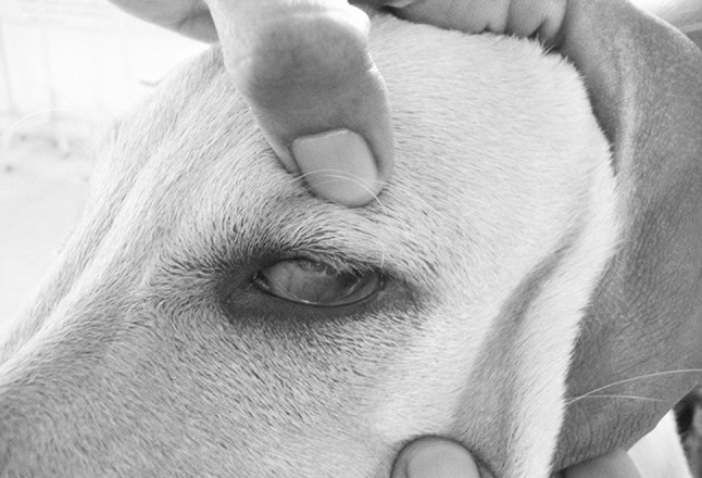
Dog showing the congested mucus membranes and suncken eyeballs
Fig. 2.

Dog showing the yellowish buccal mucus membrane
Fig. 3.

Dog showing the enlarged lymph node
Haematological and biochemical findings were incorporated in the Table 1. Significant reduction in RBC, Hb concentration, PCV percentage and platelet count (p < 0.05) were recorded among infected dogs when compared to the apparently healthy dogs. Significant decrease in WBC count and neutropenia along with lymphocytosis and monocytosis was noted. Three dogs with Babesia infection had lowered thrombocyte count (range:96,000–112,000/cumm, with mean of 105,333 ± 36,223/cumm). Thrombocyte count was compared with standard reference range mentioned by the Rizzi et al. (2010). Serum biochemical parameters revealed reduction in total protein, serum albumin, glucose levels. Increased BUN, creatinine, SGPT levels were noticed. Laboratory urinalysis was done in all the dogs. Abnormal findings were noticed in three dogs included haemoglobinuria, proteinuria, bilirubinuria, and increased amount of urobilinogen in the urine. Microscopic evaluation of the sediment revealed red blood cells as well as white blood cells, tubular epithelial cells and crystal formation.
Table 1.
Haemato-biochemical values of Babesia infected dogs (Mean ± S.E)
| Parameters | Normal group$ | Babesia infected dogs (n = 6) | Observed range | t-test | p |
|---|---|---|---|---|---|
| Hb (g/dl) | 13.03 ± 0.42 | 9.72 ± 0.32 | 10.2–9.2 | 6.84** | 0.000 |
| PCV (%) | 39.29 ± 2.14 | 30.50 ± 6.51 | 33–27 | 3.52** | 0.001 |
| TEC x106/cumm | 6.51 ± 0.19 | 4.42 ± 0.86 | 4.8–3.8 | 8.36*** | 0.000 |
| TLC/cumm | 9334.29 ± 81.34 | 8786.67 ± 71.45 | 9500–8200 | 2.62* | 0.021 |
| Neutrophils/cumm | 6698.06 ± 244.96 | 3804 ± 109.26 | 4200–3216 | 7.62*** | 0.000 |
| Lymphocytes/cumm | 2417.37 ± 119.77 | 4512 ± 289.41 | 4960–4100 | 12.37*** | 0.000 |
| Eosinophils/cumm | 135.57 ± 22.11 | 244 ± 65.74 | 312–165 | 1.54 | 0.153 |
| Monocytes/cumm | 68.36 ± 17.83 | 220 ± 88.62 | 332–140 | 3.47** | 0.005 |
| Total protein (g/dL) | 6.81 ± 0.22 | 5.48 ± 0.21 | 4.8–6.1 | 4.27* | 0.001 |
| Albumin (g/dL) | 3.25 ± 0.04 | 2.43 ± 0.09 | 2.8–2.2 | 8.29*** | 0.000 |
| Globulin (g/dL) | 3.56 ± 0.20 | 3.05 ± 0.49 | 3.9–2.4 | 1.76 NS | 0.104 |
| A:G ratio | 0.85 ± 0.11 | 0.79 ± 0.10 | 1.16–0.56 | 1.38 NS | 0.196 |
| Glucose (mg/dL) | 120.80 ± 6.22 | 88.83 ± 5.70 | 72–110 | 3.73** | 0.003 |
| SGPT (ALT) (U/L) | 18.71 ± 1.56 | 53.00 ± 8.2 | 92–38 | 3.20** | 0.008 |
| BUN (mg/dL) | 16.56 ± 0.95 | 25.67 ± 2.55 | 32–24 | 3.77** | 0.003 |
| Creatinine (mg/dL) | 0.91 ± 0.07 | 1.37 ± 0.14 | 1.8–0.8 | 2.87* | 0.015 |
$ (Reddy et al. 2014)
NS non significant (p > 0.05)
* Significant (p < 0.05)
** Highly significant (p < 0.01)
Dogs were treated with inj. Diminazene aceturate (Berenil RTU, Intervet) @ 5 mg/kg body single dose IM, inj.Nurobion forte @ 2 ml IM, inj. Meloxicam @ 0.5 mg/kg body weight IM. Symptomatic therapy along with fluids were given based on requirement of the individual case. Supportive therapy was given with four doses of iron dextron (Imferon) @ 2 ml/dog at weekly twice, daily supplementation of oral haematinics (Sharkoferrol, Alembic Ltd.). After completion of 2 weeks of therapy, cases (5/6) were responded well and attains its normal activities. But one dog was not responded and died on the 3rd day of therapy.
Discussion
Babesia normally causes massive injuries to the host depending on the virulence and pathogenicity of the parasite. The extent of the injuries also depends on the age and the hygiene of the host (Leschnik et al. 2007). Clinical signs observed in the present study includes depression, fever, tachycardia, tachypnoea, in appetence and pallor mucus membranes. It is thought that the clinical signs are the result of tissue hypoxia following the anaemia and a concomitant systemic inflammatory response syndrome caused by marked cytokine release in babesiosis (Lobetti 2006). In the severe form of the disease there is a marked haemolytic anaemia, severe acid–base abnormalities with frequent secondary multiple organ failure and complications such as acute renal failure, hepatopathy with marked icterus, hypoglycaemia (Keller et al. 2004). Schetters, et al. (2009) describes the classical presentation of Babesia infection as febrile illness with apparent anemia though the clinical presentation of canine babesiosis can be highly variable. The common laboratory abnormalities in this study was anaemia and thrombocytopenia. Babesia initiates a mechanism of antibody-mediated cytotoxic destruction of circulating erythrocytes and anemia may be more dependent on the host immune response than on the direct destruction of RBC by the piroplasm (Boozer and Macintire 2003). The mechanisms of the thrombocytopenia are not yet fully understood in babesiosis; multiple mechanisms, including platelet sequestration in the spleen, immune-mediated platelet destruction and development of disseminated intravascular coagulation are possible. Inspite of thrombocytopenia no single dog in this study was showed any hemorrhages on the surface of the body. In this study, the main biochemical changes were hypoproteinaemia, hypoalbuminemia and hypoglycemia, increased serum levels of SGPT, creatinine, BUN in majority of the infected dogs. This finding concurs with the previous reports (Furlanello et al. 2005). Increased serum activity of transaminases was suggesting that these abnormalities resulted from hepatopathy and renal failure. Centrilobular hepatitis with hypoxic liver damage could be the possible mechanism that resulted in significant changes in hepatic enzymes and decreased albumin levels (Taboada and Lobetti 2006). Renal impairment noticed due to damage of renal cells caused by inflammatory mediators, or possibly due to the development of refractory hypotension resulting in reduced renal tissue perfusion and glomerular filtration ate (Zygner and Wedrychowicz 2009). Hypoglycemia in this study was due to in appetence due to sepsis and impaired hepatic function. This finding was in consonant with the report of Amie (2009) who reported that the increased non-insulin mediated glucose consumption is believed to be induced by inflammatory mediators, more especially in macrophage-rich tissues like the spleen, liver and the lungs. Dogs with babesiosis treated with a single intramuscular injection of diminazene aceturate at the dose of 5 mg/kg (Birkenheuer et al. 1999). In dogs affected with babesiosis early diagnosis and treatment, the prognosis is good, but severely affected or untreated animals may die.
Based on the present study, dogs with fever, lymphadenopathy, inappetence, pale mucous membrane associated with anemia and thrombocytopenia can be suspected for Babesia infection. All the suspected dogs in this study were diagnosed only based on the microscopic examination of the stained peripheral blood smears. Microscopic examination may not detect low parasitemia though; it remains the most rapid confirmatory method which was carried out in the present study.
Acknowledgments
All the authors are thankful to Sri Venkateswara Veterinary University for providing facilities to carry out this work. Corresponding author would like to express his thanks to Dr. Saradamma, Assistant Director and B.Sravani, Veterinary Assistant who helped in collection of clinical samples.
References
- Amie K. Hypoglycemia. Small anim. Crit Care Med. 2009;69:295–299. [Google Scholar]
- Birkenheuer AJ, Levy MG, Savary KC, Gager RB, Breitschwerdt EB. Babesia gibsoni infections in dogs from North Carolina. J Am Anim Hosp Assoc. 1999;35:125–128. doi: 10.5326/15473317-35-2-125. [DOI] [PubMed] [Google Scholar]
- Boozer AL, Macintire DK. Canine babesiosis. Vet Clin North Am Small Anim Pract. 2003;33:885–904. doi: 10.1016/S0195-5616(03)00039-1. [DOI] [PubMed] [Google Scholar]
- Furlanello T, Fiorio F, Caldin M, Lubas G, Solano Gallego L. Clinicopathological findings in naturally occurring cases of babesiosis caused by large from Babesia from dogs of northeastern Italy. Vet Parasitol. 2005;134:77–85. doi: 10.1016/j.vetpar.2005.07.016. [DOI] [PubMed] [Google Scholar]
- Homer MJ, Aguilar-Delfin I, Telford SR, Krause PJ, Persing DH. Babesiosis. Clin Microbiol Rev. 2000;13:451–469. doi: 10.1128/CMR.13.3.451-469.2000. [DOI] [PMC free article] [PubMed] [Google Scholar]
- Irwin PJ. Canine Babesiosis. Vet Clin Small Anim. 2010;40:1141–1156. doi: 10.1016/j.cvsm.2010.08.001. [DOI] [PubMed] [Google Scholar]
- Jefferies R, Ryan UM, Jardine J, Broughton DK, Robertson ID, Irwin PJ. Blood, bull terriers and babesiosis. Further evidence for direct transmission of Babesia gibsoni in dogs. Aust Vet J. 2007;85:459–460. doi: 10.1111/j.1751-0813.2007.00220.x. [DOI] [PubMed] [Google Scholar]
- Keller N, Jacobson LS, Nel M, De Clerq M, Thompson PN, Schoeman JP. Prevalence and risk factors of hypoglycemia in virulent canine babesiosis. J Vet Intern Med. 2004;18:265–270. doi: 10.1111/j.1939-1676.2004.tb02544.x. [DOI] [PubMed] [Google Scholar]
- Leschnik M, Kirtz G, Leidinger E (2007) Seasonal occurrence of canine babesiosis is influenced by local climate conditions, In: Proceedings of the 9th International Jena Symposium on Tick Borne Diseases, Jena, Germany
- Lobetti RG. Babesiosis. In: Greene CE, editor. Infectious diseases of the dog and cat. 3. Philadelphia: W.B. Saunders; 2006. pp. 722–736. [Google Scholar]
- Matjila PT, Penzhorn BL, Bekker CP, Nijhof AM, Jongejan F. Confirmation of occurrence of Babesia vogeli in domestic dogs in South Africa. Vet Parasitol. 2004;122:119–125. doi: 10.1016/j.vetpar.2004.03.019. [DOI] [PubMed] [Google Scholar]
- Rizzi TE, Meinkoth JH, Clinkenbeard KD. Normal hematology of the dogs. In: Weiss DJ, Wardrop KJ, editors. Schalm’s Veterinary Hematology. Hoboken: Weiley-Blackwell Publishing; 2010. pp. 799–810. [Google Scholar]
- Schetters TPM, Kleuskens JAGM, van de Crommert J, de Leeuw PWJ, Finizio AL, Gorenflot A. Systemic inflammatory responses in dogs experimentally infected with Babesia canis; a haematological study. Vet Parasitol. 2009;162(1–2):7–15. doi: 10.1016/j.vetpar.2009.02.012. [DOI] [PubMed] [Google Scholar]
- Sivajothi S, Rayulu VC, Reddy BS. Haematological and biochemical changes in experimental Trypanosomaevansi infection in rabbits. J parasit dis. 2013 doi: 10.1007/s12639-013-0321-6. [DOI] [PMC free article] [PubMed] [Google Scholar]
- Reddy BS, Kumari KN, Sivajothi S. Haemato-biochemical findings and thyroxin levels in canine demodicosis. Comp Clin Pathol. 2014 [Google Scholar]
- Taboada J, Lobetti RG. Babesiosis. In: Greene C, editor. Infectious diseases of dog and cat. 3. St.Louis: W.B. Saunders Co; 2006. pp. 722–735. [Google Scholar]
- Uilenbersg G. Babesia—a historical overview. Vet Parasitol. 2006;138(1–2):3–10. doi: 10.1016/j.vetpar.2006.01.035. [DOI] [PubMed] [Google Scholar]
- Zygner W, Wedrychowicz H. Influence of anemia on azotaemia in dogs infected with Babesia canis in Poland. Bull Vet Inst Pulawy. 2009;53:663–668. [Google Scholar]