Abstract
Dirofilaria repens is a filarial nematode which cause subcutaneous dirofilariosis. Dogs, foxes and cats are the definitive hosts and principal reservoirs of the parasite. We report cases of D. repens infestation in non-descript canines from Goa, India. The nematodes were enclosed within fibrous capsule or freely present in the tunica vaginalis of the testes, in sub-cutaneous tissue of foreleg and body cavity. The parasite showed well-developed thick multilayered cuticular ridges in the outermost layer, followed by transverse smooth muscles striations.
Keywords: Dirofilaria repens, Non-descript canine, Subcutaneous dirofilariosis
Introduction
Subcutaneous dirofilariosis is caused by nematodes Dirofilaria repens Railliet and Henry (1911). This parasitosis is widely distributed in south Europe (Vakalis and Himonas 1997, Aranda et al. 1998), Asia (Harrus et al. 1999) and Africa (Kamalu 1991). Dirofilaria repens is a filarial parasite of canids transmitted by the intermediate hosts and vector, mosquitoes from genera Anopheles, Aedes, Culex Ochlerotatus and Mansonia. Adult worms locate in nodules, in subcutaneous or intramuscular connective tissue of dogs. Infection has been associated with nodular lesions (Albanese et al. 2013), skin swelling and hyperpigmentation, sub-cutaneous granulomas containing adult worms and local pruritus (Tarello 2011), allergic dermatitis (Rocconi et al. 2012) or as an incidental hematological finding (Kamalu 1991; Bredal et al. 1998; Kamalu 1986). In India, dogs are affected mainly by two filarial parasites: D. immitis (heartworm) in the cardiovascular system and D. (Nochtiella) repens in the subcutaneous connective tissue. Dogs, cats and wild carnivores are final hosts of D. repens (Megat Abd Rani et al. 2010a). Human acts as accidental host and aberrant migration of the worm can cause sub-cutaneous, conjuctival and pulmonary nodules (Pampiglione and Rivasi 2000). Here we report cases of dirofilariosis in non-descript canines.
Materials and methods
Subcutaneous cysts or nodules were found in five males and two females non-descript dogs which were brought for routine castration in the clinic, Bardez, Goa, India. In males the cysts/nodules were found sticking to the tunica vaginalis of the testes or freely as enclosed within a thin fibrous capsule and in sub-cutaneous tissue of foreleg. In females, the cyst is found rarely in the body cavity. When cyst or nodules were cut, presence of nematodes was observed. Nematode was crushed in a microscopic slide and stain with Gentian Violet to see the microfilarial larvae. In this study for histopathology, nodular mass sticking to tunica vaginalis was collected in 10 % buffered formalin. The fixed tissue was further processed and embedded with paraffin. 5-μm paraffin embedded tissue sections were stained with routine hematoxylin and eosin (H&E), and Gomori’s Trichrome for assessing fibrosis of the tissue and different layers of the parasites. Stained sections were viewed under microscope (NIKON Eclipse E600) and photomicrographs were taken. Morphological measurements (length and width) were made on seven microfilariae from each dog with the use of digitally captured images and imaging software (ImageJ).
Results and discussion
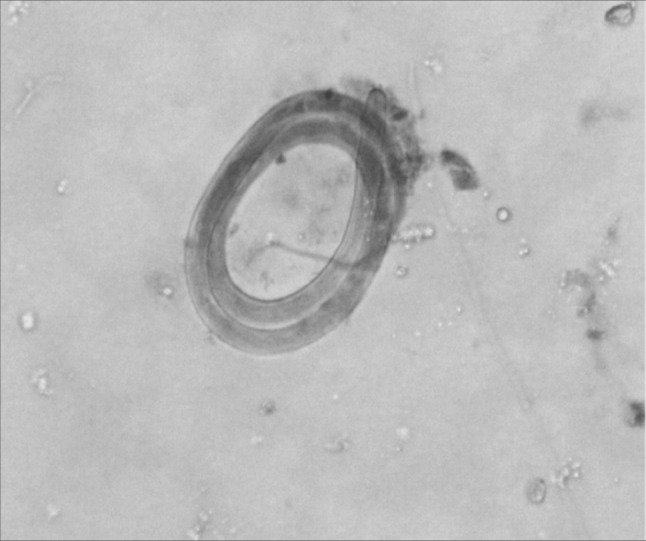
Here we report subcutaneous dirofilariosis in non-descript dogs. D. repens was first described by Railliet and Henry (1911) in which nematodes was found in the subcutaneous connective tissue of dogs in Italy. In this study, the nematods were found within a fibrous capsule in the subcutaneous tissue as well as body cavity. Grossly, the nodular masses were smooth and whitish-gray in colour measuring on average about 1 cm. When incised there were presence of nematods. Microfilarial larvae present inside the body of the nematode (Fig. 1). The microfilaria measures (mean ± SD) (374.30 ± 22.1)μm in length and (7.74 ± 0.57) μm in width. The cephalic part of the parasite was blunt and has a tapering end. It is similar to those described by Ananda et al. (2006). The mean length and width of microfilaria in this study accord well with those observed by Magnis et al. (2013) (369.44 ± 10.76 μm × 8.87 ± 0.58 μm) and (366.2 ± 12.1 μm × 6.40 ± 0.3 μm) by Cringoli et al. (2001).
Fig. 1.

Microfilaria: Microfilarial larvae are without sheath with obtuse cephalic end, sharp and filiform tail
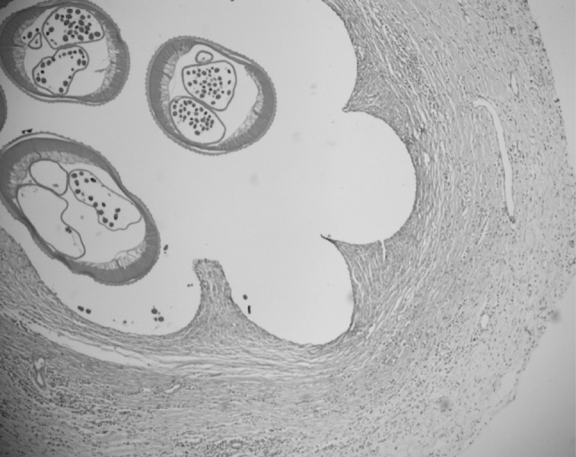
Histopathological examination of the surgically removed nematods remains the gold standard for verification of the diagnosis (Ermakovaa et al. 2014). We described the histopathology of the nodular mass found in the tunica vaginalis. The nodules showed the transverse section of 3 gravid filarial and surrounding soft tissues showed severe granulomatous inflammatory changes with no signs of malignancy. It includes infiltration of the areas around the parasite with inflammatory cells, eosinophils and mononuclear cells. Similar changes have been observed by Kamalu (1991). The microfilariae and the host immune response produce the characteristic strong fibrotic granulomatous response resulting in a well-organized nodule. The parasite and its microfilariae appear to produce toxic and immunologic actions which are responsible for many pathological lesions (Kamalu 1991).
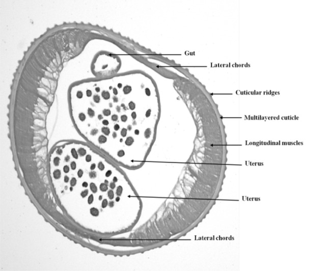
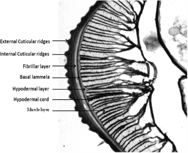
The morphology of the parasite showed well-developed thick multilayered cuticular ridges in the outermost layer (Fig. 2), followed by transverse smooth muscles striations (Fig. 3). The cuticle protruded into each lateral chord which extended into the body cavity and were divided into sublaterals. The cuticle of the nematodes is multilayered and is regarded as an extra-cellular structure (Jenkins 1969). There were presence of gravid uterus and intestine in the body cavity (Fig. 4). The worm was morphologically identified as D. repens. It is differentiated from D. Immitis as morphologically the later has smooth cuticles and lack significant longitudinal ridges (Gutierrez 2000).
Fig. 2.

Histologic section of the nodule: The worm was surrounded by fibrous connective tissue which was hyperemic and focally infiltrated by inflammatory cells including lymphocytes, eosinophils and few plasma cells. H&E, ×10
Fig. 3.

Histologic section of a female midbody: The morphology of the parasite showed well-developed thick multilayered cuticular ridges in the outermost layer, followed by transverse smooth muscles striations. The cuticle protruded into each lateral chord which extended into the body cavity and were divided into sublaterals. Two gravid uterus and intestine (gut) are present in the body cavity. H&E, ×40
Fig. 4.

Histologic section of body wall: Body wall of the parasite showing different layers starting from external cuticular ridges to muscle layers. Gomori’s Trichrome, ×60
The prevalence of dirofilariasis in domestic dogs varies by state and geographical region. Dogs living in rural areas or with access to outdoor environments are usually more affected since the risk of mosquitoes bite is higher. In general, the infection is prevalent in dogs in which prophylaxis is not routinely recommended. Reports of this infection from India are however limited (Nadgir et al. 2001). D. immitis geographically restricted to India’s north-east and D. repens to India’s south, with an overlapping area centrally (Megat Abd Rani et al. 2010b). Kerala is considered an endemic for dirofilariasis and prevalence of infection in domestic dogs has been estimated to range from 7 to 24 percent, it would definitely be much more in stray dogs (Sabu et al. 2005). Few cases were reported from the Northern (Gautam et al. 2002), Eastern (Singh et al. 2010) or Western parts (Badhe and Sane 1989) of India.
Subcutaneous dirofilariasis appears non-symptomatic in a large number of animals, defined as healthy carriers (Tarello 2002). However they cause fibrous adhesions between the scrotal sac and testicles hence during castration there are enhanced chances of subcutaneous bleeding and hematomas, sometimes it becomes difficult to retract the testicles out of the scrotum due to the adhesions. The diagnosis of this infection will continue to be the recognition and accurate identification of the parasites in excisioned or biopsy histologic sections. The morphology of the worm provides a much greater degree of accuracy (sensitivity) for diagnosis than any other serological and molecular technique.
Acknowledgments
Author acknowledged Dr. A. K. Dinda, Department of Pathology, AIIMS for providing the facility for histopathology.
References
- Albanese F, Abramo F, Braglia C, Caporali C, Venco L, Vercelli A, Ghibaudo G, Leone F, Carrani F, Giannelli A, Otranto D. Nodular lesions due to infestation by Dirofilaria repens in dogs from Italy. Vet Dermatol. 2013;24:255–256. doi: 10.1111/vde.12009. [DOI] [PubMed] [Google Scholar]
- Ananda KJ, D’Souza PE, Jagannath MS. Methods for identification of microfilaria of Dirofilaria repens and Dipetalonema reconditum. J Vet Parasitol. 2006;20:45–47. [Google Scholar]
- Aranda C, Panyella O, Eritja R, Castella J. Canine filariasis: importance and transmission in the Baix Llobregat area, Barcelona (Spain) Vet Parasitol. 1998;77:267–275. doi: 10.1016/S0304-4017(98)00109-5. [DOI] [PubMed] [Google Scholar]
- Badhe BP, Sane SY. Human pulmonary dirofilariasis in India: a case report. J Trop Med Hyg. 1989;92:425–426. [PubMed] [Google Scholar]
- Bredal WP, Gjerde B, Eberhard ML, Aleksandersen M, Wilhelmsen DK, Mansfield LS. Adult Dirofilaria repens in a sub-cutaneous granuloma on the chest of a dog. J Sm Anim Pract. 1998;39:595–597. doi: 10.1111/j.1748-5827.1998.tb03715.x. [DOI] [PubMed] [Google Scholar]
- Cringoli G, Rinaldi L, Veneziano V, Capelli G. A prevalence survey and risk analysis of filariosis in dogs from the Mt. Vesuvius area of southern Italy. Vet Parasitol. 2001;102:243–252. doi: 10.1016/S0304-4017(01)00529-5. [DOI] [PubMed] [Google Scholar]
- Ermakovaa LA, Nagornya SA, Krivorotovaa EY, Pshenichnayab NY. Dirofilaria repens in the Russian Federation: current epidemiology, diagnosis, and treatment from a federal reference center perspective. Int J Infect Dis. 2014;23:47–52. doi: 10.1016/j.ijid.2014.02.008. [DOI] [PubMed] [Google Scholar]
- Gautam V, Rustagi IM, Singh S, Arora DR. Subconjunctival infection with Dirofilaria repens. Jpn J Infect Dis. 2002;55:47–48. [PubMed] [Google Scholar]
- Gutierrez Y. Diagnostic pathology of parasitic infections with clinical correlations. 2. New York: Oxford University Press; 2000. pp. 484–485. [Google Scholar]
- Harrus S, Harmelin A, Rodrig S, Favia G. Dirofilaria repens infection in a dog in Israel. Am J Trop Med Hyg. 1999;61:639–641. doi: 10.4269/ajtmh.1999.61.639. [DOI] [PubMed] [Google Scholar]
- Jenkins T. Electron microscope observation of the body wall of T. suis Schrank 1788. (Nematoda: Trichuroidea): the cuticle and bacillary band. Z parasitenk. 1969;32:374–388. doi: 10.1007/BF00259650. [DOI] [PubMed] [Google Scholar]
- Kamalu BP. Canine filariasis in Southeastern Nigeria Bull. Anim Hlt Prod Afr. 1986;34:203–205. [Google Scholar]
- Kamalu BP. Canine filariasis caused by Dirofilaria repens in southeastern Nigeria. Vet Parasitol. 1991;40:335–338. doi: 10.1016/0304-4017(91)90113-A. [DOI] [PubMed] [Google Scholar]
- Magnis J, Lorentz S, Guardone L, Grimm F, Magi M, Naucke TJ, Deplaze P. Morphometric analyses of canine blood microfilariae isolated by the Knott’s test enables Dirofilaria immitis and D. repens species-specific and Acanthocheilonema(syn. Dipetalonema) genus-specific diagnosis. Parasit Vectors. 2013;6:48. doi: 10.1186/1756-3305-6-48. [DOI] [PMC free article] [PubMed] [Google Scholar]
- Megat Abd Rani PA, Irwin PJ, Gatne M, Coleman GT, McInnes LM, Traub RJ. A survey of canine filarial diseases of veterinary and public health significance in India. Parasit Vectors. 2010;3:30. doi: 10.1186/1756-3305-3-30. [DOI] [PMC free article] [PubMed] [Google Scholar]
- Megat Abd Rani PA, Irwin PJ, Gatne M, Coleman GT, Traub RJ. Canine vector-borne diseases in India: a review of the literature and identification of existing knowledge gaps. Parasit Vectors. 2010;3:28. doi: 10.1186/1756-3305-3-28. [DOI] [PMC free article] [PubMed] [Google Scholar]
- Nadgir S, Tallur SS, Mangoli V, Halesh LH, Krishna BV. Subconjunctival dirofilariasis in India. Southeast Asian J Trop Med Public Health. 2001;32:244–246. [PubMed] [Google Scholar]
- Pampiglione S, Rivasi F. Human dirofilariasis due to Dirofilaria (Nochtiella) repens: an update of world literature from 1995 to 2000. Parassitologia. 2000;42:231–254. [PubMed] [Google Scholar]
- Railliet A, Henry A (1911) Researches sur les ascarides des carnivores. Compted Rendus des Séances de Société Biologique de Paris 70:12–16
- Rocconi F, Tommaso MD, Traversa D, Palmieri C, Pampurini F, Boari A. Allergic dermatitis by Dirofilaria repens in a dog: clinical picture and treatment. Parasitol Res. 2012;111:493–496. doi: 10.1007/s00436-012-2833-x. [DOI] [PubMed] [Google Scholar]
- Sabu L, Devada K, Subramanian H. Dirofilariosis in dogs and humans in Kerala. Indian J Med Res. 2005;121:691–693. [PubMed] [Google Scholar]
- Singh R, Shwetha JV, Samantaray JC, Bando G. Dirofilariasis: a rare case report. Indian J Med Microbiol. 2010;28:75–77. doi: 10.4103/0255-0857.58739. [DOI] [PubMed] [Google Scholar]
- Tarello W. Cutaneous lesions in dogs with Dirofilaria (Nochtiella) repens infestation and concurrent tick-borne transmitted diseases. Vet Dermatol. 2002;13:267–274. doi: 10.1046/j.1365-3164.2002.00305.x. [DOI] [PubMed] [Google Scholar]
- Tarello W. Clinical aspects of dermatitis associated with Dirofilaria repens in pets: a review of 100 canine and 31 feline cases (1990–2010) and a report of a new clinic case imported from Italy to Dubai. J Parasitol Res. 2011 doi: 10.1155/2011/578385. [DOI] [PMC free article] [PubMed] [Google Scholar]
- Vakalis NC, Himonas CA. Human and canine dirofilariasis in Greece. Parassitologia. 1997;39:389–391. [PubMed] [Google Scholar]


