Abstract
Genetic and cytogenetic studies constitute a significant basis for understanding the biology of insect pests and the design and the construction of genetic tools for biological control strategies. Anastrepha fraterculus is an important pest of the Tephritidae family. It is distributed from southern Texas through eastern Mexico, Central America and South America causing significant crop damage and economic losses. Currently it is considered as a species complex; until now seven members have been described based on multidisciplinary approaches. Here we report the cytogenetic analysis of an Argentinian population characterized as Af. sp.1 member of the Anastrepha fraterculus species complex. The mitotic karyotype and the first detailed photographic maps of the salivary gland polytene chromosomes are presented. The mitotic metaphase complement consists of six (6) pairs of chromosomes, including one pair of heteromorphic sex chromosomes, with the male being the heterogametic sex. The analysis of the salivary gland polytene complement shows a total number of five long chromosomes that correspond to the five autosomes of the mitotic karyotype and a heterochromatic network corresponding to the sex chromosomes. Comparison of the polytene chromosome maps between this species and Anastrepha ludens shows significant similarity. The polytene maps presented here are suitable for cytogenetic studies that could shed light on the species limits within this species complex and support the development of genetic tools for sterile insect technique (SIT) applications.
Introduction
Cytogenetic analysis of Diptera species has been greatly facilitated by the existence of polytene chromosomes. Since the first publication of the chromosome maps of Drosophila [1], polytene chromosomes have proven to be an excellent genetic tool for studying chromosome structure and function, gene activity, phylogenetic relationships and have served as diagnostic tools for distinguishing members of species complexes [2–8]. Moreover, they provide means for detailed cytogenetic maps through the precise mapping by in situ hybridization [9].
For insect pest species belonging to the family of Tephritidae, advances in the field of cytogenetics contributed in understanding variation, evolution and incipient speciation phenomena as well as developing and improving pest control methods. The polytene chromosome maps of the Mediterranean fruit fly Ceratitis capitata [10,11] helped to improve the Sterile Insect Technique (SIT) by supporting the development of Genetic Sexing Strains (GSS), reviewed in [12–14]. Therefore it is considered as a tephritid model species for the development of GSS through classical genetic approaches and SIT aplications. Similarly, cytogenetic analysis of mitotic and polytene chromosome maps have helped the analysis of GSSs in other tephritid species, such as Bactrocera dorsalis, B. cucurbitae [15] and Anastrepha ludens [16].
The Tephritidae family includes five genera (Anastrepha, Bactrocera, Ceratitis, Dacus and Rhagoletis) of frugivorous species that oviposit eggs in fruits and the developing larvae feed on the mesocarp. About 100 of the tephritid species are of major economic importance. The Anastrepha genus is endemic to tropical and subtropical regions of American Continent. Currently, approximately 200 species have been identified, distributed in 17 intrageneric groups. The A. fraterculus group includes 29 species and most of them occur in Brazil [17–19].
The A. fraterculus species complex attacks more than 80 plant species, including major fruit crops [20]. It has been reported from southern Texas to Mexico, Central and South America [17,21,22]. Early studies showed differences among populations regarding morphology/morphometry [21], host preference [23,24], isozyme profiles [25] and mitotic karyotypes [26,27]. These early studies led to the assumption that the nominal A. fraterculus is a species complex. Recent studies have clearly shown that the resolution of species complexes must be based on a multidisciplinary approach, utilizing different and independent lines of evidence [28–30]. In this respect, a variety of tools have been used to shed light to the species limits among the entities of the A. fraterculus complex. These include studies on morphometrics [31–35], pre- and post-zygotic isolation [36–43] metaphase karyotypes [34,44,45], egg morphology and embryonic development [46–49], DNA markers [50–52] and pheromone profiles [53–56]. Some of the more recent studies have tried to incorporate multidisciplinary approaches for the same samples [32,57,58]. All these studies support the earlier observations about this species complex and provide insight regarding the relationships and limits among its taxa. Until now seven (7) distinct entities (Af. sp.1-7) have been identified and their geographic distribution has been described [33,37,41].
Regarding cytogenetics, different studies attribute specific mitotic karyotypes to the different entities of this complex, based on differences restricted to sex chromosomes [32,45,57,59]. In respect to the polytene chromosomes, previous efforts have presented photographs of polytene elements [60] which, however, have not provided a complete and workable polytene chromosome map. Polytene chromosomes were also used, combined with other approaches, for the analysis of two A. fraterculus populations as well as their hybrids [57]. This study revealed differences in mitotic karyotype and a high level of asynapses of polytene chromosomes in their hybrids. The cytogenetic work previously performed for this complex has been recently reviewed [61,62].
Here we present the metaphase karyotype and the first detailed photographic polytene chromosome maps from salivary glands of the Argentinian A. fraterculus Af. sp.1 member of the complex. These maps can be used as reference material for future phylogenetic studies on the A. fraterculus complex and other Anastrepha species. They can also support the construction and characterization of GSS for SIT purposes and facilitate genome mapping of the species, if coupled with in situ hybridization experiments.
Material and Methods
Anastrepha fraterculus strain
A laboratory colony of Af. sp.1 maintained at the Joint FAO/IAEA Insect Pest Control Laboratory (IPCL) was used in this study. This strain was derived from pupae sent from the Estacion Experimental AgroIndustrial Obispo Colombres, Tucuman, Argentina. The history of the strain is described in [42]. The colony is kept in standard adult (1 yeast: 3 sugar) and larval carrot diet (7% brewer’s yeast, 0.25% sodium benzoate, 0.2% methylparaben, 0/8% (v/w) HCl, 15% carrot powder, all dissolved in water).
Mitotic chromosome preparations
Spread chromosome preparations were made from brain ganglia of third–instar larvae using the method reported for C. capitata [11,63]. Brain tissue was dissected in Ringer’s solution and transferred to hypotonic solution (1% sodium citrate) on a depression slide for 10–15 min and then fixed for 3 min in freshly prepared fixative (3:1 methanol–acetic acid). During this step the fixative was changed at least two times to ensure the complete removal of the water. By the end of the fixation, the fixative was removed and a small drop of 60% acetic acid was added. Working quickly, the tissue was dispersed by drawing up into a micropipette for several times. The cell suspension was finally laid on a clean slide on a warm hotplate (40°–45°C) for drying. Chromosomes were stained with 5% Giemsa in 10mM phosphate buffer, pH 6.8. More than 15 slides prepared from about 30 larvae were analyzed in phase contrast microscope (LEIKA DMR) using 100X objective and the well spread metaphases were photographed using a digital camera (ProgResCFcool JENOPTIC/JENA).
Polytene chromosome preparations
Polytene chromosome preparations were made from well fed third-instar larvae or 1–2 days old pupae [11,63,65]. Larvae were dissected quickly in 45% acetic acid and salivary glands were carefully transferred to 3N HCl on a depression slide for 1 min. Glands were fixed in glacial acetic acid: water: lactic acid (3:2:1) for about 5 min before staining in lacto- acetic- orcein for 5–7 min. Early pupae were dissected in Ringer’s solution and the glands were transferred to 45% acetic acid for 2–3 min and then fixed in 1N HCl for 2 min. The material was passed through lacto acetic acid (80% lactic acid:60% acetic acid, 1:1) and stain in lacto acetic orcein for 10–20 min. Excess stain was removed by washing the glands in lacto-acetic acid before squashing. Chromosome slides were analyzed at 60X and 100X objectives in a phase contrast microscope (LEIKA DMR). Well spread nuclei or isolated chromosomes were photographed using a digital camera (see above). A significant number of chromosome slides were prepared from 500 larvae or pupae and the best of them with well spread nuclei (at least 200 slides) were used for analysis.
Construction of photographic polytene maps
Photographs showing well spread nuclei and/or isolated chromosomes of sufficient banding pattern quality, were selected and used. The first step was to select chromosomal regions belonging to each chromosome that: a) provided a clear banding pattern and, b) could unambiguously demonstrate the continuity of each polytene element. Afterwards, selected chromosomal regions were assembled using the Adobe Photoshop CS6 Extended Software, to construct the composite photographic map for each chromosomal element.
Results
Mitotic chromosomes
The analyzed Argentinian strain of A. fraterculus has six pairs of chromosomes including five pairs of autosomes and one pair of sex chromosomes, with the male being the heterogametic sex (XY). Fig 1 shows chromosome spreads derived from both male (1C, E) and female (1A, B, D) larvae. All the chromosomal elements are acrocentric with the exception of the Y chromosome which is probably submetacentric [45]. Two of the autosomes are longer and are easily distinguished from the rest, which are more or less of similar size. Both sex chromosomes are highly heterochromatic as shown following Giemsa (Fig 1A, 1B and 1C) and C-banding staining (Fig 1D and 1E), in accordance with previous studies based also on Giemsa staining and C banding [57,66]. From (Fig 1A, 1C, 1D and 1E) it is clear that autosomes present two chromatids, while sex chromosomes do not show two chromatids. This is probably related to the late replication of sex chromosomes, which in turn is supportive of their heterochromatic nature (Bedo 1987). The labelling system is based on that proposed by Radu and colleagues [67] for C. capitata, the first analyzed species of the Tephritidae family. The sex chromosomes are labeled as the first pair of the mitotic karyotype and the autosomes from 2–6 in order of descending size. This karyotype is in full agreement with that of the A. sp. 1 member of the complex [45,59].
Fig 1. Mitotic karyotype of A. fraterculus.
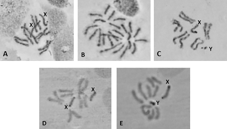
(A, B, D) female; (C, E) male. (A, B, C) Giemsa staining; (D, E) C-banding. The sex chromosomes, X and Y, are shown. The acrocentric nature of the chromosomes is evident in (B).
Polytene chromosomes
The polytene chromosomes of A. fraterculus are not an easy material to work with, due to a variety of reasons: a) polytene elements are long due to their acrocentric nature, b) the lack of a typical chromocenter complicates the location of the centromere for each element, c) the frequent chromosome fragmentation makes the analysis difficult and d) most of the chromosomal regions have a poor banding pattern and this combined with their tight coiling and twisting further compromises the identification of each element. However, these difficulties were overcome using and combining a large number of selected photographs to achieve the results presented here. The analysis showed that A. fraterculus polytene complement consists of a total of five long elements that correspond to the five autosomes, in agreement to the acrocentric nature of the mitotic complement. Sex chromosomes do not form polytene elements because of their heterochromatic nature. Their presence in polytene nuclei is evident by a heterochromatic network (Fig 2A and 2B).
Fig 2. Heterochromatic network (hn) representing the under-replicated sex chromosomes.
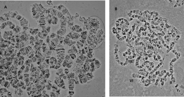
The heterochromatic network (hn) is indicated. Selected telomeres and centromeres are marked in the two nuclei.
In species lacking a chromocenter, several criteria have been used for localizing the centromeres and subsequently the free end (telomere) [10,11,68,69]. Centromeric positions usually appeared as weak points or constrictions, as well as regions (bands) with heterochromatic nature. In the case of the A. fraterculus, we observed characteristic structures that most likely represent centromeric regions, such as heterochromatic threads which are connected to some chromosome ends (Fig 3A). Moreover, there are cases where more than one chromosomes are connected to these heterochromatic structures giving the impression of a partial chromocenter (Fig 3B and 3C). An additional characteristic of the polytene chromosomes of A. fraterculus is the ectopic pairing between chromosomes ends that, interestingly, are never connected to the previous heterochromatic threads suggesting that they represent the telomeres of the chromosomes (Fig 4A–4C). Such phenomena were also observed in the analysis of A. ludens [68].
Fig 3. Centromeric regions of A. fraterculus polytene chromosomes.
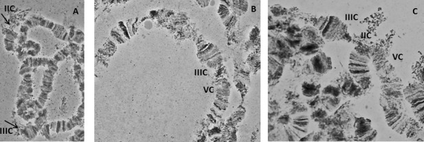
(A) Centromeres of chromosomes II and III, (B) a partial chromocenter involving chromosomes III and V, (C) a partial chromocenter involving three chromosomes, II, III and V. Arrows indicate the heterochromatic threads in (A). C indicates the centromere.
Fig 4. Ectopic pairing between telomeres of A. fraterculus polytene chromosomes.
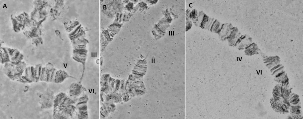
(A) a three-way pairing between telomeres of chromosomes III, V and VI, (B) II and III chromosomes, (C) IV and VI chromosomes.
The A. fraterculus polytene chromosome reference maps are shown in Figs 5–9. Chromosomes are labelled from II to VI according to their size, following the numbering system used for the first analyzed Anastrepha species, A. ludens. It is necessary to emphasize that this labeling does not imply any correlation to the mitotic karyotype. Sex chromosomes, which are not polytenized, are not represented in the polytene complement. The whole polytene complement was subdivided into 100 sections taking into account the most prominent or distinctive bands as section boundaries. The most prominent diagnostic landmarks for each element are given below.
Fig 5. Photographic map of the A. fraterculus (A.sp.1) salivary gland polytene chromosome II (sections 1–22).
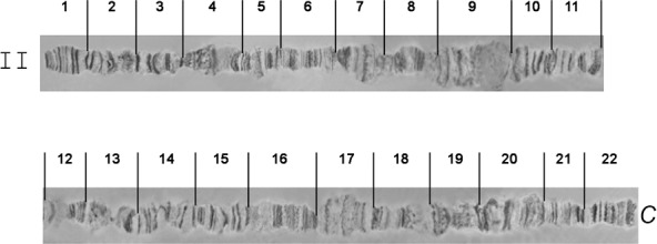
C indicates the centromere.
Fig 9. Photographic map of the A. fraterculus (A.sp.1) salivary gland polytene chromosome VI (sections 85–100).

C indicates the centromere.
Chromosome II, sections 1–22 (Fig 5)
Chromosome II is slightly longer than chromosome III and is easily identified because of the two characteristic ends, the telomere in section 1 and the proximal to the centromere region in section 22. The teleomere usually participates in ectopic pairing with other telomeres (Fig 4B). The centromere very often carries heterochromatic threads or participates in the formation of a partial chromocenter (Fig 3B and 3C). In addition, most of the regions have a clear banding pattern that helps the identification of this chromosome. Prominent landmarks of this chromosome are the characteristic constriction between sections 1 and 2, the puffs in sections 4, 7 and 17 and a series of dark bands in sections 9–11 and 13–15. These regions together with sections 1 and 22 are the most characteristic landmarks that are easily identified in well-spread nuclei (Figs 10 and 11).
Fig 10. A polytene nucleus of A. fraterculus. Characteristic landmarks of different polytene chromosome arms are shown.
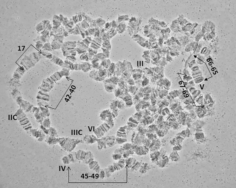
Sections 17, 40–42, 45–49 and 65–69 are indicated. Four of the five telomeres (III, IV, V, VI) and two of the five centromeres (IIC, IIIC) are also noted.
Fig 11. A polytene nucleus of A. fraterculus. Characteristic landmarks of different polytene chromosome arms are shown.
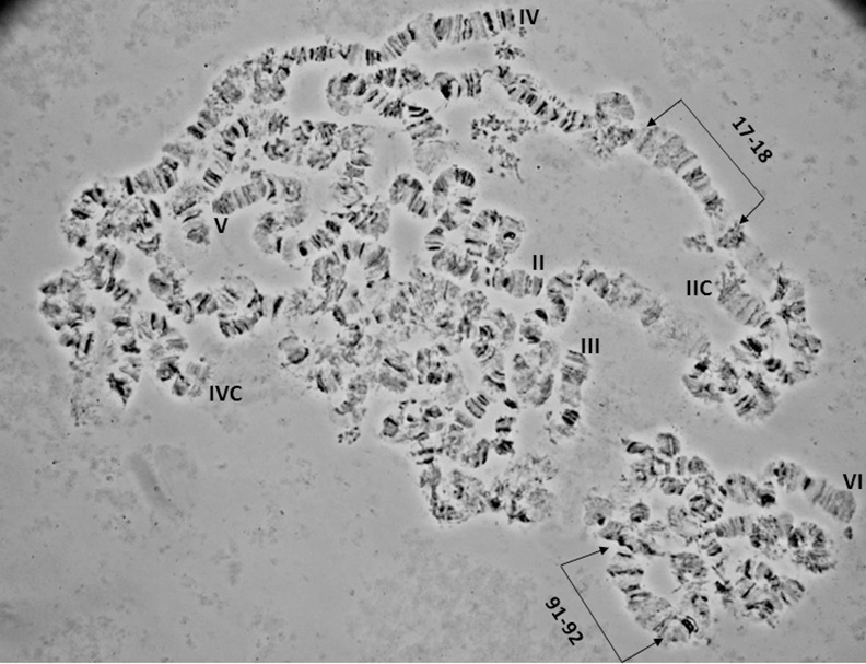
Sections 17–18 and 91–92 are indicated. The five telomeres (II, III, IV, V, VI) and two of the five centromeres (IIC, IVC) are also noted.
Chromosome III, sections 23–44 (Fig 6)
Fig 6. Photographic map of the A. fraterculus (A.sp.1) salivary gland polytene chromosome III (sections 23–44).
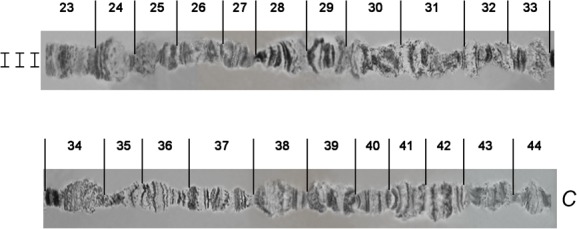
C indicates the centromere.
Chromosome III presents a poor banding pattern and numerous weak points along most of its length, especially for sections 25–34, making it thus difficult to work with. The telomere in section 23, which is very often involved in ectopic pairing, and section 24, are easily identifiable markers for this chromosome. Sections 35–44 have a better banding pattern and can serve as important landmarks for this chromosome. The end of the region in section 44 usually carries a specific heterochromatic mass that represents the centromeric region of this chromosome (Figs 10 and 12).
Fig 12. A polytene nucleus of A. fraterculus. Characteristic landmarks of different polytene chromosome arms are shown.
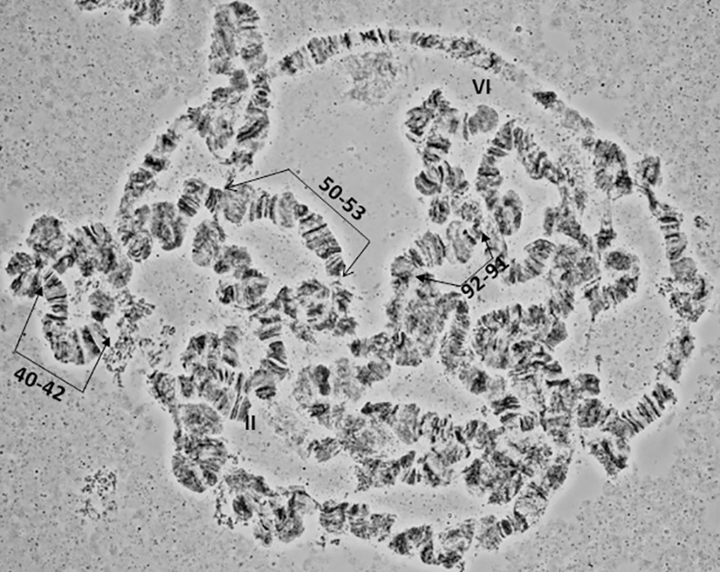
Sections 40–42, 50–53 and 91–92 are indicated. II and VI telomeres are indicated.
Chromosome IV, sections 45–64 (Fig 7)
Fig 7. Photographic map of the A. fraterculus (A.sp.1) salivary gland polytene chromosome IV (sections 45–64).
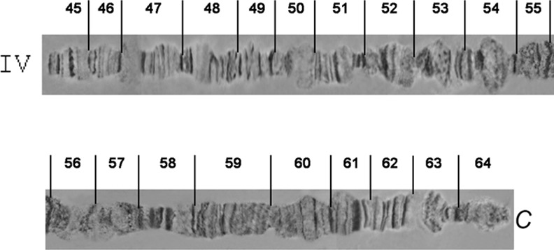
C indicates the centromere.
Chromosome IV is the most distinctive polytene element of the species with a unique banding pattern starting from the tip in section 45 to section 55 which is easily identified. The most characteristic area is the one included in sections 50–51, with two puffs and a series of bands between them. The telomere is very often taking part in ectopic pairing with other telomeres of the complement (Figs 10 and 12). The centromeric region, section 64, is very difficult to identify and can rarely be observed in spread nuclei. Similarly, difficulties exist in identifying sections 56–64.
Chromosome V, sections 65–84 (Fig 8)
Fig 8. Photographic map of the A. fraterculus (A.sp.1) salivary gland polytene chromosome V (sections 65–84).
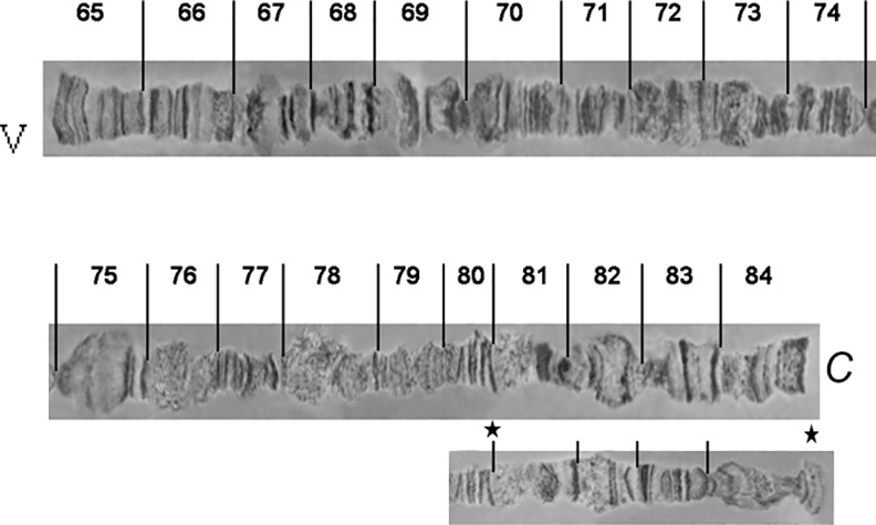
Asterisks indicate an alternative appearance of chromosomal region 81–84, due to differences in puffing pattern. C indicates the centromere.
The telomere of chromosome V, at section 65, has a unique banding pattern and it is very easily identified. Like the other telomeres of the species, it participates in ectopic pairing with other telomeres (Fig 4A). The region close to centromere, section 84, has also a characteristic banding pattern with some diffuse bands and weak points. This end is usually connected with other centromeric regions or participates in a partial chromocenter (Fig 3B). Characteristic landmarks of this chromosome are sections 66–69, section 75 where a characteristic puff is followed by three bands and the two puffs in sections 82 and 83 (Fig 10). In some of the preparations, region 81–84 presented a different banding pattern, probably to differential puffing. Although such variations are often and usually not presented, the fact that the specific one was near the centromere, which is characteristic for the chromosome, made us present this alternative configuration (Fig 8).
Chromosome VI, sections 85–100 (Fig 9)
Chromosome VI is the smallest chromosome of the complement and the most difficult to work with. It has a poor banding pattern, along with many constrictions and weak points where it is frequently broken. However, there are regions that can be used as diagnostic landmarks for this element. The telomere is localized at the beginning of section 85, based on the characteristic ectopic pairing with other tips observed in several nuclei (Fig 4A and 4C). Additional diagnostic regions of this chromosome are the puffs in sections 86 and 89 and two characteristic ones in sections 91–92 (Figs 11 and 12). It is worth saying that these two last puffs have maintained their structure in all tephritids analyzed so far.
Comparison of polytene chromosome maps between A. fraterculus and A. ludens
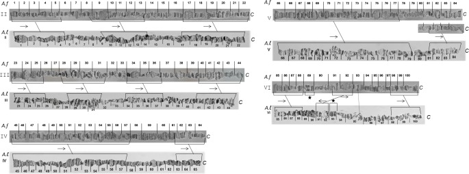
Having constructed the polytene chromosome maps of A. fraterculus we attempted their comparison with the available maps of A. ludens [68]. Both species have acrocentric chromosomes and the comparison of banding patterns of polytene elements between them revealed several similarities. The telomeres, as well as the centromeres, are either identical or similar between the two species. In both species the telomeres are participating in ectopic pairing between the chromosomes making their identification easy. Moreover, the centromeres are usually connected with heterochromatic threads and very often participate in a partial chromocenter.
The similarity of banding patterns between the two species is remarkable, especially for certain chromosomal regions distributed to all chromosomes, facilitating therefore the establishment of their homologies (Fig 13). Although this comparison is a preliminary one and also difficult due to the poor banding patterns of several chromosomal regions, differences have been observed in the VI polytene chromosome, including a transposition (A. fraterculus Af. sp.1 section 89, A.ludens section 93) and an inversion (sections 91–92) (Fig 13). It is interesting that this inversion covers a chromosomal region harboring two characteristic puffs. This chromosomal region (91–92) is found in all tephritids analyzed so far and is polymorphic regarding its position and/or direction within this chromosomal element.
Fig 13. Comparison between A. fraterculus (A. f) and A. ludens (A. l) polytene chromosome maps.
Lines connecting the chromosomes indicate sections with similar banding pattern and horizontal arrows show the relative orientation between them. C indicates the centromere. Asterisks indicate the transposition of a specific region between the two species.
Discussion
The majority of Tephritidae species analyzed so far exhibit a diploid chromosome number of 2n = 12, including a XX/XY sex chromosome pair. This is the case also for the A. fraterculus strain analyzed here (Fig 1A–1E). Sex chromosomes are easily identified based on Giemsa staining and C-banding and on the different degree of chromatid separation at metaphases in comparison to the autosomes. These characteristics support the heterochromatic nature of the sex chromosomes, a phenomenon that is common in the different genera of tephritids analyzed so far, namely Anastrepha, Bactrocera,Ceratitis, Dacus and Rhagoletis [10,65,69–76]. The heterochromatic nature of both sex chromosomes in tephritids is also evident by the abundance of highly repetitive DNA [77–79] and the limited number of genes, including the ribosomal DNA genes mapped on both sex chromosomes. This pattern of localization of the ribosomal genes is common to all tephritids analysed, such as C. capitata [80], B. oleae [81], C. rosa [77], R. pomonella [82] as well as in A. fraterculus [45,59]. Additional genes mapped on sex chromosomes include the maleness factor on Y chromosome [78] and ceratotoxins that were mapped on the X chromosome of C. capitata by in situ hybridization [83].
The karyotype presented here is in full agreement with previous studies on the Argentinian population of A. fraterculus characterized as Af. sp. 1 member of the complex [32,34,45,57,84]. The karyotypes of the seven entities (Af. sp. 1–7) identified in A. fraterculus complex, even though they present the same total number of chromosomes, they can be differentiated mainly by the size and banding pattern of the sex chromosomes [32,45]. Such differences have been reported to differentiate members of other Tephritid species complexes, such as Bactrocera tau [85] and B. dorsalis [86–89]. The size of sex chromosomes among the Tephritid species is variable [63]. This could be the result of the accumulation or loss of heterochromatin in these chromosomes. Such phenomena have been also reported in several Drosophila species, including the Hawaiian Drosophila, where species exhibit accumulation of heterochromatin on the dot chromosome (microchromosome), thus altering it to rod-shaped [90].
All the members of the A. fraterculus intrageneric group analyzed so far are characterized by the rod (acrocentric) chromosomes of their mitotic karyotype [32,34,45,57,68]. However, outside this group, there are Anastrepha species presenting: i) total chromosome number 2n = 12 with submetacentic or a combination of submetacentric and rod ones and ii) different number of total chromosomes, such as A. pickeli with 2n = 8 (XX/XY), A. leptozona with 2n = 10 (XX/XY) or different number of sex chromosomes, such as A. bistrigata and A. serpentina with a karyotype of 2n = 11 for males and 2n = 12 for females (X1X2Y/X1X2X1X2) [66,84]. Such observations are not restricted in the Anastrepha genus. Extended studies in several groups of Drosophila using comparative mitotic and polytene chromosome analysis revealed that chromosome rearrangements, such as inversions and transpositions as well as fusions/or fissions of chromosome elements have resulted in species-specific chromosomes [2]. Recently, Craddock and colleagues [91] suggested that the frequent changes on the karyotypes within the Hawaiian Drosophila species are related with the expansion of their genome size, a phenomenon that most likely has been driven by the addition of heterochromatin and satellite DNA. Such additions resulted in longer acrocentric chromosomes, changing the dot to acrocentric ones, or to metacentric by the addition of a hetrochromatic arm.
In A. fraterculus polytene nuclei, five long banded polytene chromosomes that represent the five acrocentric autosomes of the metaphase karyotype were found. This is in full agreement with the results from A. ludens [68], the phylogenetically closest species analyzed so far. In accordance with Tephritidae analyzed so far, sex chromosomes do not form polytene elements, probably due to their under-replication (reviewed in [63]). The sex chromosomes in the polytene nuclei are represented by a granular heterochromatic network (Fig 2). This correlation between sex chromosomes and the granular network in C. capitata was first suggested by Bedo [92], after analyzing polytene chromosomes of trichogen cells derived from male pupae. Later on [10] this correlation was further established through the analysis of Y–autosome translocations in medfly. More recently, Drosopoulou and her colleagues [81] proved that this network is formed by the sex chromosomes. To do so, they used FISH of sex chromosome specific probes, generated through laser microdissection of the respective mitotic sex chromosomes. Another common feature of tephritids is the absence of a typical chromocenter where all chromosomes are connected through their centromeres. This was also observed in A. fraterculus where the identification of the centromeric regions presented additional difficulties due to its acrocentric chromosomes. In some cases more than one chromosomes were connected forming a partial chromocenter (Fig 3B and 3C), a situation found also in other tephritids [68,69,76]. Telomeres show ectopic pairing (Fig 4), a phenomenon also observed in several Tephritid species [65,68,69,75,76]. This is probably related to the molecular structure and organization of the distal parts of the chromosomes in these species. In D. melanogaster, the distal parts of chromosomes consist of specific terminal repeat retrotransposons (Het-A and TART) that are arranged in tracts of variable length among several strains, resulting thus in the extension of the chromosomal ends and the frequent ectopic pairing between telomeres [5,93].
Polytene chromosomes of the two Anastrepha species studied so far, A. ludens and A. fraterculus, show significant similarities in their banding pattern. In fact, certain chromosomal regions distributed to all elements show the same banding patterns, thus allowing the establishment of chromosomal homologies between the two species (Fig 13). A previous comparative analysis of polytene elements between C. capitata and A. ludens showed that chromosome homology between them can also be established [68]. In fact, telomeres and centromeric areas, as well as specific chromosomal regions of each chromosome present the same or very similar banding patterns among the tephritids.
Polytene chromosomes have been used in many taxa to clarify either the status of species complexes or to establish phylogenetic relationships in a significant number of Diptera. The vast majority of such studies refers to Drosophila species [2,4,5,94] and mosquitos [3,95–100].
Sturtevant and Novitski [101] revealed the homology of the six chromosomal elements within Drosophila, named A-F by Muller [102]. The conservation of the basic elements between C. capitata and Drosophila [13,103] as well as between B. oleae and Drosophila [104] was shown by in situ hybridization on polytene chromosomes. Moreover, the chromosome homology between several Bactrocera species and C. capitata as well as A. ludens and C. capitata has been established based on both their polytene chromosome banding pattern similarities and/or in situ hybridization of selected probes [11,13,68–70,75,76,105–107]. These studies showed that the species are differentiated by fixed chromosomal rearrangements, mainly paracentric inversions, and are characterized by transpositions on specific chromosomes. In addition, two pericentric inversions were found to differentiate Ceratitis and Bactrocera genera, one of which differentiates Ceratitis and Dacus [105]. Recently, the genome assembly of B. tryoni confirmed the above results and showed that the Muller’s elements have maintained their essential identity in both lines of drosophilids and tephritids although a large number of intra-chromosomal rearrangements have occurred. Moreover their data support that X chromosome of Tephritid species is originated from the dot chromosome 4 (Element F) of Drosophila. These data clearly support that no new chromosomes and specifically chromosome ends have been created in these insect lineages [108]. Similar conservation of chromosome ends has been observed in mosquito Anopheles gambiae, suggesting that this is a common feature of all Diptera [109]. Mason and colleaques [110] showed that Diptera are the only group that lacks telomerase and this is a factor that contributes to their chromosome ends stability. These species protect their chromosome ends by the recruitment of retrotransposons [108].
Chromosomal rearrangements, mainly inversions, are believed to be a key player in speciation of Diptera [4,94]. The role of chromosome inversions in speciation is being discussed for decades and recent models suggest that they can promote speciation through the suppression of recombination within the inversion and near the inversion breakpoints that subsequently leads to the restriction in gene flow [111–115]. The presence of at least one fixed paracentric inversion in chromosome VI that differentiates A. fraterculus from A. ludens (Fig 13), two Anastrepha species belonging to the same intrageneric group, is in line with the aforementioned model of Diptera speciation.
Centromeres and telomeres have long been characterized as dynamic regions of chromosomal evolution. Several studies in primates indicate that the centromere position can change during short periods of evolutionary time. There are different models that try to explain the repositioning of centromeres. This can be done either through transposition of centromeric regions to new chromosomal regions or by the de novo emergence of centromeres in new regions (neocentromere emergence) [116–119]. According to the first model, this repositioning is the result of chromosomal rearrangements that could explain this change (sequential pericentric inversions, for example). All Tephritid species analyzed so far, present metacentric or submetacentric autosomes, with the exception of the Anastrephas that present acrocentric ones. Assuming that the first model of centromere repositioning applies, chromosomal changes such as transpositions or pericentric inversions should be evident and explain the transformation from acrocentric to metacentric chromosomes or vice versa. However, the comparison of the polytene chromosome banding pattern of the two Anastrepha species with acrocentric autosomes (A. fraterculus and A. ludens) to all other tephritids (with metacentric and submetacentric autosomes) does not support the presence of such extended rearrangements. The similarity in the banding pattern of chromosomal ends (meaning telomeres of the metacentric chromosomes and telomeres–centromeres of the acrocentric chromosomes) support the stability of the chromosome ends. Therefore, the de novo formation of neocentromeres in specific chromosomal regions is more compatible with our data. As discussed before, Anastrephas are variable both in chromosome number and metaphase configuration of chromosomes. The availability of so diverse chromosome configurations shows that polytene chromosome analysis of Anastrepha species with different metaphase karyotypes could shed light to the centromere evolution in Tephritidae and further elucidate their phylogenetic relationships.
Conclusions
The first polytene chromosome maps for Anastrepha fraterculus (A.sp1) presented here and their future comparison to the polytene chromosomes of other members of the complex may reveal additional structural differences among them as well as their phylogenetic relationships. The comparison with the polytene chromosome maps of A. ludens shows that these maps can be used in comparative studies with other Anastrepha species as well. Polytene chromosome analysis constitutes an important component for the development and characterization of stable GSSs of A. fraterculus towards the supporting of SIT control methods in the species. Finally, any future research on the construction of genome assemblies for A. fraterculus could benefit by in situ hybridization of unique genes or sequences on polytene chromosomes.
Acknowledgments
We gratefully acknowledge the Joint FAO/IAEA Division of Nuclear Techniques in Food and Agriculture for funding this work through CRP and SSA projects. We also thank INTA (Instituto Nacional Tecnología Agropecuaria) for allocating funds for MCG (travel and stay in Greece and Austria).
Data Availability
All data (polytene chromosome maps and comparaisons of polytene chromosome maps) are available in the paper
Funding Statement
This work was funded by 1126: Directo Council of the Natinal Institute of Agricultural Technology, Argentina (I.N.T.A.) (www.inta.gob.ar), and 16974: International Atomic Energy Agency (IAEA) (cra.iaea.org).
References
- 1.Bridges CB. Salivary chromosome maps: With a key to the banding of the chromosomes of Drosophila melanogaster. J Hered. 1935;26: 60–64. [Google Scholar]
- 2.Ashburner M, Carson HL, Thompson J. The Genetics and Biology of Drosophila. Ashburner M, Carson HL, Thompson J (eds). London: Academic press INC.; 1982. [Google Scholar]
- 3.Coluzzi M, Sabatini A, della Torre A, Di Deco MA, Petrarca V. A polytene chromosome analysis of the Anopheles gambiae species complex. Science. 2002;298: 1415–8. 10.1126/science.1077769 [DOI] [PubMed] [Google Scholar]
- 4.Krimbas CB, Powell JR. Drosophila inversion polymorphism Krimbas CB, Powell JR. Boka Raton, Florida, CRC Press, Inc; 1992. [Google Scholar]
- 5.Zhimulev IF, Belyaeva ES, Semeshin VF, Koryakov DE, Demakov SA, Demakova O V, et al. Polytene chromosomes: 70 years of genetic research. Int Rev Cytol. 2004;241: 203–75. 10.1016/S0074-7696(04)41004-3 [DOI] [PubMed] [Google Scholar]
- 6.Ayala FJ, Coluzzi M. Chromosome speciation: Humans, Drosophila and mosquitoes. Proc Natl Acad Sci U S A. 2005;102: 6535–6542. [DOI] [PMC free article] [PubMed] [Google Scholar]
- 7.Coluzzi M. Spatial distribution of chromosomal inversions and speciation in anopheline mosquitoes In: Barigozzi C (ed). Mechanisms of speciation. New York: Alan R. Liss, Inc; 1982. pp 143–153. [PubMed] [Google Scholar]
- 8.Coluzzi M, Sabatini A, Petrarca V, Di Deco MA. Chromosomal differentiation and adaptation to human environments in the Anopheles gambiae complex. Trans R Soc Trop Med Hyg. 1979;73: 483–97. [DOI] [PubMed] [Google Scholar]
- 9.Pardue ML, Gall JG. Nucleic acid hybridization to the DNA of cytological preparations. Methods in Cell Biology. 1975. bll 1–16. [DOI] [PubMed] [Google Scholar]
- 10.Bedo DG. Polytene chromosome mapping in Ceratitis capitata (Diptera: Tephritidae). Genome. 1987;29: 598–611. [Google Scholar]
- 11.Zacharopoulou A. Polytene chromosome maps in the medfly Ceratitis capitata. Genome. 1990;33: 184–197. [Google Scholar]
- 12.Franz G. Genetic sexing strains in Mediterranean fruit fly, an example for other species amenable to large-scale rearing as required for the sterile insect technique In: Dyck VA, Robinson AS, Hendrichs J (eds). Sterile Insect Technique: Principles and Practice in Area-Wide Integrated Pest Management. Dordrecht, The Netherlands: Springer; 2005. pp 427–451. [Google Scholar]
- 13.Gariou-Papalexiou A, Gourzi P, Delprat A, Kritikou D, Rapti K, Chrysanthakopoulou B, et al. Polytene chromosomes as tools in the genetic analysis of the Mediterranean fruit fly, Ceratitis capitata. Genetica. 2002;116: 59–71. [DOI] [PubMed] [Google Scholar]
- 14.Robinson AS. Development of genetic sexing strains for fruit fly sterile insect technology. Genetica. 2002;116: 1–149. [DOI] [PubMed] [Google Scholar]
- 15.Zacharopoulou A, Franz G. Genetic and cytogenetic characterization of Genetic Sexing Strains of Bactrocera dorsalis and Bactrocera cucurbitae (Diptera: Tephritidae). J Econ Entomol. 2013;106: 995–1003. [DOI] [PubMed] [Google Scholar]
- 16.Zepeda-Cisneros CS, Meza Hernández JS, García-Martínez V, Ibañez-Palacios J, Zacharopoulou A, Franz G. Development, genetic and cytogenetic analyses of genetic sexing strains of the Mexican fruit fly, Anastrepha ludens Loew (Diptera: Tephritidae). BMC Genet; 2014; Suppl 2: S1 10.1186/1471-2156-15-S2-S1 [DOI] [PMC free article] [PubMed] [Google Scholar]
- 17.Norrbom AL, Zucchi RA, H-O V. Phylogeny of the genera Anastrepha and Toxotrypana (Trypetinae: Toxotrypanini) based on morphology In: Aluja M, Norrbom A.L., (eds). Fruit flies (Tephritidae): Phylogeny and Evolution of Behavior. Boca Raton, Florida: CRC Press; 1999. pp 299–342. [Google Scholar]
- 18.White IM, Elson-Harris MM. Fruit Flies of Economic Significance: Their Identification and Bionomics. CAB International; 1992. [Google Scholar]
- 19.Zucchi RA. Lista das espécies de Anastrepha, sinonímias, plantas hospedeiras e parasitóides. In: Malavasi A, Zucchi RA (eds). Moscas-das-frutas de importância econômica no Brasil. Ribeirão Preto, Holos; 2000. pp 41–48.
- 20.Ovruski S, Schliserman P, Aluja M. Native and introduced host plants of Anastrepha fraterculus and Ceratitis capitata (Diptera: Tephritidae) in northwestern Argentina. J Econ Entomol. 2003;96: 1108–1118. [DOI] [PubMed] [Google Scholar]
- 21.Stone A. The fruit flies of the genus Anastrepha Washington, DC, USA: USDA, Misc. Publ.; 1942. [Google Scholar]
- 22.Hernández-Ortiz V, Aluja M. Lista preliminar del género neotropical Anastrepha Schiner (Diptera: Tephritidae) con notas sobre su distribución y plantas hospederas. Folia Entomológica Mex. 1993;88: 89–105. [Google Scholar]
- 23.Baker AC, Stone WE, Plummer CC, MacPhail H. A review of studies on the Mexican fruit fly and related Mexican species United States Dep. Agric. Misc. Publ; 1944. [Google Scholar]
- 24.Malavasi A, Morgante JS. Population genetics of Anastrepha fraterculus (Diptera, Tephritidae) in different hosts: Genetic differentiation and heterozygosity. Genetica. 1983;60: 207–211. [Google Scholar]
- 25.Morgante JS, Malavasi A, Bush GL. Biochemical systematics and evolutionary relationships of neotropical Anastrepha. Ann Entomol Soc Am. 1980;73: 622–630. [Google Scholar]
- 26.Bush GL. The cytotaxonomy of the larvae of some Mexican fruit flies in the genus Anastrepha. Psyche (Stuttg). 1962;68: 87–101. [Google Scholar]
- 27.Mendes LOT. Observacoes citológicas em “moscas das frutas”. Bragantia. 1958;17: 29–39. [Google Scholar]
- 28.De Queiroz K. Species concepts and species delimitation. Syst Biol. 2007;56: 879–86. [DOI] [PubMed] [Google Scholar]
- 29.Schlick-Steiner BC, Steiner FM, Seifert B, Stauffer C, Christian E, Crozier RH. Integrative taxonomy: a multisource approach to exploring biodiversity. Annu Rev Entomol. 2010;55: 421–38. 10.1146/annurev-ento-112408-085432 [DOI] [PubMed] [Google Scholar]
- 30.Schutze MK, Aketarawong N, Amornsak W, Armstrong KF, Augustinos AA, Barr N, et al. Synonymization of key pest species within the Bactrocera dorsalis species complex (Diptera: Tephritidae): taxonomic changes based on a review of 20 years of integrative morphological, molecular, cytogenetic, behavioural and chemoecological data. Syst Entomol. 2014; [Google Scholar]
- 31.Hernández-Ortiz V, Gómez-Anaya JA, Sánchez A, McPheron BA, Aluja M. Morphometric analysis of Mexican and South American populations of the Anastrepha fraterculus complex (Diptera: Tephritidae) and recognition of a distinct Mexican morphotype. Bull Entomol Res. 2007;94: 487–499. [DOI] [PubMed] [Google Scholar]
- 32.Hernández-Ortiz V, Bartolucci AF, Morales-Valles P, Frías D, Selivon D. Cryptic Species of the Anastrepha fraterculus Complex (Diptera: Tephritidae): A Multivariate Approach for the Recognition of South American Morphotypes. Ann Entomol Soc Am. 2012;105: 305–318. [Google Scholar]
- 33.Hernández-Ortiz V, Canal NA, Salas JOT, Ruíz-Hurtado FM, Dzul-Cauich JF. Taxonomy and phenotypic relationships of the Anastrepha fraterculus complex in the Mesoamerican and Pacific Neotropical dominions (Diptera, Tephritidae). Zookeys; 2015;540: 95–124. 10.3897/zookeys.540.6027 [DOI] [PMC free article] [PubMed] [Google Scholar]
- 34.Selivon D, Perondini ALP, Morgante JS. A genetic-morphological characterization of two cryptic species of the Anastrepha fraterculus complex (Diptera: Tephritidae). Ann Entomol Soc Am. 2005;98: 367–381. [Google Scholar]
- 35.Canal NA, Hernández-Ortiz V, Salas JOT, Selivon D. Morphometric study of third-instar larvae from five morphotypes of the Anastrepha fraterculus cryptic species complex (Diptera, Tephritidae). Zookeys. 2015;540: 41–59. 10.3897/zookeys.540.6012 [DOI] [PMC free article] [PubMed] [Google Scholar]
- 36.Allinghi A, Calcagno G, Petit-Marty N, Gómez Cendra P, Segura D, Vera T, et al. Compatibillity and competitiveness of a laboratory strain of Anastrepha fraterculus (Diptera: Tephritidae) after irradiation treatment. Florida Entomol. 2007;90: 27–32. [Google Scholar]
- 37.Devescovi F, Abraham S, Roriz AKP, Nolazco N, Castañeda R, Tadeo E, et al. Ongoing speciation within the Anastrepha fraterculus cryptic species complex: the case of the Andean morphotype. Entomol Exp Appl. 2014;152: 238–247. [Google Scholar]
- 38.Juárez ML, Devescovi F, Břízová R, Bachmann G, Segura DF, Kalinová B, et al. Evaluating mating compatibility within fruit fly cryptic species complexes and the potential role of sex pheromones in pre-mating isolation. Zookeys. 2015;540: 125–55. 10.3897/zookeys.540.6133 [DOI] [PMC free article] [PubMed] [Google Scholar]
- 39.Petit-Marty N, Vera MT, Calcagno G, Cladera JL, Segura DF, Allinghi A, et al. Sexual behavior and mating compatibility among four populations of Anastrepha fraterculus (Diptera: Tephritidae) from Argentina. Ann Entomol Soc Am. 2004;97: 1320–1327. [Google Scholar]
- 40.Rull J, Abraham S, Kovaleski A, Segura DF, Islam A, Wornoayporn V, et al. Random mating and reproductive compatibility among Argentinean and southern Brazilian populations of Anastrepha fraterculus (Diptera: Tephritidae). Bull Entomol Res. 2012;102: 435–43. 10.1017/S0007485312000016 [DOI] [PubMed] [Google Scholar]
- 41.Rull J, Abraham S, Kovaleski A, Segura DF, Mendoza M, Liendo MC, et al. Evolution of pre-zygotic and post-zygotic barriers to gene flow among three cryptic species within the Anastrepha fraterculus complex. Entomol Exp Appl. 2013;148: 213–222. [Google Scholar]
- 42.Vera MT, Cáceres C, Wornoayporn V, Islam A, Robinson AS, De La Vega MH, et al. Mating Incompatibility Among Populations of the South American Fruit Fly Anastrepha fraterculus (Diptera: Tephritidae). Ann Entomol Soc Am. 2006;99: 387–397. [Google Scholar]
- 43.Segura DF, Vera MT, Rull J, Wornoayporn V, Islam A, Robinson AS. Assortative mating among Anastrepha fraterculus (Diptera: Tephritidae) hybrids as a possible route to radiation of the fraterculus cryptic species complex. Biol J Linn Soc. 2011;102: 346–354. [Google Scholar]
- 44.Basso A, Sonvico A, Quesada-Allue LA, Manso F. Karyotypic and molecular identification of laboratory stocks of the South American fruit fly Anastrepha fraterculus (Wied) (Diptera: Ephritidae). J Econ Entomol. 2003;96: 1237–1244. [DOI] [PubMed] [Google Scholar]
- 45.Goday C, Selivon D, Perondini ALP, Greciano PG, Ruiz MF. Cytological characterization of sex chromosomes and ribosomal DNA location in Anastrepha species (Diptera, Tephritidae). Cytogenet Genome Res. 2006;114: 70–6. [DOI] [PubMed] [Google Scholar]
- 46.Dutra VS, Ronchi-Teles B, Steck GJ, Silva JG. Egg morphology of Anastrepha spp. (Diptera: Tephritidae) in the fraterculus group using scanning electron microscopy. Ann Entomol Soc Am. 2011;104: 16–24. [Google Scholar]
- 47.Dutra VS, Ronchi-Teles B, Steck GJ, Silva JG. Description of larvae of Anastrepha spp. (Diptera: Tephritidae) in the fraterculus group. Ann Entomol Soc Am. 2012;105: 529–538. [Google Scholar]
- 48.Selivon D, Morgante JS, Perondini ALP. Egg size, yolk mass extrusion and hatching behavior in two cryptic species of Anastrepha fraterculus (Wiedemann) (Diptera, Tephritidae). Brazilian J Genet. 1997;20: 587–594. [Google Scholar]
- 49.Selivon D, Perondini ALP. Egg shell morphology in two cryptic species of the Anastrepha fraterculus complex (Diptera: Tephritidae). Ann Entomol Soc Am. 1998;91: 473–478. [Google Scholar]
- 50.Manni M, Lima KM, Guglielmino CR, Lanzavecchia SB, Juri M, Vera T, et al. Relevant genetic differentiation among Brazilian populations of Anastrepha fraterculus (Diptera, Tephritidae). Zookeys. 2015;540: 157–73. 10.3897/zookeys.540.6713 [DOI] [PMC free article] [PubMed] [Google Scholar]
- 51.Sutton BD, Steck GJ, Norrbom AL, Rodriguez EJ, Srivastava P, Alvarado NN, et al. Nuclear ribosomal internal transcribed spacer 1 (ITS1) variation in the Anastrepha fraterculus cryptic species complex (Diptera, Tephritidae) of the Andean region. Zookeys. 2015;540: 175–91. 10.3897/zookeys.540.6147 [DOI] [PMC free article] [PubMed] [Google Scholar]
- 52.Smith-Caldas MRB, McPheron BA, Silva JG, Zucchi RA. Phylogenetic relationships among species of the fraterculus group (Anastrepha: Diptera: Tephritidae) inferred from DNA sequences of mitochondrial cytochrome oxidase I. Neotrop Entomol. 2001;30: 565–573. [Google Scholar]
- 53.Břízová R, Mendonça AL, Vanícková L, Mendonça AL, Eduardo Da Silva C, Tomčala A, et al. Pheromone analyses of the Anastrepha fraterculus (Diptera: Tephritidae) cryptic species complex. Florida Entomol. 2013;96: 1107–1115. [Google Scholar]
- 54.Vaníčková L, Svatoš A, Kroiss J, Kaltenpoth M, Do Nascimento RR, Hoskovec M, et al. Cuticular hydrocarbons of the South American fruit fly Anastrepha fraterculus: variability with sex and age. J Chem Ecol. 2012;38: 1133–42. 10.1007/s10886-012-0177-8 [DOI] [PubMed] [Google Scholar]
- 55.Vaníčková L, Břízová R, Pompeiano A, Ferreira LL, de Aquino NC, Tavares R de F, et al. Characterisation of the chemical profiles of Brazilian and Andean morphotypes belonging to the Anastrepha fraterculus complex (Diptera, Tephritidae). Zookeys. 2015;540: 193–209. 10.3897/zookeys.540.9649 [DOI] [PMC free article] [PubMed] [Google Scholar]
- 56.Vaníčková L, Břízová R, Mendonça AL, Pompeiano A, Do Nascimento RR. Intraspecific variation of cuticular hydrocarbon profiles in the Anastrepha fraterculus (Diptera: Tephritidae) species complex. J Appl Entomol. 2015;139: 679–689. [Google Scholar]
- 57.Caceres C, Segura DF, Vera MT, Wornoayporn V, Cladera JL, Teal P, et al. Incipient speciation revealed in Anastrepha fraterculus (Diptera; Tephritidae) by studies on mating compatibility, sex pheromones, hybridization, and cytology. Biol J Linn Soc. 2009;97: 152–165. [Google Scholar]
- 58.Dias VS, Silva JG, Lima KM, Petitinga CSCD, Hernández-Ortiz V, Laumann RA, et al. An integrative multidisciplinary approach to understanding cryptic divergence in Brazilian species of the Anastrepha fraterculus complex (Diptera: Tephritidae). Biol J Linn Soc. 2015; 725–746. [Google Scholar]
- 59.Giardini MC, Milla FH, Lanzavecchia S, Nieves M, Cladera JL. Sex chromosomes in mitotic and polytene tissues of Anastrepha fraterculus (Diptera, Tephritidae) from Argentina: a review. Zookeys. 2015;540: 83–94. 10.3897/zookeys.540.6058 [DOI] [PMC free article] [PubMed] [Google Scholar]
- 60.Giardini MC, Milla F, Manso FC. Structural map of the polytene chromosomes from the salivary glands of South American fruit fly Anastrepha fraterculus Wied (Diptera, Tephritidae). Caryologia. 2009;62: 204–212. [Google Scholar]
- 61.Cladera JL, Vilardi JC, Juri M, Paulin LE, Giardini MC, Gómez Cendra P V, et al. Genetics and biology of Anastrepha fraterculus: research supporting the use of the sterile insect technique (SIT) to control this pest in Argentina. BMC Genet. 2014;15 Suppl 2: S12 10.1186/1471-2156-15-S2-S12 [DOI] [PMC free article] [PubMed] [Google Scholar]
- 62.Vaníčková L, Hernández-Ortiz V, Bravo ISJ, Dias V, Roriz AKP, Laumann RA, et al. Current knowledge of the species complex Anastrepha fraterculus (Diptera, Tephritidae) in Brazil. Zookeys. 2015;540: 211–37. 10.3897/zookeys.540.9791 [DOI] [PMC free article] [PubMed] [Google Scholar]
- 63.Mavragani-Tsipidou P, Zacharopoulou A, Drosopoulou E, Augustinos AA, Bourtzis K, Marec F. Tephritid Fruit Flies Sharakhov I (ed). Protocols for cytogenetic mapping of insect genomes. CRC Press, Taylor and Francis Group, LLC; 2014. pp 1–62. [Google Scholar]
- 64.Selivon D, Perondini ALP. Evaluation of techniques for C and ASG banding of the mitotic chromosomes of Anastrepha species (Diptera, Tephritidae). Brazilian J Genet. 1997;20: 651–653. [Google Scholar]
- 65.Mavragani-Tsipidou P, Karamanlidou G, Zacharopoulou A, Koliais S, Kastritsis C. Mitotic and polytene chromosome analysis in Dacus oleae (Diptera: Tephritidae). Genome. 1992;35: 373–378. [DOI] [PubMed] [Google Scholar]
- 66.Selivon D, Perondini ALP, Rocha LS. Karyotype characterization of Anastrepha fruit flies (Diptera: Tephritidae). Neotrop Entomol. 2005;34: 273–279. [Google Scholar]
- 67.Radu M, Rossler Y, Koltin Y. The chromosomes of the Mediterranean fruit fly Ceratitis capitata (Wied): Karyotype and chromosomal organization. Cytologia. 1975;40: 823–828. [Google Scholar]
- 68.Garcia-Martinez V, Hernandez-Ortiz E, Zepeta-Cisneros CS, Robinson AS, Zacharopoulou A, Franz G. Mitotic and polytene chromosome analysis in the Mexican fruit fly, Anastrepha ludens (Loew) (Diptera: Tephritidae). Genome. 2009;52: 20–30. 10.1139/G08-099 [DOI] [PubMed] [Google Scholar]
- 69.Zhao JT, Frommer M, Sved JA, Zacharopoulou A. Mitotic and polytene chromosome analyses in the Queensland fruit fly, Bactrocera tryoni (Diptera: Tephritidae). Genome. 1998;41: 510–526. [PubMed] [Google Scholar]
- 70.Augustinos A, Drosopoulou E, Gariou-Papalexiou A, Bourtzis K, Mavragani-Tsipidou P, Zacharopoulou A. The Bactrocera dorsalis species complex: comparative cytogenetic analysis in support of Sterile Insect Technique applications. BMC Genet. 2014;15 Suppl 2: S16 10.1186/1471-2156-15-S2-S16 [DOI] [PMC free article] [PubMed] [Google Scholar]
- 71.Drosopoulou E, Augustinos AA, Nakou I, Koeppler K, Kounatidis I, Vogt H, et al. Genetic and cytogenetic analysis of the American cherry fruit fly, Rhagoletis cingulata (Diptera: Tephritidae). Genetica. 2011;139: 1449–1464. 10.1007/s10709-012-9644-y [DOI] [PubMed] [Google Scholar]
- 72.Drosopoulou E, Nestel D, Nakou I, Kounatidis I, Papadopoulos NT, Bourtzis K, et al. Cytogenetic analysis of the Ethiopian fruit fly Dacus ciliatus (Diptera: Tephritidae). Genetica. 2011;139: 723–732. 10.1007/s10709-011-9575-z [DOI] [PubMed] [Google Scholar]
- 73.Drosopoulou E, Koeppler K, Kounatidis I, Nakou I, Papadopoulos NT, Bourtzis K, et al. Genetic and cytogenetic analysis of the walnut-husk fly (Diptera: Tephritidae). Ann Entomol Soc Am. 2010;103: 1003–1011. [Google Scholar]
- 74.Kounatidis I, Papadopoulos N, Bourtzis K, Mavragani-Tsipidou P. Genetic and cytogenetic analysis of the fruit fly Rhagoletis cerasi (Diptera: Tephritidae). Genome. 2008;51: 479–491. 10.1139/G08-032 [DOI] [PubMed] [Google Scholar]
- 75.Zacharopoulou A, Augustinos AA, Sayed WAA, Robinson AS, Franz G. Mitotic and polytene chromosomes analysis of the oriental fruit fly, Bactrocera dorsalis (Hendel) (Diptera: Tephritidae). Genetica. 2011;139: 79–90. 10.1007/s10709-010-9495-3 [DOI] [PubMed] [Google Scholar]
- 76.Zacharopoulou A, Sayed WAA, Augustinos AA, Yesmin F, Robinson AS, Franz G. Analysis of mitotic and polytene chromosomes and photographic polytene chromosome maps in Bactrocera cucurbitae (Diptera: Tephritidae). Ann Entomol Soc Am. 2011;104: 306–318. [Google Scholar]
- 77.Willhoeft U, Franz G. Comparison of the mitotic karyotypes of Ceratitis capitata, Ceratitis rosa, and Trirhithrum coffeae (Diptera: Tephritidae) by C-banding and FISH. Genome. 1996;39: 884–889. [DOI] [PubMed] [Google Scholar]
- 78.Willhoeft U, Franz G. Identification of the sex-determining region of the Ceratitis capitata Y chromosome by deletion mapping. Genetics. 1996;144: 737–745. [DOI] [PMC free article] [PubMed] [Google Scholar]
- 79.Tsoumani KT, Drosopoulou E, Bourtzis K, Gariou-Papalexiou A, Mavragani-Tsipidou P, Zacharopoulou A, et al. Achilles, a new family of transcriptionally active retrotransposons from the olive fruit fly, with Y chromosome preferential distribution. PLoS One. 2015;10: e0137050 10.1371/journal.pone.0137050 [DOI] [PMC free article] [PubMed] [Google Scholar]
- 80.Bedo DG, Webb GC. Conservation of nucleolar structure in polytene tissues of Ceratitis capitata (Diptera: Tephritidae). Chromosoma. 1989;98: 443–449. [Google Scholar]
- 81.Drosopoulou E, Nakou I, Síchová J, Kubíčková S, Marec F, Mavragani-Tsipidou P. Sex chromosomes and associated rDNA form a heterochromatic network in the polytene nuclei of Bactrocera oleae (Diptera: Tephritidae). Genetica. 2012;140: 169–80. [DOI] [PubMed] [Google Scholar]
- 82.Procunier WS, Smith JJ. Localization of ribosomal DNA in Rhagoletis pomonella (Diptera: Tephritidae) by in situ hybridization. Insect Mol Biol. 1993;2: 163–174. [DOI] [PubMed] [Google Scholar]
- 83.Rosetto M, De Filippis T, Mandrioli M, Zacharopoulou A, Gourzi P, Manetti AGO, et al. Ceratotoxins: Female-specific X-linked genes from the medfly, Ceratitis capitata. Genome. 2000;43: 707–711. [PubMed] [Google Scholar]
- 84.Selivon D, Sipula FM, Rocha LS, Perondini ALP. Karyotype relationships among Anastrepha bistrigata, A. striata and A. serpentina (Diptera, tephritidae). Genet Mol Biol. 2007;30: 1082–1088. [Google Scholar]
- 85.Baimai V. Cytological evidence for a complex of species within the taxon Bactrocera tau (Diptera: Tephritidae) in Thailand. Biol J Linn Soc. 2000;69: 399–409. [Google Scholar]
- 86.Baimai V, Sumrandee C, Tigvattananont S, Trinachartvanit W. Metaphase karyotypes of fruit flies of Thailand. V. Cytotaxonomy of ten additional new species of the Bactrocera dorsalis complex. Cytologia. 2000;65: 409–417. [Google Scholar]
- 87.Baimai V, Trinachartvanit W, Tigvattananont S, Grote PJ, Poramarcom R, Kijchalao U. Metaphase karyotypes of fruit flies of Thailand. I. Five sibling species of the Bactrocera dorsalis complex. Genome. 1995;38: 1015–1022. [DOI] [PubMed] [Google Scholar]
- 88.Baimai V, Phinchongsakuldit J, Trinachartvanit W. Metaphase karyotypes of fruit flies of Thailand (III): Six members of the Bactrocera dorsalis complex. Zool Stud. 1999;38: 110–118. [DOI] [PubMed] [Google Scholar]
- 89.Hunwattanakul N, Baimai V. Mitotic karyotypes of four species of fruit flies (Bactrocera) in Thailand. Kasetsart J(natSci). 2008;28: 142–148. [Google Scholar]
- 90.Yoon JS, Resch K, Wheeler MR, Richardson RH. Evolution in Hawaiian Drosophilidae: chromosomal phylogeny of the Drosophila crassifemur complex. Evolution. 1975;29: 249–256. [DOI] [PubMed] [Google Scholar]
- 91.Craddock EM, Gall JG, Jonas M. Hawaiian Drosophila genomes: size variation and evolutionary expansions. Genetica. 2016;144: 107–24. 10.1007/s10709-016-9882-5 [DOI] [PubMed] [Google Scholar]
- 92.Bedo DG. Polytene and mitotic chromosome analysis in Ceratitis capitata (Diptera; Tephritidae). Can J Genet Cytol. 1986;28: 180–188. [Google Scholar]
- 93.Lefevre G. A photographic representation and interpretation of the polytene chromosomes of Drosophila melanogaster salivary glands In: Ashburner M, Novitski E (eds). The Genetics and Biology of Drosophila. London: Academic Press; 1976. [Google Scholar]
- 94.Carson HL. Inversions in Hawaiian Drosophila In: Krimbas CB, Powell J (eds). Drosophila inversion polymorphisms. Boca Raton, Florida: CRC Press; 1992. pp 407–439. [Google Scholar]
- 95.Coluzzi M, Petrarca V, di Deco MA. Chromosomal inversion intergradation and incipient speciation in Anopheles gambiae. Bolletino di Zool. 2009;52: 45–63. [Google Scholar]
- 96.Lee Y, Collier TC, Sanford MR, Marsden CD, Fofana A, Cornel AJ, et al. Chromosome inversions, genomic differentiation and speciation in the African Malaria mosquito Anopheles gambiae. PLoS One. 2013;8. [DOI] [PMC free article] [PubMed] [Google Scholar]
- 97.Sharakhova M V, Antonio-Nkondjio C, Xia A, Ndo C, Awono-Ambene P, Simard F, et al. Polymorphic chromosomal inversions in Anopheles moucheti, a major malaria vector in Central Africa. Med Vet Entomol. 2014;28: 337–40. 10.1111/mve.12037 [DOI] [PMC free article] [PubMed] [Google Scholar]
- 98.Sharakhova M V., Xia A, Mcalister SI, Sharakhov I V. A standard cytogenetic photomap for the mosquito Anopheles stephensi (Diptera: Culicidae): Application for physical mapping. J Med Entomol. 2006;43: 861–866. [DOI] [PubMed] [Google Scholar]
- 99.Sharakhova M V, Antonio-Nkondjio C, Xia A, Ndo C, Awono-Ambene P, Simard F, et al. Cytogenetic map for Anopheles nili: application for population genetics and comparative physical mapping. Infect Genet Evol. 2011;11: 746–54. 10.1016/j.meegid.2010.06.015 [DOI] [PMC free article] [PubMed] [Google Scholar]
- 100.Petrarca V, Nugud AD, Ahmed MAE, Haridi AM, Di Deco MA, Coluzzi M. Cytogenetics of the Anopheles gambiae complex in Sudan, with special reference to An. arabiensis: relationships with East and West African populations. Med Vet Entomol. 2000;14: 149–164. [DOI] [PubMed] [Google Scholar]
- 101.Sturtevant AH, Novitski E. The homologies of the chromosome elements in the genus Drosophila. Genetics. 1941;26: 517–541. [DOI] [PMC free article] [PubMed] [Google Scholar]
- 102.Muller HJ. Bearings of the Drosophila work on systematics In: Huxley J (ed). The New Systematics. Oxford University Press; 1940. pp 185–268. [Google Scholar]
- 103.Zacharopoulou A, Frisardi M, Savakis C, Robinson AS, Tolias P, Konsolaki M, et al. The genome of the Mediterranean fruitfly Ceratitis capitata: localization of molecular markers by in situ hybridization to salivary gland polytene chromosomes. Chromosoma. 1992;101: 448–455. [DOI] [PubMed] [Google Scholar]
- 104.Tsoumani KT, Augustinos AA, Kakani EG, Drosopoulou E, Mavragani-Tsipidou P, Mathiopoulos KD. Isolation, annotation and applications of expressed sequence tags from the olive fly, Bactrocera oleae. Mol Genet Genomics. 2011;285: 33–45. 10.1007/s00438-010-0583-y [DOI] [PubMed] [Google Scholar]
- 105.Augustinos AA, Drosopoulou E, Gariou-Papalexiou A, Asimakis ED, Cáceres C, Tsiamis G, et al. Cytogenetic and symbiont analysis of five members of the B. dorsalis complex (Diptera, Tephritidae): no evidence of chromosomal or symbiont-based speciation events. Zookeys. 2015;540: 273–98. 10.3897/zookeys.540.9857 [DOI] [PMC free article] [PubMed] [Google Scholar]
- 106.Drosopoulou E, Nakou I, Mavragani-Tsipidou P. The Bactrocera oleae genome: localization of nine genes on the polytene chromosomes of the olive fruit fly (Diptera: Tephritidae). Genome. 2014;57: 573–576. 10.1139/gen-2014-0172 [DOI] [PubMed] [Google Scholar]
- 107.Mavragani-Tsipidou P. Genetic and cytogenetic analysis of the olive fruit fly Bactrocera oleae (Diptera: Tephritidae). Genetica. 2002;116: 45–57. [DOI] [PubMed] [Google Scholar]
- 108.Sved JA, Chen Y, Shearman D, Frommer M, Gilchrist AS, Sherwin WB. Extraordinary conservation of entire chromosomes in insects over long evolutionary periods. Evolution. 2016;70: 229–34. 10.1111/evo.12831 [DOI] [PubMed] [Google Scholar]
- 109.Zdobnov EM, von Mering C, Letunic I, Torrents D, Suyama M, Copley RR, et al. Comparative genome and proteome analysis of Anopheles gambiae and Drosophila melanogaster. Science. 2002;298: 149–59. [DOI] [PubMed] [Google Scholar]
- 110.Mason JM, Reddy HM, Frydrychova-Capkova R. Telomere maintenance in organisms without telomerase In: Herve Seligmann (ed). DNA Replication-Current Advances. InTech; 2010. [Google Scholar]
- 111.Faria R, Navarro A. Chromosomal speciation revisited: rearranging theory with pieces of evidence. Trends Ecol Evol. 2010;25: 660–669. 10.1016/j.tree.2010.07.008 [DOI] [PubMed] [Google Scholar]
- 112.Kirkpatrick M, Barton N. Chromosome inversions, local adaptation and speciation. Genetics. 2006;173: 419–434. [DOI] [PMC free article] [PubMed] [Google Scholar]
- 113.Kulathinal RJ, Stevison LS, Noor MAF. The genomics of speciation in Drosophila: diversity, divergence, and introgression estimated using low-coverage genome sequencing. Plos Genet. 2009;5. [DOI] [PMC free article] [PubMed] [Google Scholar]
- 114.Noor MAF, Grams KL, Bertucci LA, Reiland J. Chromosomal inversions and the reproductive isolation of species. Proc Natl Acad Sci U S A. 2001;98: 12084–12088. [DOI] [PMC free article] [PubMed] [Google Scholar]
- 115.Rieseberg LH. Chromosomal rearrangements and speciation. Trends Ecol Evol. 2001;16: 351–358. [DOI] [PubMed] [Google Scholar]
- 116.Stanyon R, Rocchi M, Capozzi O, Roberto R, Misceo D, Ventura M, et al. Primate chromosome evolution: ancestral karyotypes, marker order and neocentromeres. Chromosome Res. 2008;16: 17–39. 10.1007/s10577-007-1209-z [DOI] [PubMed] [Google Scholar]
- 117.Ventura M, Archidiacono N, Rocchi M. Centromere emergence in evolution. Genome Res. 2001;11: 595–9. [DOI] [PMC free article] [PubMed] [Google Scholar]
- 118.Ventura M, Weigl S, Carbone L, Cardone MF, Misceo D, Teti M, et al. Recurrent sites for new centromere seeding. Genome Res. 2004;14: 1696–703. [DOI] [PMC free article] [PubMed] [Google Scholar]
- 119.Williams BC, Murphy TD, Goldberg ML, Karpen GH. Neocentromere activity of structurally acentric mini-chromosomes in Drosophila. Nat Genet. 1998;18: 30–7. [DOI] [PubMed] [Google Scholar]
Associated Data
This section collects any data citations, data availability statements, or supplementary materials included in this article.
Data Availability Statement
All data (polytene chromosome maps and comparaisons of polytene chromosome maps) are available in the paper


