Abstract
Dual hereditary jaundice, a combination of Dubin–Johnson and Gilbert's syndromes, is a rare clinical entity resulting from the compound defects of bilirubin conjugation and transport. We aimed to study the hereditary jaundice in 56 members from seven seemingly unrelated Roma families, to find the causal genetic defect and to estimate its origin in Roma population. On the basis of biochemical results of total and conjugated serum bilirubin and clinical observations, ABCC2 gene, TATA box and phenobarbital enhancer (PBREM) of UGT1A1 gene were analyzed by sequencing, RFLP and fragment analysis. We found a novel variant c.1013_1014delTG in the eighth exon of ABCC2 gene in 17 individuals in homozygous state. Dual defect NG_011798.1:c.[1013_1014delTG] NG_002601.2:g.[175492_175493insTA] in homozygous state was found in four subjects. Biochemical analyses of porphyrins and coproporphyrin isomers in urine performed by HPLC showed inverted ratio of excreted coproporphyrin, with the predominance of coproporphyrin I (up to 100%), typical for patients with Dubin–Johnson syndrome. Pursuant cultural and social specifics of the population led us to suspect a founder effect; therefore, we performed a haplotype study using genotyping data from Affymetrix Genome-Wide Human SNP Array 6.0. As a result, we detected a common 86 kbp haplotype encompassing promoter and part of the ABCC2 coding region among all families, and estimated the age of the ancestral variant to 178–185 years. In this study, we found a novel deletion in ABCC2 gene, described genetic and biochemical features of dual hereditary jaundice and confirmed the existence of founder effect and common haplotype among seven Roma families.
Introduction
Bilirubin excretion involves bilirubin conjugation with glucuronic acid (mediated via uridine diphosphate glucuronosyltransferase 1A1–UGT1A1) followed by the transport of bilirubin conjugates from hepatocytes into the bile by MRP2 (multidrug resistance-associated protein 2, coded for by ABCC2 gene). Genetic defects in bilirubin elimination pathway clinically manifested by jaundice are known as hereditary hyperbilirubinemias. Dubin–Johnson syndrome (DJS; OMIM #237500) is a rare autosomal recessive hyperbilirubinemia characterized by the absence of functional MRP2 protein at the canalicular membrane of hepatocytes.1, 2, 3, 4 MRP2 protects organisms from the toxic compounds of their catabolism and its intact function is a pitfall in anticancer therapy.5, 6 To date, more than 40 variants in ABCC2 causing DJS have been described (Human Genome Mutation Database) and several SNPs with pharmacogenomics importance.7, 8
The clinical profile of DJS comprises of: (1) conjugated hyperbilirubinemia in blood (>7 μmol/l); (2) inverted ratio of excreted coproporphyrin isomers I to III in urine (>80% of isomer I); (3) prolonged visualization of liver by technetium (Tc-99)-labeled hepatobiliary iminodiacetic acid 99mTc-HIDA; and (4) abnormal deposits of melanin-like pigment in lysosomes of hepatocytes. Liver function remains otherwise normal.9 The rare DJS is highly prevalent among small populations in Iranian (1:20), Moroccan (frequency of carriers 1:100), or Ashkenazi Jews (www.health.gov.il).3, 10 Such a founder effect reflects the deviation from the natural selection and the population isolation, potentiated by consanguineous marriages, resulting in a loss of genetic variability over generations.
Gilbert's syndrome (GS; OMIM #143500) is relatively common among the general population. The frequency of TA insertion in TATA box (rs8175347) of UGT1A1 gene (Ref. Seq. NG_002601.2) in Caucasians is around 30%, with the 5% manifestation by jaundice.11 Reduced transcription of UGT1A1 gene with seven TA repeats (NG_002601.2:g.[175492_175493insTA]) compared with wild-type six TA repeats. The reduction is often potentiated by simultaneous presence of variant c.-3279T>G (rs4124874) in PBREM, located in a promoter region of UGT1A1. Finally, it results in lower glucuronidation causing unconjugated hyperbilirubinemia with the total serum bilirubin >20 μmol/l and usually <80 μmol/l.12, 13
Dual hereditary jaundice, a compound defect of bilirubin conjugation (GS) and transport (DJS), was first described by Cebecauerova et al.14 Biochemical characteristics of dual hereditary jaundice correspond to predominant defect of bilirubin glucuronides transport, including conjugated and total bilirubin (TBi) elevation, and urinary coproporphyrin isomer I increase as mentioned above.
The population of Roma (Gypsies) is the largest minority in Europe of about 11 millions of individuals. A great majority of European Roma shares the same haplotypes – haplogroup H-M82 of chromosome Y and mitochondrial haplogroup M. Their common ancestors originate 1500 years ago from Northern India.15, 16, 17
In this work, we investigated the prevalence of dual hereditary jaundice among 56 members from seven seemingly unrelated Roma families. Here we report the identification of a novel variant in ABCC2 gene together with a compound defect in ABCC2 and UGT1A1 genes and a shared haplotype among all seven families. These findings demonstrate the highest prevalence of DJS in Europe thus far. Moreover, this is the first study to describe the biochemical and molecular genetic defects of bilirubin transport and coproporphyrin excretion in several individuals with dual hereditary jaundice.
Patients and Methods
Patients
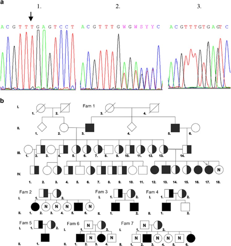
A total of 56 individuals from seven unrelated Slovak Roma families were investigated for suspected hereditary hyperbilirubinemia. All participants signed the informed consent approved by the Ethics Committee of General University Hospital in Prague. The first proband was a girl from a large family with a history of noninfection jaundice, born from a consanguineous marriage of two cousins (Figure 1). Her biochemical tests showed TBi of 35–70 μmol/l, conjugated bilirubin (cBi) of 31–54 μmol/l (F1.IV/17) and normal levels of alanine aminotransferase (ALT) and aspartate aminotransferase (AST). She was hospitalized at the age of 9 months due to severe bilirubinemia, acute respiratory infection and suspected anemia, and treated with Unasyn, Dithiaden, Halixol, folic acid, Maltofer and glucose. The performed 99mTc-HIDA cholescintigraphy of liver and biliary tract revealed delayed visualization of liver and gallbladder, typical for DJS. Similarly, other individuals were tested for TBi, cBi (standard diazo- reaction), ALT and AST.
Figure 1.
(a) Sequences of deletion in ABCC2 c.1013_1014delTG (Ref. Seq. NG_011798.1; NM_000392.4). 1, homozygous variant c.1013_1014delTG in patient with DJS; 2, heterozygous variant in carrier; 3, wild type. (b) Pedigree of the families with DJS: ▪ homozygote;  heterozygote; □ healthy; □ male; ○ female; ◊ unknown, abortion; N, not investigated; arrow, proband; double bond indicates consanguinity.
Genetic analyses
Genomic DNA was extracted from 7 mL whole blood in EDTA using standard procedures. All 32 exons and adjacent intron regions of ABCC2 gene (NM_000392.4, exons were numbered according Ref. Seq. NG_011798.1, Supplementary Table 1) were amplified using PCR, PP Master Mix polymerase with buffer (Top-Bio, Prague, Czech Republic) and 2.5 pM of primer; purified (Promega A9282 kit, Madison, WI, USA); and sequenced by ABI PRISM 3100 AVANT (Applied Biosystems (Foster City, CA, USA), Big Dye Terminator 3.1 Sequencing Kit). Fragment analysis of TA insertion of TATA box of UGT1A1 gene was performed (rs8175347, chr2:hg19:g.175492_175493insTA) (see Supplementary Table 1). PBREM enhancer variant c.-3279T>G (rs4124874; chr2:hg19:g.172270T>G) was amplified and assessed with RFLP as described previously.12 All sequence variants were annotated according to reference sequences (NG_011798.1 for ABCC2 and NG_002601.2 for UGT1A1), and the findings were submitted to ClinVar database (http://www.ncbi.nlm.nih.gov/clinvar).
Coproporphyrin isomer analysis
Urine samples from eight patients with jaundice were collected (100 ml per patient), and stored in dark at −80 °C. Porphyrins and coproporphyrin isomers I and III were analyzed by HPLC with a fluorescence detector (excitation 405 nm, emission 620 nm), Chromsystems column (# 44100) and kit Porphyrins in Urine (# 44000, Chromsystems, Grafelfing, Germany).
Microarray, haplotyping and variant's age estimation
To assess a common haplotype, genomic DNA from 40 individuals was hybridized to Affymetrix (Santa Clara, CA, USA) Genome-Wide Human SNP Array 6.0 according to the manufacturer's recommendation and further analyzed. Genotype calling and loss of heterozygosity in 10q24 region was determined with Affymetrix Genotyping Console version 4.1. The genotyping data were entered in PLINK 1.0718 and SNPs present on chromosome 10 were selected for further analyses. After performing standard quality checks, unphased genotype data of the whole cohort were entered into the Genotype Visualization and Algorithmic Tool (GEVALT) software, version 2.19 Phasing, linkage disequilibrium analysis and estimation of the haplotype block structure was done utilizing the Genotype Resolution and Block Identification using Likelihood (GERBILT) algorithm.20 A haplotype block encompassing the site of c.1013_1014delTG variant in ABCC2 gene included 21 SNPs. The age of the ancestral variant c.1013_1014delTG in Slovakian Roma population was estimated using a standard Luria–Delbrück algorithm modified by Austerlitz21 in Mathematica 8 using the Mathematica notebook kindly provided by F. Austerlitz. Variant's age was estimated for a current population size of 400 000–450 000 Roma.22
Results
Biochemical blood tests included total and conjugated serum bilirubin, ALT and AST. ALT and AST ranged within normal levels, indicating the absence of liver damage. TBi in DJS patients was 18.8–72.2 μmol/l with cBi 9.5–55.2 μmol/l (Table 1). The percentage of cBi out of TBi in patients with dual defect DJS+GS were 28, 38, 43 and 89% (see Table 2). The last value 89% was found in proband F1.IV/17 on medication with following TBi/cBi: 38/33; 70/55; and 35/31 μmol/l. Values of TBi and cBi of patients with the same UGT1A1 and ABCC2 haplotype varied (Table 1).
Table 1. Total and cBi (in μmol/l) and genotype of patients with/out DJSa and GSb.
| Family | Genealogy | DJSa | GSb | PBREM | cBi | Tbi | % cBi of Tbi |
|---|---|---|---|---|---|---|---|
| F1 | II/2 | −/− | 6/6 | T/T | 1.9 | 5.7 | 33.3 |
| II/3 | +/+ | 6/7 | T/G | 28.7 | 46.5 | 61.7 | |
| II/5 | +/+ | 6/7 | T/G | 55.2 | 72.2 | 76.5 | |
| II/6 | −/− | 6/7 | T/G | 2.3 | 7.6 | 30.3 | |
| III/1 | −/− | 6/7 | T/G | 1.8 | 3.6 | 50.0 | |
| III/2 | +/− | 6/7 | T/G | 2.0 | 12.3 | 16.3 | |
| III/3 | −/− | 6/6 | T/T | 0.1 | 4.6 | 2.2 | |
| III/4 | +/− | 6/7 | T/G | 2.8 | 7.2 | 38.9 | |
| III/5 | +/− | 6/6 | T/T | 2.3 | 6.5 | 35.4 | |
| III/6 | +/− | 6/7 | T/T | 1.8 | 5.1 | 35.3 | |
| III/7 | +/− | 6/6 | T/T | 1.0 | 2.8 | 35.7 | |
| III/8 | −/− | 6/6 | T/T | 2.4 | 9.5 | 25.3 | |
| III/9 | +/− | 6/7 | T/G | 0.3 | 9.0 | 3.3 | |
| III/10 | +/− | 6/7 | T/G | 1.2 | 1.5 | 80.0 | |
| III/11 | +/− | 6/6 | T/T | 0.7 | 5.7 | 12.3 | |
| III/12 | +/− | 6/6 | T/T | 0.2 | 3.7 | 5.4 | |
| III/13 | +/− | 6/7 | T/G | 0.3 | 5.0 | 6.0 | |
| III/14 | +/− | 6/7 | T/G | 0.2 | 5.5 | 3.6 | |
| IV/1 | +/− | 7/7 | G/G | 1.5 | 6.8 | 22.1 | |
| IV/2 | −/− | 6/7 | T/G | 3.0 | 3.8 | 78.9 | |
| IV/3 | +/− | 6/6 | T/T | 0.2 | 4.7 | 4.3 | |
| IV/4 | +/− | 6/7 | T/G | — | — | — | |
| IV/5 | +/− | 6/7 | T/G | — | — | — | |
| IV/6 | +/− | 6/7 | G/T | — | — | — | |
| IV/7 | +/− | 6/7 | T/G | — | — | — | |
| IV/8 | +/− | 6/7 | T/G | 1.8 | 5.8 | 31.0 | |
| IV/9 | +/− | 6/7 | T/G | — | — | — | |
| IV/10 | −/− | 6/7 | T/G | 3.0 | 5.5 | 54.5 | |
| IV/11 | −/− | 6/7 | T/G | — | — | — | |
| IV/12 | +/+ | 6/6 | T/T | 16.7 | 28.1 | 59.4 | |
| IV/13 | +/+ | 7/7 | G/G | 17.7 | 46.7 | 37.9 | |
| IV/14 | +/− | 6/6 | T/T | 1.3 | 4.5 | 28.9 | |
| IV/15 | +/− | 6/7 | T/G | 3.2 | 12.7 | 25.2 | |
| IV/16 | +/+ | 6/7 | T/G | 9.5 | 18.8 | 50.5 | |
| IV/17 | +/+ | 7/7 | G/G | 31.0 | 35.0 | 89.0 | |
| F2 | I/1 | +/− | 7/7 | G/G | — | — | — |
| I/2 | +/− | 6/7 | T/G | — | — | — | |
| II/1 | +/+ | 6/7 | T/G | 11.4 | 32.4 | 35.2 | |
| II/5 | +/+ | 7/7 | G/G | 16.9 | 60.4 | 28.0 | |
| F3 | I/1 | +/− | 6/7 | T/G | — | — | — |
| I/2 | +/− | 6/7 | T/G | — | — | — | |
| II/1 | +/+ | 7/7 | G/G | 21.4 | 49.5 | 43.2 | |
| II/2 | +/+ | 6/6 | T/T | — | — | — | |
| F4 | I/1 | +/− | 6/7 | G/G | — | — | — |
| I/2 | +/− | 6/7 | G/G | — | — | — | |
| II/1 | +/+ | 6/6 | T/G | 28.0 | 42.0 | 66.7 | |
| II/2 | −/− | 6/7 | T/G | — | — | — | |
| F5 | I/1 | +/+ | 6/6 | T/G | 32.7 | 39.9 | 82.0 |
| I/2 | +/− | 6/7 | T/G | — | — | — | |
| II/1 | +/+ | 6/7 | T/G | — | — | — | |
| F6 | I/1 | +/− | 6/6 | T/T | — | — | — |
| II/1 | +/+ | 6/7 | T/G | — | — | — | |
| II/3 | +/+ | 6/7 | T/G | 17.6 | 43.5 | 40.5 | |
| F7 | I/2 | +/− | 6/6 | T/G | — | — | — |
| II/2 | +/+ | 6/6 | T/T | 32.4 | 57.4 | 56.4 | |
| II/6 | +/+ | 6/6 | T/T | — | — | — |
+/+ homozygote for c.1013_1014delTG in ABCC2
6/6 healthy, 6/7 heterozygote, 7/7 homozygote for g.175492_175493insTA in UGT1A1;
Bold, double homozygotes.
Table 2. Excretion of coproporphyrin isomers I and III in urine and serum bilirubin in patients with DJS (+/+) and GS (7/7) and in healthy carriers (+/-) or (6/7).
| F1.IV/13 | F1.IV/17 | F2.II/5 | F3.II/1 | F1.II/5 | F1.IV/16 | F2.II/1 | F3.I/1 | F4.I/1 | F4.I/2 | F4.II/1 | |
|---|---|---|---|---|---|---|---|---|---|---|---|
| Total porphyrins (nmol/l) | 212 | 348 | 258 | 78 | 587 | 230 | 205 | 91 | 93 | 58 | 133 |
| Uroporphyrin (nmol/l) | 88 | 80 | 78 | 13 | 118 | 74 | 84 | 10 | 6 | 6 | 19 |
| Coproporphyrin (nmol/l) | 124 | 268 | 180 | 63 | 469 | 156 | 121 | 78 | 75 | 52 | 110 |
| Isomer I (%) | 99 | 97 | 92 | 100 | 98 | 96 | 93 | 43 | 53 | 57 | 89 |
| Isomer III (%) | 1 | 3 | 8 | — | 2 | 4 | 7 | 57 | 47 | 43 | 11 |
| Conjugated bilirubin (μmol/l) | 17.7 | 31.0 | 16.9 | 21.4 | 55.2 | 9.5 | 11.4 | — | — | — | 28.0 |
| Total bilirubin (μmol/l) | 46.7 | 35.0 | 60.4 | 49.5 | 72.2 | 18.8 | 32.4 | — | — | — | 42.0 |
| DJS | +/+ | +/+ | +/+ | +/+ | +/+ | +/+ | +/+ | +/− | +/− | +/− | +/+ |
| GS | 7/7 | 7/7 | 7/7 | 7/7 | 6/7 | 6/7 | 6/7 | 6/7 | 6/7 | 6/7 | 6/6 |
Reference ranges: total porphyrins <300 nmol/l; uroporphyrin <40 nmol/l; coproporphyrin <150 nmol/l;
Conjugated bilirubin <7 μmol/l, total bilirubin <20 μmol/l.
Molecular genetic analysis of the proband revealed the deletion in ABCC2 gene c.1013_1014delTG (http://www.ncbi.nlm.nih.gov/clinvar/?term=SCV000195771), responsible for DJS (Figure 1). The causal variant is located in the eighth exon and, via a frameshift, it leads to a premature stop codon p.(Val338Glufs14*) resulting in a shortened protein product. Investigation of TATA box of UGT1A1 of the same proband showed also NG_002601.2:g.[175492_175493insTA] causing GS. Next, family members and probands were investigated for the defect in ABCC2 gene with the total result of 17 homozygous patients with the deletion c.1013_1014delTG and 30 heterozygous carriers as evident from Figure 1. The presence of the same genetic defect in all seven seemingly unrelated families suggested a founder effect of c.1013_1014delTG among the investigated Roma families. Moreover, four patients with homozygous variant in ABCC2 gene were also homozygous for the TA insertion in TATA box of UGT1A1 gene with a genotype NG_011798.1:c.[1013_1014delTG] NG_002601.2:g.[175492_175493insTA] (Table 1). All patients with homozygous TA insertion were also homozygotes for c.-3279T>G variant in PBREM (Table 1 and Supplementary Table 2).
Pedigrees from seven investigated families are depicted on Figure 1. Consanguineous marriages between cousins were found in three of them (F 1, 4 and 5). In five families, both parents were heterozygous carriers of variant c.1013_1014delTG. In next two families, fathers' samples were not available, however the genotypes of offspring suggest heterozygous or homozygous occurrence of the mutated allele in both fathers.
Coproporphyrin isomer excretion
We analyzed coproporphyrin isomers in urine samples of 11 individuals: in 8 patients with DJS caused by c.1013_1014delTG in ABCC2 gene and in 3 heterozygous carriers. Significantly increased proportion of excreted coproporphyrin isomer I was observed in all DJS patients with values ranging from 89 to 100% (Table 2). In heterozygous carriers, the ratio of excreted isomer I was 43–57%, compared with about 25–30% in healthy controls (control data from literature).23, 24 No differences were found in excrection of coproporphyrin isomers in patients with DJS compared with those with dual defect of DJS+GS.
Microarrays and haplotyping
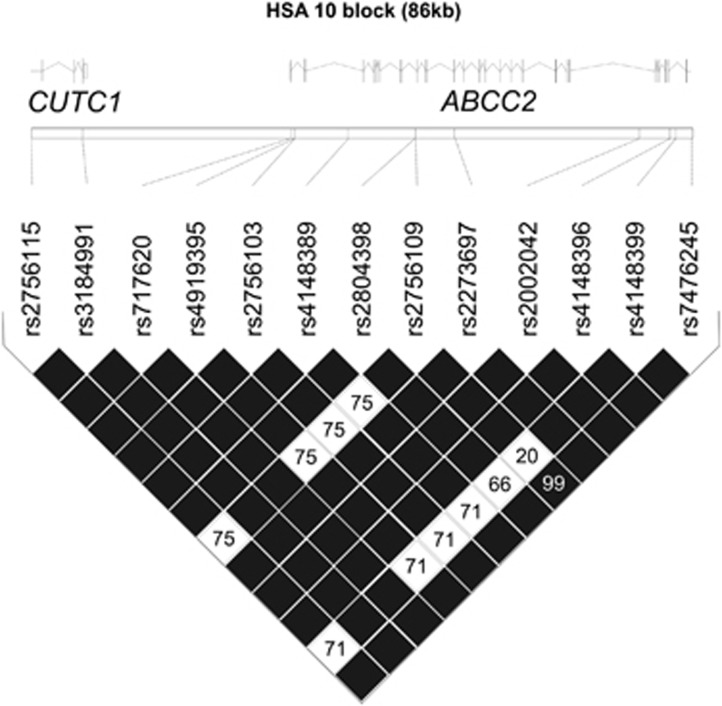
The analysis of chromosome 10 genotypes in ABCC2 region revealed a shared 86-kbp haplotype upstream and within ABCC2 gene, transmitted together with the deletion c.1013_1014delTG (Figure 2,Table 3). Furthermore, the loss of heterozygosity was found in all homozygous patients with DJS with use of CNV analysis from Affymetrix Genotyping Console. Loss of heterozygosity in DJS patients was in all cases in concordance with both genetic and biochemical data.
Figure 2.
The span of a common haplotype in families with DJS encompassing c.1013_1014delTG in ABCC2 gene with indication of linkage disequilibrium (r2). Black diamonds without numerical annotation correspond to complete linkage disequilibrium between the SNPs. Only polymorphic SNPs are shown.
Table 3. A shared haplotype in family 2.
| I/1 father | I/2 mother | II/1 daughter | II/5 son | |||||
|---|---|---|---|---|---|---|---|---|
| rs2756115 | T | C | T | C | C | C | C | C |
| rs17112219 | G | G | G | G | G | G | G | G |
| rs3184991 | T | T | T | T | T | T | T | T |
| rs2093354 | A | A | A | A | A | A | A | A |
| rs2804412 | G | G | G | G | G | G | G | G |
| rs2804409 | T | T | T | T | T | T | T | T |
| rs2756095 | A | A | A | A | A | A | A | A |
| rs7176201 | C | C | C | C | C | C | C | C |
| rs4919395 | G | A | G | A | A | A | A | A |
| rs2756103 | A | C | A | C | C | C | C | C |
| rs4148389 | A | G | A | G | G | G | G | G |
| rs17222744 | A | A | A | A | A | A | A | A |
| rs2804398 | A | T | A | T | T | T | T | T |
| rs2756109 | T | T | T | T | T | T | T | T |
| rs11816875 | G | G | G | G | G | G | G | G |
| rs2804397 | G | G | G | G | G | G | G | G |
| c.1013_1014delTG | W | * | W | * | * | * | * | * |
| rs2273697 | A | G | A | G | G | G | G | G |
| rs2002042 | C | C | C | C | C | C | C | C |
| rs4148396 | C | T | C | T | T | T | T | T |
| rs4148399 | T | G | T | G | G | G | G | G |
| rs7476245 | G | G | G | G | G | G | G | G |
Estimation of variant's age
Age of ancestral variant c.1013_1014delTG was estimated using the Luria–Delbrück algorithm modified by Austerlitz.20, 21 Population size of Slovakian Roma inhabitants was set at 400 000–450 00022 and the frequency of disease allele was estimated between 0.01 and 0.001. Using these data, our results show that the variant originated 7.1–7.4 generations ago, which corresponds to 177.5–185 years ago (Supplementary Table 3).
Discussion
Our study describes compound defect of ABCC2 and UGT1A genes, causing DJS and GS. The frequency of TA insertion in UGT1A1 in Caucasians is about 15%.25 Similar distribution may be assumed in the Roma population, although the accurate frequency has not been established yet. The new variant c.1013_1014delTG described herein is located in the eighth exon of ABCC2 gene. A 2-bp deletion leads to frameshift and truncation of the encoded MRP2 protein, from original size of 1545 amino acids to the reduced size of 352 residues. Deletion truncates protein's two of total three membrane spanning domains (MSD2 and MSD3) and one ATP-binding site, and newly formed mRNA probably subjects to a rapid degradation.4 The excretion of cBi into the bile is performed mainly via ABCC2 efflux pump with minor elimination via ABCG2.26, 27 However, ABCG2 does not provide sufficient substitution of impaired ABCC2, and bilirubin glucuronides are transported by upregulated basolateral MRP3 into the blood.
Biochemistry
Results of total bilirubin and its conjugated fraction correspond to the presence/absence of genetic defect in bilirubin pathway (Table 1). All DJS patients with homozygous presence of c.1013_1014delTG had elevated cBi (9–55 μmol/l) as well as TBi (19–72 μmol/l). Both TBi and cBi showed high variability within the same genotype. Fluctuating bilirubin levels are present in both DJS and GS. Many factors affecting hyperbilirubinemia have been described with the most important TA insertion found in four our patients with DJS and in two healthy DJS carriers.
Effect of a double defect
To date, dual hereditary jaundice has been described only in a single case by Czech authors Cebecauerova et al. with no additional studies up today.15 Our study describes the coincidental defects in two genes of bilirubin metabolism in another four patients. The percentages of cBi out of TBi in our DJS+GS patients were 28%, 38%, 43% and 89%, respectively (Table 1). We suppose that coincidental defects of bilirubin conjugation and transport result in decreased amount of cBi available for its transport, therefore the percentage of cBi from TBi is <50% in DJS+GS compared with >50% in DJS only (Table 1,Supplementary Table 2). Except the last value of 89% (baby girl proband, data collected under medication), our findings are in agreement with the previous study. Both proteins UGT1A1 and MRP2 are responsible for the drug's biotransformation, which may explain a higher cBi and TBi in patient with dual defect under medication. Certain drug precautions should be taken in advance of pharmacotherapy in patients with dual hereditary jaundice including irinotecan, atazanavir, cisplatin, vincristine or doxorubicin to prevent its toxicity or sensitivity.28, 29
Coproporphyrin
Urinary excretion of coproporphyrin reflects the heme degradation and excreted isomers are presented as isomers I–IV with the most significant isomers I and III. The proportion of coproporphyrin isomer III in urine samples of healthy adults is 70–80%, compared with ~20–30% in DJS patients, who excrete about 80–97% of coproporphyrin I. Under normal conditions, excretion of coproporphyrin isomers depends on maturation of hepatobiliary system and changes within the first days of newborn's life.30, 31
We have investigated coproporphyrin in eight patients with DJS caused by c.1013_1014delTG in ABCC2 and all of them showed shifted ratio excreted coproporphyrin isomers in urine (89–100%, Table 2). Several studies show >90% predominance of coproporphyrin I in urine of patients with DJS, but to our knowledge, no data show results of 100%.32, 33, 34 Our patient with 100% excretion of isomer I was 1-year-old boy with total serum bilirubin 50 μmol/l and its conjugated fraction 21 μmol/l. Coproporphyrin isomers were analyzed from two independent samples with the same result. Before the era of molecular genetics, heterozygous carriers of DJS were only detectable by changed isomers of coproporphyrin in urine. Compared with 20–30% in healthy controls, heterozygotes secrete about 40% of isomer I. Average percentage of isomer I in our DJS carriers was 43–57%, which is similar to data from literature.23, 24, 35 Excretion pattern of coproporphyrin isomers in four patients with dual hereditary jaundice was the same as in other DJS patients with no defect in UGT1A1 (see Table 2). Our findings for coproporphyrin I was 92, 97, 99 and 100% in patients with dual defect compared with 89, 93, 96 and 98% in DJS patients with heterozygous NG_002601.2:g.175492_175493insTA and 89% in DJS patient with the wild-type UGT1A gene.
Haplotyping and dating an ancestral variant
After the finding of an identical genetic defect, we have suspected the founder effect of c.1013_1014delTG variant among Roma families. The consanguinity was observed in three of seven families and all families lived within tens of kilometers of one another (Supplementary Figure 1). We have identified a common haplotype spanning 86 kbp segregating with the deletion in ABCC2 and a loss of heterozygosity in ABCC2 region in patients with DJS (Figure 2). Several haplotype analyses of ABCC2 have been performed due to its pharmacogenomic impact, even one concerning the impact of ABCC2 haplotype on coproporphyrin excretion, but only using SNPs with proven pharmacogenomics effect.36, 37 There are more than 130 SNPs within ABCC2 and their importance in vitro has mostly not been described yet. Our study clearly shows, that in addition to the ABCC2 haplotype, which is shared in our DJS patients, other factors may also influence coproporphyrin excretion in urine, such as SNPs in other genes or undiscovered environmental factors.
Dating the ancestral variant c.1013_1014delTG in Slovakian Roma population revealed the probable origin of variant 178–185 years ago. Allelic association estimates the age of the ancestral variant from haplotypic data, without specification of the population growth rate.21
Conclusion
Our study describes defects in UGT1A1 and ABCC2 genes corroborating genetic findings with biochemical results in more individuals with dual hereditary jaundice. In 17 Roma patients with DJS we have detected a new deletion NG_011798.1:c.[1013_1014delTG], which represents the highest prevalence of DJS in Europe. Four of our patients were homozygous for a dual defect NG_011798.1:c.[1013_1014delTG] NG_002601.2:g.[175492_175493insTA]. We also uncovered the existence of the founder effect of ABCC2 variant in all patients from seven families and a shared haplotype 86 kbp and estimated the age of the deletion to 178–185 years.
Acknowledgments
We would like to thank Hynek Strnad for his assistance with the raw data, Frederic Austerlitz for his Mathematica notebook and Sudha Veeraraghavan for the critical reading of the manuscript. This work was financially supported by grants UNCE 204011, PRVOUK P24/LF1/3, RVO-VFN 641652012 and further by BIOCEV (CZ.1.05/1.1.00/02.0109). OS is supported by MSMT project LK11217.
The authors declare no conflict of interest.
Footnotes
Supplementary Information accompanies this paper on European Journal of Human Genetics website (http://www.nature.com/ejhg)
Supplementary Material
References
- Dubin I, Johnson FB: Chronic idiopathic jaundice with unidentified pigment in liver cells: a new clinicopathologic entity with report of 12 cases. Medicine 1954; 33: 155. [DOI] [PubMed] [Google Scholar]
- Kitamura T, Hardenbrook C, Kamimoto Y et al: Defective ATP-dependent bile canalicular transport of organic anions in mutant (TR2) rats with conjugated hyperbilirubinemia. Proc Natl Acad Sci USA 1990; 87: 3557–3561. [DOI] [PMC free article] [PubMed] [Google Scholar]
- Mor-Cohen R, Zivelin A, Rosenberg N et al: Identification and functional analysis of two novel mutations in the multidrug resistance protein 2 gene in Israeli patients with Dubin-Johnson syndrome. J Biol Chem 2001; 276: 36923–36930. [DOI] [PubMed] [Google Scholar]
- Keitel V, Nies AT, Brom M et al: A common Dubin-Johnson syndrome mutation impairs protein maturation and transport activity of MRP2 (ABCC2). Am J Physiol Gastrointest Liver Physiol 2003; 284: G165–G174. [DOI] [PubMed] [Google Scholar]
- Elferink O, Ottenhoff R, Liefting WG et al: ATP-dependent efflux of GSSG and GS-conjugate from isolated rat hepatocytes. Am J Physiol 1990; 258: G669–G706. [DOI] [PubMed] [Google Scholar]
- Chen ZS, Kawabe T, Ono M et al: Effect of multidrug resistance-reversing agents on transorting activity of human canalicular resistance multispecific organic anion transporter. Mol Pharmacol 1999; 56: 1219–1228. [DOI] [PubMed] [Google Scholar]
- Evers R, de Haas M, Sparidans R et al: Vinblastine and sulfinpyrazone export by the multidrug resistance protein MRP2 is associated with glutathione export. Br J Cancer 2000; 83: 375–383. [DOI] [PMC free article] [PubMed] [Google Scholar]
- van Zanden JJ, de Mul A, Wortelboer HM et al: Reversal of in vitro cellular MRP1 and MRP2 mediated vincristine resistance by the flavonoid myricetin. Biochem Pharmacol 2005; 69: 1657–1665. [DOI] [PubMed] [Google Scholar]
- Wada M, Toh S, Taniguchi K et al: Mutations in the canalicular multispecific organic anion transporter (cMOAT) gene, a novel ABC transporter, in patients with hyperbilirubinemia II/Dubin-Johnson syndrome. Hum Mol Genet 1998; 7: 203–207. [DOI] [PubMed] [Google Scholar]
- Mor-Cohen R, Zivelin A, Rosenberg N, Goldberg I, Seligsohn U: A novel ancestral splicing mutation in the multidrug resistance protein 2 gene causes Dubin-Johnson syndrome in Ashkenazi Jewish patients. Hepatol Res 2005; 31: 104–111. [DOI] [PubMed] [Google Scholar]
- Bosma PJ, Chowdhury A, Bakker C et al: Genetic basis of reduced expression of bilirubin UDP-glucuronosyltransferase 1 in Gilbert's syndrome. N Engl J Med 1995; 333: 1171–1175. [DOI] [PubMed] [Google Scholar]
- Sugatani J, Yamakawa K, Yoshinari K et al: Identification of a defect in the UGT1A1 gene promoter and its association with hyperbilirubinemia. Biochem Biophys Res Com 2002; 292: 492–497. [DOI] [PubMed] [Google Scholar]
- Arias IM, London IM: Bilirubin glucuronide formation in vitro; demonstration of a defect in Gilbert's disease. Science 1957; 126: 563–564. [DOI] [PubMed] [Google Scholar]
- Cebecauerova D, Jirasek T, Budisova L et al: Dual hereditary Jaundice: simultaneous occurence of mutations causing Gilbert's and Dubin-Johnson syndrome. Gastroenterology 2005; 129: 315–320. [DOI] [PubMed] [Google Scholar]
- Gresham D, Morar B, Underhill PA et al: Origins and divergene of Roma (gypsies). Am J Hum Genet 2001; 69: 1314–1331. [DOI] [PMC free article] [PubMed] [Google Scholar]
- Mendizabal I, Lao O, Marigorta UM et al: Reconstructing the population history of European Romani from genome-wide data. Curr Biol 2012; 22: 2342–2349. [DOI] [PubMed] [Google Scholar]
- Kalaydjieva L, Morar B, Chaix R, Tang H: A newly discovered founder population: the Roma/Gypsies. Bioessays 2005; 27: 1084–1094. [DOI] [PubMed] [Google Scholar]
- Purcell S, Neale B, Todd-Brown K et al: PLINK: a toolset for whole-genome association and population-based linkage analysis. Am J Hum Genet 2007; 81: 559–575. [DOI] [PMC free article] [PubMed] [Google Scholar]
- Kimmel G, Shamir R Maximum Likelihood Resolution of Multi-block Genotypes. Proceedings of the Eighth Annual International Conference on Research in Computational Molecular Biology (RECOMB); New York, NY, USA, 2004, pp 2–6.
- Kimmel G, Shamir R: GERBIL: genotype resolution and block identification using likelihood. Proc Natl Acad Sci USA 2005; 102: 158–162. [DOI] [PMC free article] [PubMed] [Google Scholar]
- Austerlitz F, Kalaydjieva L, Heyer E: Detecting population growth, selection and inherited fertility from haplotypic data in humans. Genetics 2003; 165: 1579–1586. [DOI] [PMC free article] [PubMed] [Google Scholar]
- Sprocha B: Prognosis of Roma population in Slovakia to year 2013. Slovak Stat Demogr 2011; 21: 22–46. [Google Scholar]
- Koskelo P, Toivonen I, Adlercreutz H: Urinary coproporphyrin isomer distribution in the Dubin-Johnson syndrome. Clin Chem 1967; 13: 1006–1009. [PubMed] [Google Scholar]
- Kondo T, Kuchiba K, Shimizu Y: Coporporphyrin isomers in Dubin-Johnson syndrome. Gastroenterology 1976; 70: 1117–1120. [PubMed] [Google Scholar]
- Owens D, Evans J: Population studies on Gilbert's syndrome. J Med Genet 1975; 12: 152–156. [DOI] [PMC free article] [PubMed] [Google Scholar]
- Cherrington NJ, Hartley DP, Li N et al: Organ distribution of multidrug resistence proteins 1, 2 and 3 (MRP1, 2 and 3) mRNA and hepatic induction of Mrp3 by constitutive androstane receptor activators in rats. J Pharmacol Exp Ther 2002; 300: 97–104. [DOI] [PubMed] [Google Scholar]
- van de Steeg E, Stranecky V, Hartmannova H et al: Complete OATP1B and OTP1B3 deficiency causes human Rotor syndrome by interrupting conjugated bilirubin reuptake into the liver. J Clin Invest 2012; 122: 519–528. [DOI] [PMC free article] [PubMed] [Google Scholar]
- Lankish TO, Schultz C, Zwingers T et al: Gilbert's syndrome and irinotecan toxicity: combination with UDP-glucuronosyltransferase 1A7 variants increases risk. Cancer Epidemiol Biomarkers Prev 2008; 17: 695–701. [DOI] [PubMed] [Google Scholar]
- Koike K, Kawabe T, Tanaka T et al: Multispecific organic anion transporter (cMOAT) antisence cDNA enhanes drug sensitivity in human hepatic cancer cells. Cancer Res 1997; 57: 5475–5479. [PubMed] [Google Scholar]
- Ben-Ezzer J, Blonder J, Shani M et al: Abnormal excretion of the isomers of urinary coproporphyrin by clinically unaffected family members. Isr J Med Sci 1973; 9: 1431–1436. [PubMed] [Google Scholar]
- Rocchi E, Balli F, Gibertini P et al: Coproporphyrin excretion in healthy newborn babies. J Pediatr Gastroenterol Nutr 1984; 3: 402–407. [DOI] [PubMed] [Google Scholar]
- Kimura A, Yuge K, Kosai KI et al: Neonatal cholestasis in two siblings: a variant of Dubin-Johnson syndrome? J Paediatr Child Health 1995; 6: 557–560. [DOI] [PubMed] [Google Scholar]
- Nakata F, Oyanagi K, Fujiwara M et al: Dubin-Johnson syndrome in a neonate. Eur J Pediatr 1979; 4: 299–301. [DOI] [PubMed] [Google Scholar]
- Wolkoff AW, Cohen LE, Arias IM.: Inheritance of the Dubin-Johnson syndrome. N Engl J Med 1973; 288: 113–117. [DOI] [PubMed] [Google Scholar]
- Benz-de Bretagne I, Respaud R, Vourc'h P et al: Urinary elimination of coproporphyrins is dependent on ABCC2 polymorphisms and represents a potential biomarker of MRP2 activity in humans. J Biomed Biotechnol 2011; 2011: 498757. [DOI] [PMC free article] [PubMed] [Google Scholar]
- Laechelt S, Turrini E, Ruehmkorf A et al: Impact of ABCC2 haplotypes on transcriptional and posttranscriptional gene regulation and function. Pharmacogenomics 2011; 11: 25–34. [DOI] [PubMed] [Google Scholar]
- Kwan P, Wong V, Ng PW et al: Gene-wide tagging study of the association between ABCC2, ABCC5 and ABCG2 genetic polymorphisms and multidrug resistence in epilepsy. Pharmacogenomics 2011; 3: 319. [DOI] [PubMed] [Google Scholar]
Associated Data
This section collects any data citations, data availability statements, or supplementary materials included in this article.


