Abstract
Many rationally engineered DNA nanostructures use mechanically interlocked topologies to connect individual DNA components, and their physical connectivity is achieved through the formation of a strong linking duplex. The existence of such a structural element also poses a significant topological constraint on functions of component rings. Herein, we hypothesize and confirm that DNA catenanes with a strong linking duplex prevent component rings from acting as the template for rolling circle amplification (RCA). However, by using an RNA-containing DNA [2] catenane with a strong linking duplex, we show that a stimuli-responsive RNA-cleaving DNAzyme can linearize one component ring, and thus enable RCA, producing an ultra-sensitive biosensing system. As an example, a DNA catenane biosensor is engineered to detect the model bacterial pathogen Escherichia coli through binding of a secreted protein, with a detection limit of 10 cells ml−1, thus establishing a new platform for further applications of mechanically interlocked DNA nanostructures.
 DNA nanostructures with interlocked topologies will tend to display different behaviour to the linear counterparts. Here, the authors show a DNA catenane that is inactive for rolling circle amplification but is activated upon cleavage of one ring, and exploit this for the development of a biosensing system.
DNA is not only important in biological systems as genetic material, it has also become a key player in synthetic biology. DNA can be engineered into catalysts (DNAzymes) and molecular receptors (DNA aptamers), making DNA a functionally versatile polymer. DNA, as a highly programmable material based on predictable Watson–Crick base-pairing interactions, has become a valuable macromolecule for rational engineering of molecular machines for potential nanotechnological applications.
In recent years, tremendous progress has been made toward building DNA-based nanodevices with increasing structural complexity and functional capabilities1,2,3,4,5,6,7,8,9,10,11,12. One important feature of many reported DNA nanostructures, such as DNA Borromean rings1 and DNA catenanes3, is the use of mechanically interlocked topologies to connect individual DNA components. The mechanical interlocking between DNA strands can be easily achieved in the case of DNA through the formation of a linking duplex between partner rings before ring closure. The existence of a linking duplex is not only essential to the creation of a strong connectivity between partner rings but also necessary for the stability of these well-defined structures.
As we will show in this work, the linking-duplex feature also enables the use of topologically interlocked architectures, such as DNA catenanes, for the design of amplified biosensors for bioanalytical applications. The biosensing strategy is based on the following idea: the strong physical engagement of two mechanically interlocked single-stranded DNA rings in a DNA [2] catenane (termed D2C in this study for simplicity) with a strong linking duplex makes the component rings unsuitable as the template for rolling circle amplification (RCA), an isothermal DNA amplification technique13,14,15. However, when one of the component rings is engineered to be a substrate of a stimuli-responsive RNA-cleaving DNAzyme (RCD), the system can be programmed into a biosensor that is capable of reporting a target of interest in three sequential reactions: target-induced RNA cleavage, nucleolytic conversion of the cleavage product into a DNA primer, and DNA amplification via RCA. By this approach, we establish an amplified biosensing system that is capable of achieving ultra-sensitive detection of Escherichia coli (E. coli) at a concentration as low as 10 cells ml−1 without cell culture.
Results
Inability of a D2C with a strong linking duplex to undergo RCA
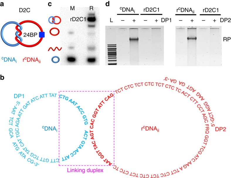
The conceptual cornerstone of this work is the assumption that the components of a D2C with a strong linking duplex are unable to undergo RCA. To test this idea, we synthesized a D2C (Fig. 1a) consisting of two component single-stranded DNA rings, named CDNAi and rCDNAii (Fig. 1b; r stands for the single ribonucleotide, ribo-A, in the sequence of CDNAii). Briefly, the linear DNA rLDNAii was circularized into rCDNAii using CDNAi as the ligation template (sequences of all DNA species are provided in Supplementary Table 1). The resultant D2C, denoted rD2C1, contains a strong linking duplex of 24 bp, which translates into two helical turns (boxed nucleotides in Fig. 1b). The reaction yield of rD2C1 was determined to be 58% (Fig. 1c).
Figure 1. Inability of a D2C to undergo RCA.
(a) Schematic illustration of a D2C made of CDNAi and rCDNAii, with a linking duplex of 24 bp. (b) Sequences of rD2C1, DP1 and DP2. Boxed nucleotides represent the 24-bp linking duplex. F: fluorescein-dT; R, adenosine ribonucleotide; Q, dabcyl-dT. (c) Synthesis of rD2C1 by circularizing linear DNAii over CDNAi as the template. Lane M, markers made of LDNAii, CDNAi and rCDNAii. Lane R: circularization mixture. (d) RCA reactions with gel-purified CDNAi, rCDNAii and rD2C1 using DP1 and DP2 as primers. Lane L, DNA ladders ranging from 1 to 10 kbp; RP, RCA product.
We then performed RCA reactions with gel-purified CDNAi, rCDNAii and rD2C1. Agarose gel analysis indicated that RCA products were produced with CDNAi (using DP1 as the primer; Fig. 1d) and rCDNAii (using DP2 as the primer; Fig. 1d). In contrast, no RCA products were observed for rD2C1 using the same set of primers (Fig. 1d). This experiment shows that the topological constraint imposed by a strong linking duplex indeed prevents ϕ29 DNA polymerase (ϕ29DP) from replicating interlocked circular templates. In a control experiment, it was found that RCA was not inhibited when the linking duplex of the DNA catenane was made of 9 bp (Supplementary Fig. 1).
Enabling RCA via cleavage of a component ring by a DNAzyme
The inability of ϕ29DP to carry out RCA with topologically constrained DNA catenanes provides a novel avenue to explore these intricate DNA assemblies for practical applications. Given our interest in DNAzymes and biosensing, we turned our attention next to the engineering of a highly unique biosensing system that takes advantage of topologically constrained DNA catenanes, DNAzymes and RCA.
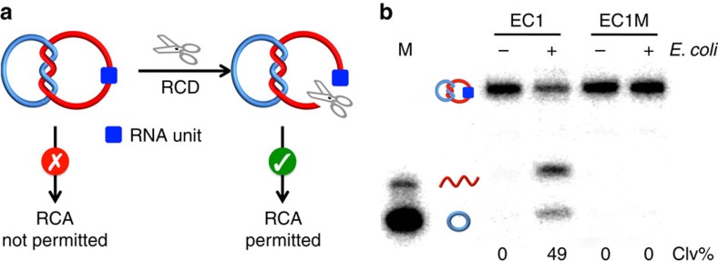
The working principle of our biosensing system is shown in Fig. 2a. It uses an RCD to cleave the embedded RNA linkage within rD2C1 (note that the rCDNAii was designed to contain a single ribonucleotide; Fig. 1b). It is expected that the cleavage and linearization of rCDNAii by the RCD will release the topological constraint on the DNA assembly, which converts CDNAi into a suitable template for RCA.
Figure 2. Cleavage of an RNA-containing D2C by an RCD.
(a) Restoration of RCA compatibility of an rD2C using an RCD. (b) Cleavage of rD2C1 by EC1, an E. coli-responsive DNAzyme. Concentration of E. coli: 105 cells ml−1. Reaction mixtures were analyzed by 10% denaturing PAGE. EC1M: a mutant EC1 that cannot be activated by E. coli. Both rCDNAii and CDNAi in rD2C1 were radioactively labelled with 32P to facilitate DNA visualization on the gel. Clv%: per cent cleavage.
In theory, our approach should be compatible with any RCD, making it a platform for detection of any species recognized by an allosteric DNAzyme. For this work, we employed EC1, which was previously isolated by us from a random-sequence DNA pool using in vitro selection for specific detection of E. coli, a model bacterial pathogen16,17. EC1 was found to be activated by a protein molecule secreted specifically by E. coli cells (illustrated in Supplementary Fig. 2). Therefore, the use of EC1 enables the detection of this pathogen. As illustrated in Fig. 2b, EC1 was indeed able to cleave the rCDNAii present in rD2C1 in an E. coli-dependent manner, resulting in CDNAi and a linear DNAii. An inactive DNAzyme mutant, EC1M, was also tested as a control. No cleavage product was observed for EC1M, indicating that the cleavage reaction is highly dependent on the DNAzyme sequence. We also examined the cleavage activity at different reaction times and found that the cleavage activity reached a plateau in 1 h (Supplementary Fig. 3). Thus, this reaction time was used for the remaining experiments.
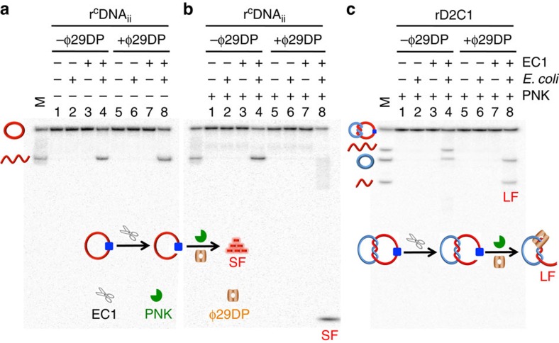
Upon demonstrating EC1-mediated cleavage of rCDNAii in rD2C1, we examined the use of the cleavage reaction mixture for initiating the RCA reaction with ϕ29DP. Because ϕ29DP has 3′–5′ exonucleolytic activity that can degrade single-stranded DNA from the 3′-end but does not digest double-stranded DNA18,19, we speculated that the system should not require an external primer, as ϕ29DP should be able to convert the linearized DNAii into a primer for RCA. To evaluate this hypothesis, we determined whether ϕ29DP could digest EC1-linerized DNAii. From the data presented in Fig. 3a, it is clear that ϕ29DP could not digest linearized DNAii (comparing lanes 4 and 8).
Figure 3. 3′–5′ exonucleolytic activity of ϕ29DP on rCDNAii and rD2C1.
Degradation of EC1-mediated cleavage product of rCDNAii (a,b) and rD2C1 (c) by ϕ29DP and PNK. Concentration of E. coli: 105 cells ml−1. Reaction mixtures were analyzed by 20% denaturing PAGE. LF, large DNA fragment; SF, small DNA fragment. M lanes contain various DNA markers as indicated. rCDNAii, both rCDNAii and CDNAi in rD2C1 were radioactively labelled with 32P to facilitate DNA visualization on the gel.
There are two possible reasons for this finding. The first possibility is that ϕ29DP is incapable of digesting an RNA-terminated DNA molecule. However, testing with an RNA-terminated oligonucleotide ruled out this possibility (Supplementary Fig. 4). The second scenario is that ϕ29DP is not able to digest an RNA-terminated DNA molecule containing a 2′,3′-cyclic phosphate on the RNA moiety, which is a common product of RNA cleavage20. To test this, we treated the reaction mixture with T4 polynucleotide kinase (PNK), which is known to be capable of removing the terminal 2′,3′-cyclic phosphate in RNA21. As shown in Fig. 3b, treatment with PNK indeed facilitated the digestion of EC1-linearized rDNAii, as evidenced by the accumulation of small cleavage fragments (labelled SF in lane 8).
We next investigated the combined action of ϕ29DP and PNK on EC1-linearized rDNAii within rD2C1. As shown in Fig. 3c, ϕ29DP degraded complexed DNAii to a product of ∼60 nt (labelled LF, representing long fragment; lane 8). We also conducted the digestion assays using different incubation times (Supplementary Fig. 5). The progressive accumulation of LF and disappearance of the cleaved rDNAii was observed. These experiments allow us to conclude that the combination of ϕ29DP and PNK can remove the single-stranded fragment at the 3′-end of the EC1-linearized DNAii from rD2C1. It is expected that the trimmed DNAii can now function as a primer to initiate RCA over the complexed CDNAi template.
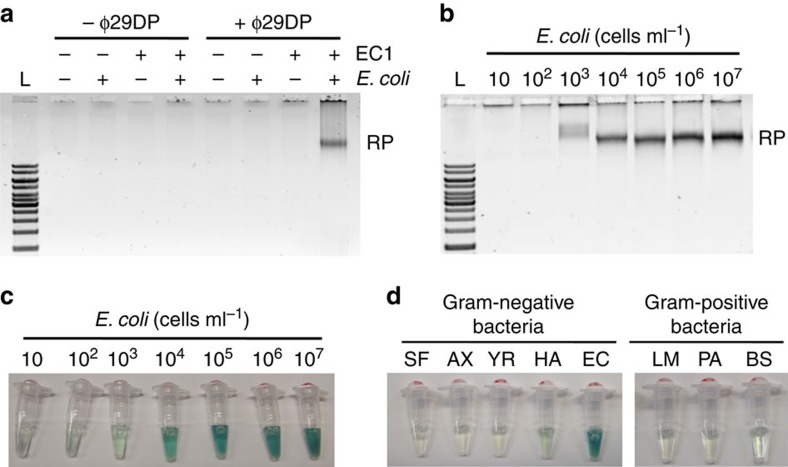
To verify the point above, we carried out the RCA reaction with the rD2C1 assembly. The reaction was performed in two sequential steps: activation of EC1 by E. coli and PNK treatment, followed by the addition of ϕ29DP and dNTPs. As expected, RCA products were indeed observed following this procedure (the last lane of Fig. 4a; the other lanes represent various controls). The RCA products were further analyzed through partial digestion with EcoRV (Supplementary Fig. 6) as the sequence of CDNAi was designed to contain a recognition sequence for this restriction enzyme. The appearance of the expected characteristic DNA banding pattern on the gel, which consists of monomeric, dimeric and other higher-ordered DNA amplicons, verified that the RCA products indeed contained the correct repetitive sequences.
Figure 4. E. coli-dependent RCA reaction.
(a) RCA reactions of rD2C1 in the presence of E. coli (105 cells ml−1) analyzed using 0.6% agarose gel electrophoresis. Note every reaction also contained PNK and dNTPs. L, DNA ladders ranging from 1 to 10 kbp; RP, RCA product. (b) Determination of detection sensitivity through analysis of RP using 0.6% agarose gel electrophoresis. (c) Determination of detection sensitivity via the colourimetric assay enabled by PW17 peroxidase DNAzyme. (d) Analysis of assay specificity using the colourimetric assay. The gram-negative bacteria used were Serratia fonticola (SF), Achromobacter xylosoxidans (AX), Yersinia ruckeri (YR) and Hafnia alvei (HA). The gram-positive bacteria used were Leuconostoc mesenteroides (LM) and Pediococcus acidilactici (PA).
Quantitative detection of E. coli using the DNA catenane sensor
We then investigated the feasibility of performing quantitative analysis using the DNA catenane sensor. Samples containing at E. coli 10–107 cells ml−1 were assessed for RCA amplified detection using a gel-staining method. By this method, we were able to detect as low as 103 cells ml−1 (Fig. 4b). Although gel-based RP analysis can perform quantitative detection of E. coli, the procedure is extremely inconvenient. To overcome this issue, we developed a colorimetric assay by modifying the sequence of CDNAi (the new sequence is named CDNAiCD) such that the RCA product contained a repetitive sequence of PW17, a peroxidase-like DNAzyme capable of generating a colourimetric signal22,23,24,25,26. In the presence of hemin, PW17 catalyzes the H2O2-mediated oxidation of 2,2′-azino-bis(3-ethylbenzthiazoline-6-sulphonic acid) (ABTS) into a coloured product. As shown in Fig. 4c, this colourimetric method was indeed able to detect E. coli in a concentration-dependent manner and registered a detection sensitivity of 103 cells ml−1, similar to what was observed with the gel-based method.
We also evaluated the bacterial detection specificity using the colourimetric assay. We selected four other Gram-negative and three Gram-positive bacteria that were previously tested for EC1-based detection. It was observed that none of these bacteria were able to produce a positive signal, indicating that the rD2C1/EC1 system retained the high recognition specificity for E. coli (Fig. 4d). To further evaluate the specificity, we checked the potential influence of small RNAs on E. coli detection due to the fact that small RNAs (for example, microRNA) are suitable primers for RCA. For this experiment, we used the total small RNAs extracted from breast cancer cell line MCF-7. Agarose gel and colourimetric results indicated that the small RNAs were not able to induce the RCA reaction (Supplementary Fig. 7). This high specificity is attributed to the unique topologically constrained structure of the DNA catenane.
Enhancing detection sensitivity using hyperbranched RCA
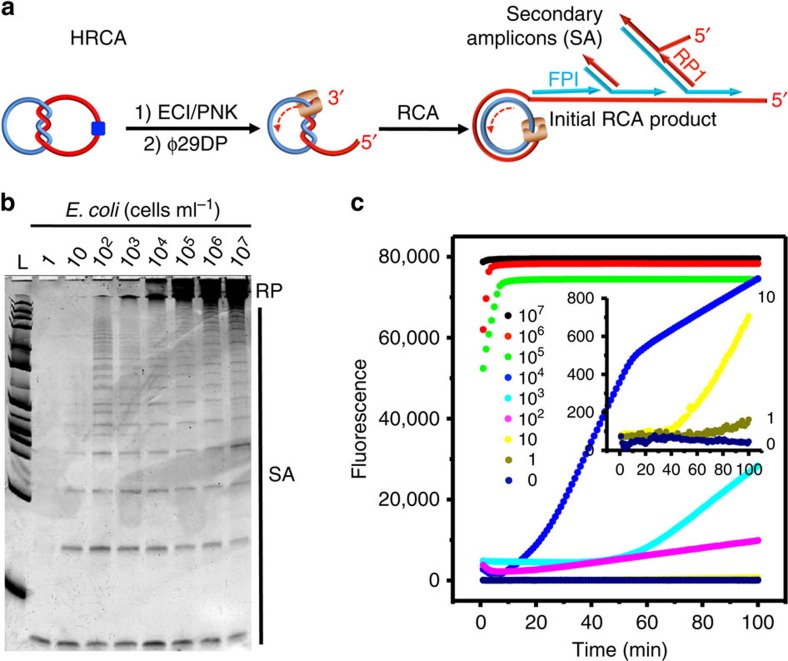
Finally we examined the possibility of performing a double-primed hyperbranched RCA (HRCA)27 reaction with the rD2C1/EC1 system to further increase the detection sensitivity. In HRCA (Fig. 5a), multiple priming events can be continuously initiated by a forward primer (FP1) and a reverse primer (RP1) as the original RCA product strand elongates, resulting in an exponential amplification28.
Figure 5. E. coli-dependent HRCA reaction.
(a) Schematic illustration of HRCA. FP1, forward primer; RP1, reverse primer. (b) Denaturing PAGE analysis of HRCA products. RP, RCA products; SA, secondary amplicons produced from the initial RCA products. (c) Real-time monitoring of HRCA reactions at various E. coli concentrations (cells ml−1).
We found that HRCA was indeed functional with the rD2C1/EC1 system: as shown in Fig. 5b, in addition to the observation of the RCA products on denaturing polyacrylamide gel electrophoresis (PAGE), a series of shorter DNA molecules were also produced, representing various secondary amplicons produced from the primary amplicons (that is, initial RCA products). The HRCA reactions, in response to varying concentrations of E. coli, were also monitored in real time through the use of EvaGreen, a DNA-binding dye (Fig. 5c). We found that this method exhibited much enhanced detection sensitivity, as it was able to detect E. coli at a concentration of as low as 10 cells ml−1 without cell culture.
It is interesting to note that RCA reactions with ϕ29DP remain functional even in the presence of 50% human blood (Supplementary Fig. 8). In comparison, PCR with Taq DNA polymerase was completely inhibited by less than 0.2% human blood29,30,31. To demonstrate the performance of the assay when using more complex samples, we spiked whole blood with E. coli and demonstrated that under these conditions the DNA catenane sensor was still able to detect E. coli at a concentration of 10 cells ml−1 (Supplementary Fig. 9). This observation is consistent with a recent report where EC1 was used to detect E. coli cells in human blood32.
The ability of the DNA catenane sensor to distinguish between live and dead E. coli cells was also investigated. For this experiment, we used lysozyme to kill E. coli (105 cells ml−1) and compared the signal responses of the catenane sensor in the presence of live and dead bacteria using HRCA. Negligible signal was observed with dead cells while a high activity was seen with live E. coli cells (Supplementary Fig. 10).
We also performed an E. coli detection experiment with an enzyme-linked immunosorbent assay (ELISA) using a commercial ELISA kit designed to detect E. coli host cell protein (Supplementary Fig. 11). It was found that the ELISA method was able to detect E. coli at a concentration of 103 cells ml−1. Therefore, our amplified DNA catenane sensor offers a detection sensitivity that is 100 times better than the ELISA method, while the test time for both methods are similar (∼3 h; see Supplementary Table 2 for additional information).
Discussion
We have shown for the first time that mechanically interlocked DNA [2] catenanes with a strong linking duplex impose a significant topological constraint on their component DNA rings, making them unsuitable as the template for RCA. We have further demonstrated that such DNA nanostructures can be uniquely exploited for the design of a biosensing system where the elimination of the topological engagement, achieved simply through the cleavage of one interlocked DNA ring in an analyte-dependent manner, frees up the other ring for the RCA reaction. As an example, we have produced one of the two interlocked DNA rings to contain a RNA linkage so that an RCD can be used to cleave one interlocked ring. Through the use of an RCD whose activity is specifically triggered by a secreted protein in E. coli, we have shown that the featured biosensing system is capable of achieving ultra-sensitive detection of this bacterial pathogen.
The biosensing system featured here offers some distinct advantages over existing detection methods for E. coli, such as cell culturing, PCR and ELISA (see Supplementary Table 2 for additional information). The use of RCA and HRCA for signal amplification makes this system extremely sensitive for bacterial detection, which can achieve the detection of as low as 10 cells ml−1 without a cell-culturing step. The assay is also more compatible with point-of-care or field applications because RCA is an isothermal process and there is no need for DNA extraction (as in the case of PCR). In addition, the system functions well with biological samples (no interferences from small RNAs and compatibility with blood samples).
It is conceivable that the same design can be extended to other RCDs, DNA-cleaving DNAzymes, as well as ribozymes and protein enzymes that have DNA or RNA-cleaving activities. Although rolling circle amplification was exploited to achieve signal amplification in this study, it should be feasible to take advantage of other signal amplification strategies, such as the DNAzyme cross-amplification system developed by Levy and Ellington that does not need a DNA polymerase33. We envision that the concept presented in this study should open up new opportunities for exploring mechanically interlocked DNA architectures for many potential applications in chemical biology, medical diagnostics, and environmental monitoring.
Methods
Preparation of rD2C1
A total of 100 pmol of rLDNAii was first labelled with γ-[32P]ATP at the 5′ end using T4 PNK according to the manufacturer's protocol. To ensure that all DNA molecules contain the 5′ phosphate required for the subsequent ligation reaction, PNK-mediated end-labelling solution containing 5′-32P labelled rLDNAii was further incubated with 2 mM non-radioactive ATP at 37 °C for 30 min. Then 120 pmol of CDNAi, prepared using the procedure detailed in Supplementary Methods, was added and heated to 90 °C for 30 s. After cooling to room temperature and leaving the solution for 15 min, 10 μl of 10 × T4 DNA ligase buffer and 10 U T4 DNA ligase were added (total 100 μl) and incubated at room temperature for 2 h. The obtained rD2C1 molecules were concentrated by standard ethanol precipitation and purified by 10% dPAGE.
E. coli-dependent RCA reaction
A single colony of E. coli K12 freshly grown on Luria broth (LB) agar plate was taken and used to inoculate 2 ml of LB. After shaking at 37 °C for 14 h at 250 r.p.m., the bacterial culture was serially diluted in 10-fold intervals. Overall, 100 μl of each diluted solution was plated onto LB agar plate (done in triplicate) and cultured at 37 °C for 15 h to obtain the cell counts. Colonies in each plate were counted; the average number of colonies from the three plates was taken as the number of E. coli cells for this dilution. This number was then used to calculate the number of cells for the other dilutions. A total of 500 μl of each dilution was centrifuged at 13,000g for 20 min at 4 °C and re-suspended in 100 μl of 1 × RB (50 mM HEPES, 150 mM NaCl, 15 mM MgCl2, pH 7.5). After being frozen at −20 °C, E. coli cells were sonicated for 1 min and put on the ice for 5 min. This process was repeated three times. Then the cell suspension containing different numbers of E. coli cells were centrifuged at 13,000g for 10 min at 4 °C. The obtained crude intracellular mixture produced by E. coli cells (CIM-EC) in the supernatant was used for the following experiment.
A cleavage reaction mixture containing 5 μl of CIM-EC, 1 μl of rD2C1 (5 μM), 4 μl of EC1 (50 μM) and 10 μl of 2 × RB was incubated at room temperature for 60 min. Then 1 μl of PNK (10 U μl−1) was added and incubated at 37 °C for 30 min. The RCA reaction was initiated by the addition of 1 μl of ϕ29DP (10 U μl−1), 1 μl of dNTPs (50 mM), 5 μl of 10 × RCA reaction buffer and 22 μl of water. The reaction mixtures were incubated at 30 °C for 60 min before heating at 90 °C for 5 min. The resultant RCA products were analyzed by 0.6% agarose gel electrophoresis.
Colourimetric detection
rD2C1 used for the colourimetric detection of E. coli was made of rCDNAii and CDNAiCD, prepared using the procedure described in Supplementary Methods. After the cleavage reaction described above, 1 μl of PNK (10 U μl−1) was added and incubated at 37 °C for 30 min. The RCA reaction was then initiated by the addition of 1 μl of ϕ29DP (10 U μl−1), 1 μl of dNTPs (50 mM), 2 μl of hemin (100 μM), 5 μl of 10 × RCA reaction buffer and 20 μl of water. The reaction mixtures were incubated at 30 °C for 60 min before heating at 65 °C for 20 min. After cooling to room temperature, 2 μl of ABTS (50 mM) and 1 μl of H2O2 (8.8 mM) were added, and the colourimetric result was recorded immediately using a digital camera.
HRCA reaction
Following the cleavage reaction, 1 μl of PNK (10 U μl−1) was added and incubated at 37 °C for 30 min. The HRCA reaction was then initiated by the addition of 1 μl of ϕ29DP (10 U μl−1), 1 μl of dNTPs (50 mM), 1 μl of FP1 (50 μM), 1 μl of RP1 (50 μM), 5 μl of 10 × RCA reaction buffer, 2.5 μl of 20 × EvaGreen and 17.5 μl of water. These reactions were carried out in BioRad CFX96 qPCR system set to a constant temperature of 30 °C, and the fluorescence intensity was recorded in 1 min intervals.
Other experiments
Details for the following experiments are provided in Supplementary Methods: preparation of rLDNA′ii and rCDNA′ii, and rD2C1′; comparison of the cleavage activity of EC1 and EC1M in the presence of E. coli; degradation of rD2C1 by ϕ29DP in the presence of EC1, E. coli and PNK; cell culture and miRNA extraction; restriction digestion of RCA products; effect of blood on RCA reactions; detection of E. coli in blood samples; and detection of E. coli using an ELISA kit.
Data availability
The authors declare that the data supporting the findings of this study are available within the article and its Supplementary Information files.
Additional information
How to cite this article: Liu, M. et al. Programming a topologically constrained DNA nanostructure into a sensor. Nat. Commun. 7:12074 doi: 10.1038/ncomms12074 (2016).
Supplementary Material
Supplementary Figures 1-11, Supplementary Tables 1-2 and Supplementary Methods
Acknowledgments
Funding for this work was provided by Natural Sciences and Engineering Research Council of Canada (NSERC) Discovery Grants (Y.L. and J.D.B.), Pro-Lab Developments Inc., Collaborative Health Research Projects (CHRP) Initiative from Canadian Institutes of Health Research (CIHR), the Canada Foundation for Innovation (CFI) and the Ministry for Research and Innovation (MRI) of Ontario. Part of the work was conducted at the McMaster Biointerfaces Institute. J.D.B. holds the Canada Research Chair in Bioanalytical Chemistry and Biointerfaces.
Footnotes
Author contributions M.L., Q.Z., J.D.B. and Y.L. designed the research project; M.L. and Q.Z. performed the experiments and discussed the experimental results; Z.L. and J.G. performed some experiments; M.L., J.D.B., and Y.L. analyzed the data and wrote the paper.
References
- Mao C., Sun W. & Seeman N. C. Assembly of Borromean rings from DNA. Nature 386, 137–138 (1997). [DOI] [PubMed] [Google Scholar]
- Wang H., Du S. M. & Seeman N. C. Tight single-stranded DNA knots. J. Biomol. Struct. Dyn. 10, 853–863 (1993). [DOI] [PubMed] [Google Scholar]
- Schmidt T. L. & Heckel A. Construction of a structurally defined double-stranded DNA catenane. Nano Lett. 11, 1739–1742 (2011). [DOI] [PubMed] [Google Scholar]
- Ackermann D. et al. A double-stranded DNA rotaxane. Nat. Nanotechnol. 5, 436–442 (2010). [DOI] [PubMed] [Google Scholar]
- Zhang F., Nangreave J., Liu Y. & Yan H. Structural DNA nanotechnology: state of the art and future perspective. J. Am. Chem. Soc. 136, 11198–11211 (2014). [DOI] [PMC free article] [PubMed] [Google Scholar]
- Chen Y. J., Groves B., Muscat R. A. & Seelig G. DNA nanotechnology from the test tube to the cell. Nat. Nanotechnol. 10, 748–760 (2015). [DOI] [PubMed] [Google Scholar]
- Pinheiro A. V., Han D., Shih W. M. & Yan H. Challenges and opportunities for structural DNA nanotechnology. Nat. Nanotechnol. 6, 763–772 (2011). [DOI] [PMC free article] [PubMed] [Google Scholar]
- Elbaz J., Cecconello A., Fan Z., Govorov A. O. & Willner I. Powering the programmed nanostructure and function of gold nanoparticles with catenated DNA machines. Nat. Commun. 4, 2000 (2013). [DOI] [PMC free article] [PubMed] [Google Scholar]
- Liu X. Q., Lu C. H. & Willner I. Switchable reconfiguration of nucleic acid nanostructures by stimuli-responsive DNA machines. Acc. Chem. Res. 47, 1673–1680 (2014). [DOI] [PubMed] [Google Scholar]
- Li T., Lohmann F. & Famulok M. Interlocked DNA nanostructures controlled by a reversible logic circuit. Nat. Commun. 5, 4940 (2013). [DOI] [PMC free article] [PubMed] [Google Scholar]
- Zhang D. Y. & Seelig G. Dynamic DNA nanotechnology using strand-displacement reactions. Nat. Chem. 3, 103–113 (2011). [DOI] [PubMed] [Google Scholar]
- Wu Z. S., Shen Z., Tram K. & Li Y. Engineering interlocking DNA rings with weak physical interactions. Nat. Commun. 5, 4279 (2014). [DOI] [PubMed] [Google Scholar]
- Fire A. & Xu S. Q. Rolling replication of short DNA circles. Proc. Natl Acad. Sci. USA 92, 4641–4645 (1995). [DOI] [PMC free article] [PubMed] [Google Scholar]
- Liu D., Daubendiek S. L., Zillman M. A., Ryan K. & Kool E. T. Rolling circle DNA synthesis: small circular oligonucleotides as efficient templates for DNA polymerases. J. Am. Chem. Soc. 118, 1587–1594 (1996). [DOI] [PMC free article] [PubMed] [Google Scholar]
- Zhao W., Ali M. M., Brook M. A. & Li Y. Rolling circle amplification: applications in nanotechnology and biodetection with functional nucleic acids. Angew. Chem. Int. Ed. 47, 6330–6337 (2008). [DOI] [PubMed] [Google Scholar]
- Ali M. M., Aguirre S. D., Lazim H. & Li Y. Fluorogenic DNAzyme probes as bacterial indicators. Angew. Chem. Int. Ed. 50, 3751–3754 (2011). [DOI] [PubMed] [Google Scholar]
- Aguirre S. D., Ali M. M., Salena B. J. & Li Y. A sensitive DNA enzyme-based fluorescent assay for bacterial detection. Biomolecules 3, 563–577 (2013). [DOI] [PMC free article] [PubMed] [Google Scholar]
- Liu M., Zhang W., Zhang Q., Brennan J. D. & Li Y. Biosensing by tandem reactions of structure switching, nucleolytic digestion, and DNA amplification of a DNA assembly. Angew. Chem. Int. Ed. 54, 9637–9641 (2015). [DOI] [PubMed] [Google Scholar]
- Blanco L. & Salas M. Characterization of a 3′→5′ exonuclease activity in the phage ϕ29-encoded DNA polymerase. Nucleic Acids Res. 13, 1239–1249 (1985). [DOI] [PMC free article] [PubMed] [Google Scholar]
- Silverman S. K. In vitro selection, characterization, and application of deoxyribozymes that cleave RNA. Nucleic Acids Res. 33, 6151–6163 (2015). [DOI] [PMC free article] [PubMed] [Google Scholar]
- Schurer H., Lang K., Schuster J. & Morl M. A universal method to produce in vitro transcripts with homogeneous 3′ ends. Nucleic Acids Res. 30, e56 (2002). [DOI] [PMC free article] [PubMed] [Google Scholar]
- Li Y. & Sen D. A catalytic DNA for porphyrin metallation. Nat. Struct. Biol 3, 743–747 (1996). [DOI] [PubMed] [Google Scholar]
- Travascio P., Li Y. & Sen D. DNA-enhanced peroxidase activity of a DNA aptamer-hemin complex. Chem. Biol. 5, 505–517 (1998). [DOI] [PubMed] [Google Scholar]
- Travascio P., Witting P. K., Mauk A. G. & Sen D. The peroxidase activity of a hemin-DNA oligonucleotide complex: free radical damage to specific guanine bases of the DNA. J. Am. Chem. Soc. 123, 1337–1348 (2001). [DOI] [PubMed] [Google Scholar]
- Cheglakov Z., Weizmann Y., Basnar B. & Willner I. Diagnosing viruses by the rolling circle amplified synthesis of DNAzymes. Org. Biomol. Chem. 5, 223–225 (2007). [DOI] [PubMed] [Google Scholar]
- Tian Y., He Y. & Mao C. Cascade signal amplification for DNA detection. ChemBioChem. 7, 1862–1864 (2006). [DOI] [PubMed] [Google Scholar]
- Zhang D., Brandwein M., Hsuih T. & Li H. Amplification of target-specific, ligation-dependent circular probe. Gene 211, 277–285 (1998). [DOI] [PubMed] [Google Scholar]
- Lizardi P. M. et al. Mutation detection and single-molecule counting using isothermal rolling-circle amplification. Nat. Genet. 19, 225–232 (1998). [DOI] [PubMed] [Google Scholar]
- Al-Soud W. A. & Radstrom P. Effect of amplification facilitators on diagnostic PCR in the presence of blood, feces and meat. J. Clin. Microbiol. 38, 4463–4470 (2000). [DOI] [PMC free article] [PubMed] [Google Scholar]
- Al-Soud A. W. & Radstrom P. Capacity of nine thermo-stable DNA polymerases to mediate DNA amplification in the presence of PCR-inhibiting samples. Appl. Environ. Microbiol. 64, 3748–3753 (1998). [DOI] [PMC free article] [PubMed] [Google Scholar]
- Schrader C., Schielke A., Ellerbroek L. & Johne R. PCR inhibitors—occurrence, properties and removal. J. Appl. Microbiol. 113, 1014–1026 (2012). [DOI] [PubMed] [Google Scholar]
- Kang D. K. et al. Rapid detection of single bacteria in unprocessed blood using integrated comprehensive droplet digital detection. Nat. Commun. 5, 5427 (2014). [DOI] [PMC free article] [PubMed] [Google Scholar]
- Levy M. & Ellington A. D. Exponential growth by cross-catalytic cleavage of deoxyribozymogens. Proc. Natl Acad. Sci. USA 100, 6416–6421 (2003). [DOI] [PMC free article] [PubMed] [Google Scholar]
Associated Data
This section collects any data citations, data availability statements, or supplementary materials included in this article.
Supplementary Materials
Supplementary Figures 1-11, Supplementary Tables 1-2 and Supplementary Methods
Data Availability Statement
The authors declare that the data supporting the findings of this study are available within the article and its Supplementary Information files.


