Abstract
The implantation of objects in the penis for aesthetic reasons or sexual pleasure is becoming more popular among specific socioethnic groups within both, non-Western and Western countries. The implantation and removal of penile implants is currently often performed unanaesthetised, and in unsterile conditions, putting men who undergo the practice at risk of complications. This paper describes a patient with an infection of the penis after self-removal of a penile implant, requiring urgent medical treatment.
Background
Artificial penile nodules are foreign bodies that are implanted in the subcutis of the penis, commonly in the prepuce or the dorsum of the penile shaft. Men implant one or more of these objects to increase the sexual pleasure of their partners during intercourse,1–3 but also for aesthetic reasons.4 Unlike during the process of implantation, when complications arise, men are likely to seek professional medical help. Unfamiliarity with these implants can cause confusion among doctors.5 6 The aim of this case report is to increase awareness of this increasingly common cultural phenomenon by demonstrating one of the potential complications of implantation and self-removal of an artificial penile nodule.
Case presentation
A 26-year-old man of Maroon origin reported to have placed multiple artificial penile nodules about 10 years ago as a ‘wicked act’ during adolescence. He had removed most of the nodules during the years, but a last nodule of about a cm in diameter was still left in the prepuce and had started to hurt during intercourse a month prior. The patient had noticed an increasing swelling of the prepuce and skin surrounding the artificial penile nodule. Consequently, he had removed the last nodule at home with a razor blade 2 weeks before presentation. Afterwards, he presented at a missionary hospital located in a rural area because of an unfavourable trend in wound healing. He was placed on oral antibiotics, however, during the following days the wound deteriorated and caused an inflammation of the penis. He therefore revisited the missionary hospital and was referred to the academic hospital for further treatment.
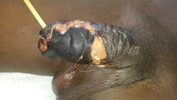
The patient presented at our hospital, with severe pain that had been gradual in onset. He also had symptoms of dysuria and obstructive lower urinary tract symptoms caused by the inflammatory swelling of the penis. He had no fever, an unremarkable medical history, and reported to be compliant to the prescribed antibiotics. Physical examination showed a swollen penis with black necrotic plaques over an almost circular area covering about 60% of the penis, and a foul odour (figure 1). Palpation of the penis caused an exudation of a relatively large amount of purulent exudate from underneath the necrotic plaques but did not reveal any crepitus. There was no tenderness and no signs of inflammation at the perianal area and testicles. A reactively enlarged lymph node was seen in the left groin.
Figure 1.

Penis of the patient on day 1. A urine catheter was placed at admission to help alleviate obstructive lower urinary tract symptoms and facilitate adequate wound hygiene.
Investigations
Initial laboratory investigation showed a C reactive protein of 122 (n=0–5), leucocytes of 6.0 (n=4.0–11.0), haemoglobin of 8.4 (n=8.7–11.2), sodium of 136 (n=132–148), creatinine of 87 (60–110) and glucose of 12.5 (4.0–6.5). The calculated Laboratory Risk Indicator for Necrotizing Fasciitis (LRINEC) score was 1, indicating a <5% risk of necrotising fasciitis.7 The patient had, furthermore, been found to be HIV negative on admission.
Differential diagnosis
The differential diagnosis included a necrotising fasciitis and cellulites. Although there was extensive skin necrosis of the penis, the latter was considered more likely as the LRINEC score was 1.
Treatment
Inpatient care
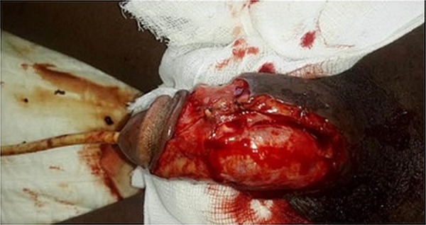
On admission, a urinary catheter was inserted to alleviate lower urinary tract symptoms and avoid contact of the infected wound with urine. Necrotectomy was performed under local anaesthesia (lidocaine 1%). Owing to necrosis, the whole prepuce was excised, taking along a major part of the necrotic skin of the shaft (figure 2). Wound cultures were taken for microbiological analyses. The patient was placed on intravenous antibiotics (augmentin 1.2 g and metronidazol 500 mg 3 times a day), which were switched to oral antibiotics after 3 days (augmentin 625 mg and metronidazol 500 mg 3 times a day). Oral pain medication was given in the form of paracetamol 1 g 4 times a day and tramadol 50 mg 3 times a day, and was stopped after 2 weeks. The wound was treated with baneomycine ointment two times daily after flushing with isotonic saline and Betadine solution. Wound cultures later showed Escherichia coli, Staphylococcus aureus and Streptococcus agalactiae (table 1). Antibiotics were not adjusted as clinical infection control was already established; the reactive oedema as a part of the inflammatory phase resolved and granulation tissue appeared as the proliferation phase of wound healing continued. After removal of the urinary catheter after 2 weeks, no lower urinary tract symptoms were observed and the patient could be discharged. On discharge, the patient gave informed consent for publication.
Figure 2.

Penis of the patient on day 1 after wound debridement.
Table 1.
Culture and sensitivity analysis of the wound culture
| Antibiotic | Escherichia coli | Staphylococcus aureus | Streptococcus agalactiae |
|---|---|---|---|
| Amoxicillin | 0 | + | |
| Cefotaxime | + | ||
| Ciprofloxacin | + | ||
| Doxycycline | 0 | + | + |
| Gentamycin | + | + | 0 |
| Trimethoprim/sulfamethoxazole | 0 | + | 0 |
| Amoxicillin/clavulanic acid | 0 | ||
| Methicillin | + | ||
| Clindamycin | + | ||
| Erythromycin | + | + | |
| Benzylpenicillin | 0 | + |
0, Resistant; +, Susceptible.
Outpatient care
Two weeks after discharge, new wound cultures were taken, which were negative for growth of bacteria. Consequently, the patient was referred to a plastic surgeon for a skin mesh graft. However, the patient refused the placement of a skin graft for unknown reasons and a wait-and-see policy was applied.
Outcome and follow-up
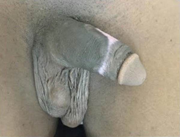
The wound healing continued uncomplicated with good granulation (figure 3). During the following months, there were no symptoms regarding micturition, erections and intercourse.
Figure 3.

Penis of the patient after 2 months. There were no symptoms regarding micturition, erections and intercourse.
Discussion
Although the practice was described in the Kama Sutra,3 the first medical case reports of artificial penile nodules surfaced during the second half of the 20th century, from Southeast Asia, mainly from Thailand.1 2 8 The implantation of penile objects has been linked to the delinquent behaviour of prisoners in Australia, and reported among Japanese racketeers (Yakuza).9 10 In the 90s, similar cases were reported in Saudi,11 Fijian12 and Russian men.5 13 It was not until the beginning of the 21st century that penile implants were reported in Westerners.4 6 10 14 15 Although this is the first case report from Suriname, the practice of implanting artificial penile nodules has probably been around in this country for about a decade.
Owing to the widespread use of penile implants, they have gotten several names. Besides ‘bugru’, meaning ‘bullet’ in Surinamese Creole, the implants have been named ‘Tancho nodules’,1 ‘pearls’,2 ‘Yakuza balls’,8 ‘Fijian penis marbles’,12 ‘fang muk’ and ‘chagan balls’.6
Penile implants are mostly seen in men from low socioeconomic groups, and commonly carried out in prisons. In an interview among 2018 Australian prisoners, by Yap et al,10 5.8% of the respondents reported to have implanted an object under the skin on their penis; 73% did so in prison. Most implants are placed without or with minimal medical knowledge, sometimes by the wearer himself. Commonly, the unanaesthetised skin is cut with a sharp object, such as a part of an aluminium can or a pen.4 The materials used vary from plastic beads, plastic tops of drawing pins,2 glass5 11 and ivory,11 to carved domino fragments4 15 or parts of a plastic toothbrush. In Suriname, the most used material is the glass ball from the pourer on top of a whiskey bottle. The upcoming popularity of artificial penile nodules has also led to the emergence of professional services for the implantation of artificial penile nodules. For instance, a specialised medical centre for artificial penile nodule placement has been founded in Suriname, and there are increasing numbers of Dutch tattoo shops that provide services for the implantation of penile nodules to their customers.16
Although artificial penile nodules can be in situ for over 10 years without giving any problems,8 10 14 there are several reasons for having to remove them. Among these are discomfort,11 complications such as infections (in Suriname referred to as ‘bugritis’)2 4 11 and erosion through the skin.15 In women, the beads have reported to cause abrasions and post-coital vaginal pain.3 However, a recent systematic review found a relatively low prevalence of these complications in men and women, most likely as a consequence of under-reporting bias.14
Learning points.
Artificial penile nodules are penile foreign bodies implanted for aesthetic reasons or sexual pleasure enhancement and are becoming increasingly prevalent within non-Western as well as Western communities.
The full extent of the potential complications of placing foreign bodies in the penile shaft or prepuce is currently under-reported in the literature.
Our case demonstrates that soft-tissue infections requiring urgent medical treatment can occur after self-removal of an artificial penile nodule.
We strongly recommend that the removal of these foreign bodies be performed by medically trained personnel.
Footnotes
Contributors: AJ supervised the medical treatment, proposed the case for a case report and reviewed the final version of the article. MJ wrote the introduction and discussion and reviewed the final version of the article. KHK wrote the case presentation and learning points, and reviewed the final version of the article. SB wrote the treatment, outcome and follow-up, and reviewed the final version of the article.
Competing interests: None declared.
Patient consent: Obtained.
Provenance and peer review: Not commissioned; externally peer reviewed.
References
- 1.Bork K, Bräuninger W [Artificial penile nodules (tancho nodules) in Southeast Asian men]. Hautarzt 1985;36:354–5. [PubMed] [Google Scholar]
- 2.Lim KB, Seow CS, Tulip T et al. . Artificial penile nodules: case reports. Genitourin Med 1986;62:123–5. [DOI] [PMC free article] [PubMed] [Google Scholar]
- 3.Stankov O, Ivanovski O, Popov Z. Artificial penile bodies-from Kama sutra to modern times. J Sex Med 2009;6:1543–8. 10.1111/j.1743-6109.2009.01230.x [DOI] [PubMed] [Google Scholar]
- 4.Hudak SJ, McGeady J, Shindel AW et al. . Subcutaneous penile insertion of domino fragments by incarcerated males in southwest United States prisons: a report of three cases. J Sex Med 2012;9:632–4. 10.1111/j.1743-6109.2011.02551.x [DOI] [PMC free article] [PubMed] [Google Scholar]
- 5.Levy G, Mercer D, Amosi D et al. . Self-implanted artificial nodules: a computed tomography mimic of penile pathology. Acta Radiol 2008;49:236–8. 10.1080/02841850701675685 [DOI] [PubMed] [Google Scholar]
- 6.Wilcher G. Artificial penile nodules—a forensic pathosociology perspective: four case reports. Med Sci Law 2006;46:349–56. 10.1258/rsmmsl.46.4.349 [DOI] [PubMed] [Google Scholar]
- 7.Wong CH, Khin LW, Heng KS et al. . The LRINEC (Laboratory Risk Indicator for Necrotizing Fasciitis) score: a tool for distinguishing necrotizing fasciitis from other soft tissue infections. Crit Care Med 2004;32:1535–41. 10.1097/01.CCM.0000129486.35458.7D [DOI] [PubMed] [Google Scholar]
- 8.Nitidandhaprabhas P. Artificial penile nodules: case reports from Thailand. Br J Urol 1975;47:463 10.1111/j.1464-410X.1975.tb04009.x [DOI] [PubMed] [Google Scholar]
- 9.Tsunenari S, Idaka T, Kanda M et al. . Self-mutilation. Plastic spherules in penile skin in yakuza, Japan's racketeers. Am J Forensic Med Pathol 1981;2:203–7. 10.1097/00000433-198109000-00003 [DOI] [PubMed] [Google Scholar]
- 10.Yap L, Butler T, Richters J et al. . Penile implants among prisoners-a cause for concern? PLoS ONE 2013;8:e53065 10.1371/journal.pone.0053065 [DOI] [PMC free article] [PubMed] [Google Scholar]
- 11.Marzouk E. Implantation of beads into the penile skin and its complications. Scand J Urol Nephrol 1990;24:167–9. 10.3109/00365599009180852 [DOI] [PubMed] [Google Scholar]
- 12.Norton SA. Fijian penis marbles: an example of artificial penile nodules. Cutis 1993;51:295–7. [PubMed] [Google Scholar]
- 13.Serour F. Artificial nodules of the penis. Report of six cases among Russian immigrants in Israel. Sex Transm Dis 1993;20:192–3. [PubMed] [Google Scholar]
- 14.Fischer N, Hauser S, Brede O et al. . Implantation of artificial penile nodules—a review of literature. J Sex Med 2010;7:3565–71. 10.1111/j.1743-6109.2009.01659.x [DOI] [PubMed] [Google Scholar]
- 15.Flynn RM, Jain S. A domino effect? The spread of implantation of penile foreign bodies in the prison system. Urol Case Rep 2014;2:63–4. 10.1016/j.eucr.2014.01.006 [DOI] [PMC free article] [PubMed] [Google Scholar]
- 16.Natascha F. Balletjes van seksueel genot: een kijkje in de boegroepraktijken. [Balls of sexual pleasure: a look into bugru practice.] Retreived on 24 September 2015. http://www.elinea.nl/artikel/balletjes-van-seksueel-genot-een-kijkje-in-de-boegroepraktijken-2.


