
Figure 3.
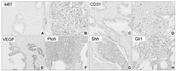
A–E, Expression of ki67, CD31 and vascular endothelial growth factor (VEGF) in non-neoplastic peripheral zone (PZ) and primary prostate carcinoma. Expression of Ki67 is lower in non-neoplastic PZ (A) compared to prostate carcinoma (B). Number of CD31-positive vessels increases from non-neoplastic prostate (C) to prostate carcinoma (D). Low expression levels of VEGF in adjacent non-neoplastic epithelium compared to the malignant cells (E). F–H, Expression of Ptch (F), Shh (G) and Gli1 (H) in bone marrow metastases. Note that Ptch expression in epithelial cells of a metastatic tumour is higher than that in primary carcinomas in Figure 2H and 2I.