ABSTRACT
We previously identified three noncontiguous regions on Bacillus anthracis plasmid pXO1 that comprise a system for accurate plasmid partitioning and maintenance. However, deletion of these regions did not decrease retention of certain shortened pXO1 plasmids during vegetative growth. Using two genetic tools developed for DNA manipulation in B. anthracis (the Cre-loxP and Flp-FRT systems), we found two other noncontiguous pXO1 regions that together are sufficient for plasmid stability. This second pXO1 maintenance system includes the tubZ and tubR genes, characteristic of a type III partitioning system, and the IntXO recombinase gene (GBAA_RS29165), encoding a tyrosine recombinase, along with its adjacent 37-bp perfect stem-loop (PSL) target. Insertion of either the tubZ and tubR genes or the IntXO-PSL system into an unstable mini-pXO1 plasmid did not restore plasmid stability. The need for the two components of the second pXO1 maintenance system follows from the sequential roles of IntXO-PSL in generating monomeric circular daughter pXO1 molecules (thereby presumably preventing dimer catastrophe) and of TubZ/TubR in partitioning the monomers during cell division. We show that the IntXO recombinase deletes DNA regions located between two PSL sites in a manner similar to the actions of the Cre-loxP and Flp-FRT systems.
IMPORTANCE Tyrosine recombinases catalyze cutting and joining reactions between short specific DNA sequences. Three types of reactions occur: integration and excision of DNA segments, inversion of DNA segments, and separation of monomeric forms from replicating circular DNA molecules. Here we show that the newly discovered site-specific IntXO-PSL recombinase system that contributes to the maintenance of the B. anthracis plasmid pXO1 can be used for genome engineering in a manner similar to that of the Cre-loxP or Flp-FRT system.
INTRODUCTION
The large low-copy-number pXO1 plasmid (181,677 bp) of Bacillus anthracis encodes the anthrax toxin proteins and other virulence-related factors. A pXO1 minireplicon plasmid (pMR) comprised of two essential open reading frames (ORFs) (GBAA_RS28535 and GBAA_RS28545) (Fig. 1A) replicates but is not stably maintained in B. anthracis, whereas the full-size parent pXO1 plasmid (carrying 217 ORFs) is extremely stable under the same growth conditions (1). (Table S1 in the supplemental material lists all genes and proteins discussed in this work.) Recently we found that retention of pMR can be stabilized by insertion of several noncontiguous pXO1 regions containing three genes which work cooperatively to achieve plasmid maintenance: amsP (GBAA_RS28725), minP (GBAA_RS28775), and sojP (GBAA_RS28785) (2). The minP and sojP genes encode proteins belonging to the ParA/MinD family described by Lutkenhaus (3). MinD is involved in spatial regulation of the cytokinetic Z ring, and ParA proteins are involved in chromosome and plasmid segregation (3).
FIG 1.
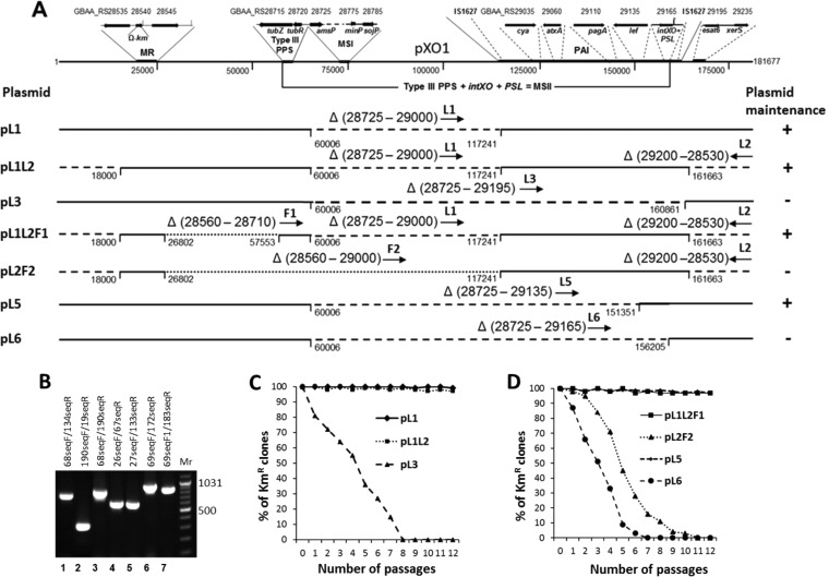
Effects of large deletions within pXO1 on plasmid stability. (A) pXO1 structure, with the minireplicon (MR), type III plasmid partitioning system (type III PPS), first pXO1 maintenance system (MSI), newly discovered second pXO1 maintenance system (MSII), and pathogenicity island (PAI) indicated. The MR structure was adapted from reference 1 and shown with the Ω Kmr cassette inserted into pXO1 GBAA_RS28540 to produce pXO1K (2). The type III PPS structure is based on data from reference 36 and does not include the tubY gene, which was characterized as an essential part of the MSI (amsP) (2). The MSII system, identified in this study, includes the type III PPS combined with the IntXO tyrosine recombinase gene and the recombinase target PSL. The PAI structure, with the IS1627 insertion sequence and B. anthracis toxin components indicated, as well as all other genes indicated on the pXO1 scheme, were prepared according to the Ames Ancestor plasmid pXO1 complete sequence (NCBI GenBank accession no. NC_007322.2). The numbers located below and on both sides of pXO1 represent nucleotide numbers in the complete sequence. The GBAA_RS symbols at the top of the pXO1 diagram are locus tag numbers for the Ames Ancestor plasmid pXO1. The pXO1 regions deleted in each construct are shown in parentheses following the Greek letter Δ (the numbers are for the ORFs in the deleted area). The orientations of loxP (deletions produced with the Cre-loxP system are indicated with dashed lines) and FRT (deletions produced with the Flp-FRT system are indicated with dotted lines) sites that replaced the deleted regions are indicated by arrows and L and F designations, respectively. The numbers located below the deletions are the first and last base pairs included in the deletions. (B) PCR verification of deletions. Primers used to verify the retention of specific segments are indicated at the top of the gel. Lanes: 1, Ames 35K(pL1); 2, Ames 35K(pL2); 3, Ames 35K (pL3); 4, Ames 35K(pL1L2F1); 5, Ames 35K(pL2F2); 6, Ames 35K(pL5); 7, Ames 35K(pL6); Mr, GeneRuler DNA ladder mix for size determination (e.g., 1,031 bp). (C) Percentages of kanamycin-resistant (i.e., plasmid-containing) bacteria in cultures of B. anthracis Ames 35 containing the pXO1K variants with deletions identifying the PAI role in pXO1 maintenance. Bacteria were subcultured every 12 h on LB agar without kanamycin and grown at 37°C. Bacteria from each subculture were diluted and plated on LB agar, with and without kanamycin (20 μg/ml), to determine the fractions retaining the plasmids. Maximum root mean square deviations in the percentage of kanamycin-resistant colonies did not exceed 10%. (D) Percentages of kanamycin-resistant (i.e., plasmid-containing) bacteria in cultures of B. anthracis Ames 35 containing the pXO1K variants with deletions identifying the type III PPS and IntXO involvement in pXO1 maintenance. Maximum root mean square deviations in the percentage of kanamycin-resistant colonies did not exceed 10%.
The amsP-, minP-, and sojP-encoded proteins, comprising the plasmid maintenance system (maintenance system I [MSI]) identified in our prior work (2), differ from the proteins comprising the type III plasmid partitioning system (PPS) found in the well-characterized large Bacillus thuringiensis virulence plasmid pBtoxis (4). This type III PPS includes the DNA-binding protein TubR, which interacts with the centromeric tubC site of pBtoxis, and the polymer-forming protein TubZ. Both proteins are involved in promoting stability of pBtoxis in B. thuringiensis (5). Aylett and Lowe identified the tubC sequence and described the structure of the centromeric complex of TubZRC involved in pBtoxis partitioning (6). Hoshino et al. showed that the corresponding TubZ protein encoded on pXO1 also assembles into polymers, both in vivo and in vitro, via association with the DNA-binding protein TubR (7, 8). Although another large Bacillus plasmid, pBsph from Bacillus sphaericus, was also shown to contain an active type III PPS (9, 10), we could not confirm that a type III system operates in B. anthracis to maintain plasmid pXO1 during vegetative cell division. This was despite the presence in pXO1 of a tubZ/tubR gene cluster having the same configuration as that in pBtoxis (2). To resolve this apparent contradiction, we attempted to find factors on pXO1 that could activate the putative type III PPS and play a role in plasmid stabilization.
In this study, we describe work to localize components of a second maintenance system (MSII) of B. anthracis plasmid pXO1. Two genetic tools developed for DNA manipulation in B. anthracis (the Cre-loxP and Flp-FRT systems) were employed to delete pXO1 regions upstream and downstream of the MR region (Fig. 1A) (1, 2). This approach identified a second, independent pXO1 maintenance system (MSII) located in two regions: one containing the tubZ and tubR genes that are characteristic of a type III PPS and another carrying a tyrosine recombinase gene (GBAA_RS29165; encodes a protein we designate the IntXO recombinase) along with its adjacent 37-bp stem-loop target (perfect stem-loop [PSL]). The requirement for multiple components in the second pXO1 maintenance system suggests a two-step process: first, IntXO-PSL ensures conversion of pXO1 replicative forms to monomeric circular plasmids, thereby preventing dimer catastrophe (11); and second, the TubZ/TubR system promotes partitioning of the monomers to the daughter cells during mother cell segregation (12). The existence of two independent plasmid maintenance systems seems redundant but may serve to ensure plasmid retention.
MATERIALS AND METHODS
Bacterial growth conditions and phenotypic characterization.
Escherichia coli strains were grown in Luria-Bertani (LB) broth and used as hosts for cloning. LB agar was used for selection of transformants (13). B. anthracis strains were also grown in LB medium. Antibiotics (Sigma-Aldrich, St. Louis, MO) were added to the medium when appropriate to give the following final concentrations: ampicillin (Ap), 100 μg/ml (only for E. coli); erythromycin (Em), 400 μg/ml for E. coli and 10 μg/ml for B. anthracis; spectinomycin (Sp), 150 μg/ml for both E. coli and B. anthracis; and kanamycin (Km), 20 μg/ml for B. anthracis. SOC medium (Quality Biologicals, Inc., Gaithersburg, MD) was used for outgrowth of transformation mixtures prior to plating on selective media to isolate transformants. B. anthracis spores were prepared by growth on NBY-Mn agar (nutrient broth, 8 g/liter; yeast extract, 3 g/liter; MnSO4·H2O, 25 mg/liter; agar, 15 g/liter) at 30°C for 5 days (14). Spores and vegetative cells of B. anthracis were visualized with a Nikon Eclipse E600W light microscope.
DNA isolation and manipulation.
Preparation of plasmid DNA from E. coli, transformation of E. coli, and recombinant DNA techniques were carried out by standard procedures (13). E. coli SCS110 competent cells were purchased from Agilent Technologies (Santa Clara, CA), and E. coli TOP10 competent cells were purchased from Life Technologies (Grand Island, NY). Recombinant plasmid construction was carried out in E. coli TOP10 cells. Plasmid DNA from B. anthracis was isolated according to the Qiagen protocol for the purification of plasmid DNA from Bacillus subtilis (Qiagen, Germantown, MD). Chromosomal DNA from B. anthracis was isolated with a Wizard genomic purification kit (Promega, Madison, WI) in accordance with the protocol for isolation of genomic DNA from Gram-positive bacteria. B. anthracis was electroporated with unmethylated plasmid DNA isolated from E. coli SCS110 (dam dcm). Electroporation-competent B. anthracis cells were prepared and transformed as previously described (1).
Restriction enzymes, T4 ligase, T4 DNA polymerase, and alkaline phosphatase were purchased from New England BioLabs (Ipswich, MA). The pGEM-T Easy vector system (Promega) was applied for PCR fragment cloning. Phusion high-fidelity DNA polymerase from New England BioLabs was used for fragment PCR. Ready-To-Go PCR beads (GE Healthcare UK Limited) were used for routine DNA rearrangement analysis as described previously (1). All constructs were verified by DNA sequencing and/or restriction enzyme digestion. The oligonucleotides, plasmids, and strains used in this study are listed in Tables 1 to 3, respectively.
TABLE 1.
Oligonucleotides used in this study
| Oligonucleotide pair | Sequences (5′–3′)a | Relevant property | Restriction sites |
|---|---|---|---|
| 46f/68r | ACTGCTCGAGTGCTCCCTCAAGTCGTGCAGTCA/ACTGACTAGTCCATCGCTTAACTGCTCATCAATC | Primer pair to amplify left fragment of L1, L5, L6, and L3 deletions | XhoI/SpeI |
| 133F/137R | ACTGCTCGAGGGTGTTTTTCGTTTGAAGAC/ACTGACTAGTTACTTTTCGGCCTCTTCCTG | Primer pair to amplify right fragment of L1 and F2 deletions | XhoI/SpeI |
| 68seqF/134seqR | TACTTACGAAATATAACCCG/TGTTCCAATTCACTTAGCTC | Primer pair to verify L1 deletion | |
| 190F/191R | ACTGACTAGTCAGCTTATGGTATAGCAACG/ACTGCTCGAGGTCTTGGATAACCACTATCATA | Primer pair to amplify left fragment of L2 deletion | SpeI/XhoI |
| 19F/19R | ACTGACTAGTTCAGGATCAAAGAAACCAGC/ACTGCTCGAGGCAGAGTTCTGAAACAAAGGTATT | Primer pair to amplify right fragment of L2 deletion | SpeI/XhoI |
| 190seqF/19seqR | GGGGAAAGACAGCGATAAGT/GTTAATGGAAAGAGATGGCT | Primer pair to verify L2 deletion | |
| 190F1/191R1 | ACTGCTCGAGCAGCTTATGGTATAGCAACG/ACTGACTAGTGTCTTGGATAACCACTATCATA | Primer pair to amplify right fragment of L3 deletion | XhoI/SpeI |
| 68seqF/190seqR | TACTTACGAAATATAACCCG/TGGCCCCTCATAAGTATATG | Primer pair to verify L3 deletion | |
| 26F/29R | ACTGCTCGAGCATTTTAATTGTTCCAGGGG/ACTGACTAGTATTGCCCGTCACTCCTTTCT | Primer pair to amplify left fragment of F1 and F2 deletions | XhoI/SpeI |
| 66F/67R | ACTGCTCGAGGTTCGGACAGCCTAACAATTTA/ACTGACTAGTGTGCTTTAGCAAACGAATATAC | Primer pair to amplify right fragment of F1 deletion | XhoI/SpeI |
| 26seqF/67seqR | TCCTTAATTCCGGATCAATCAC/CATTCTAAACAAAAGACCGACA | Primer pair to verify F1 deletion | |
| 27seqF/133seqR | CTATCTTTTTAATTGCAGCT/ATTGTTTTCCTACAGTTTGT | Primer pair to verify F2 deletion | |
| 172F/172R | ACGTCTCGAGAAGCATCTTTCCCATAAATGTC/ACGTACTAGTCCAAAAAAATCACACTGTCAAG | Primer pair to amplify right fragment of L5 deletion | XhoI/SpeI |
| 69seqF/172seqR | AATATTACGGAATTCAGTCCCGCTATATATGC/GGAAGAGCATTTAAAGGAAATCATGAAACAC | Primer pair to verify L5 deletion | |
| 182F/184R | ACGTCTCGAGGCCCATGCCCATAATTTTTTCGAAATGACT/ACTGTCTAGAAACGATATCCGCCATCTGCTTTCCA | Primer pair to amplify right fragment of L6 deletion | XhoI/XbaI |
| 69seqF1/183seqR | CTTACGAAATATAACCCGTAATAACAACCGT/TCCTCCTACCGCATTTGGATTGAAA | Primer pair to verify L6 deletion | |
| 67F/69R | ACGTCGATCGGTTCGGACAGCCTAACAATTTATTG/ACGTACCGGCGTCATCAATCTCTTCAGCATAAACG | Primer pair to amplify tubZR gene cluster | PvuI/BsrFI |
| IntF/IntR | ACGTCATATGAATATTATAAATTACGAACA/ACGTGATATCGCATCAAATCGAGAATCTTC | Primer pair to amplify IntXO recombinase gene | NdeI/EcoRV |
| 183F/181R | ACGTCCCGGGGAGTACTCCTTATACTGAAGATAC/ACGTCCCGGGAAAATAGGAAGAAACCGATG | Primer pair to amplify IntXO recombinase gene along with PSL sequence | SmaI/SmaI |
| ESATF/ESATR | ACGTACTAGTCGGACAGGATGAAAATGTATTTAGTAAGCTT/ACGTCTCGAGAGAAGAACCTGGATCAATGGAGAAA | Primer pair to amplify DNA region downstream of esat6 gene | SpeI/XhoI |
| seqFESAT/seqRESAT | ACATTCAACTTCTACTCTCAAACAACTC/GGATAACTCCTTAATGATTCCGATTGT | Primer pair to verify esat6 gene deletion | |
| PSCP1F/PSCPR | CGGGAGGAAATAATTCTATG/GCTTGGTTTAGTTGTCAAGAGAGAATAACT | Primer pair to verify Ω Kmr cassette deletion |
Restriction enzyme recognition sites are underlined.
TABLE 2.
Plasmids used in this study
| Plasmid | Relevant characteristic(s) | Source or reference |
|---|---|---|
| pGEM-T Easy | Cloning vector for PCR products; Apr in E. coli | Promega |
| pUC4-ΩKM2 | pUC4 carrying an Ω element with a kanamycin resistance marker; Kmr in E. coli and B. anthracis | 2 |
| pSW4 | Carries promoter of anthrax protective antigen gene (pag) in a shuttle plasmid; Apr in E. coli; Kmr in B. anthracis | 35 |
| pSW4-IntXO | Nde-EcoRV PCR fragment amplified with IntF/IntR primers and inserted into pSW4 between NdeI and SmaI sites | This work |
| pCrePAS2 | Contains entire Cre recombinase gene under the control of the pagA promoter; has permissive and restrictive temperatures of 30°C and 37°C, respectively, for B. anthracis and can easily be isolated from E. coli strains grown at 37°C | 2 |
| pFPAS | Contains Flp recombinase gene under the control of the pagA promoter; shuttle vector based on plasmids pHY304 and pUC18; contains Ω Spr cassette, conferring Spr in both E. coli and B. anthracis; has permissive and restrictive temperatures of 30°C and 37°C, respectively, for B. anthracis and can easily be isolated from E. coli grown at 37°C | 2 |
| pIntPAS | Contains entire IntXO recombinase gene under the control of the pagA promoter; has permissive and restrictive temperatures of 30°C and 37°C, respectively, for B. anthracis and can easily be isolated from E. coli strains grown at 37°C; Spr in both E. coli and B. anthracis | This work |
| pSC | Contains multiple restriction sites flanked by two direct loxP sites; has permissive and restrictive temperatures of 30°C and 37°C, respectively, for B. anthracis; Apr in E. coli; Emr in both E. coli and B. anthracis | 1 |
| pSCF | Contains multiple restriction sites flanked by two direct FRT sites; has permissive and restrictive temperatures of 30°C and 37°C, respectively, for B. anthracis and can easily be isolated from E. coli strains grown at 37°C; Apr in E. coli; Emr in both E. coli and B. anthracis | 2 |
| pAM-PSL | Contains a multiple-cloning site between two directly repeated FRT sequences, a ColE1 origin of replication, and an Apr gene for selection in E. coli | GeneArt |
| pSCP | Contains multiple-restriction site flanked by two direct PSL sites; has permissive and restrictive temperatures of 30°C and 37°C, respectively, for B. anthracis and can easily be isolated from E. coli strains grown at 37°C; Apr in E. coli; Emr in both E. coli and B. anthracis | This work |
| pSCP-ESAT | pSCP with ESATF/ESATR PCR fragment inserted into the pSCP multiple-restriction site | This work |
| pSCP(Ω-km) | pSCP with the Ω Kmr cassette from pUC4-ΩKM2 inserted into the pSCP site | This work |
| pSCPΔ(Ω-km) | pSCP(Ω-km) with Ω Kmr cassette deleted; contains one PSL site, in contrast to the two PSL sites in pSCP | This work |
| pMR | pXO1 minimal replicon; Spr in both E. coli and B. anthracis | 1 |
| pZR | A BsrFI-PvuI fragment containing the tubZ-tubR cluster was inserted into pMR, replacing the BsrFI-PvuI fragment within the Apr gene | This work |
| pZR1 | A SmaI fragment containing the IntXO recombinase gene and its upstream PSL sequence was inserted into pZR, replacing the AfeI fragment within the GBAA_RS28540 ORF | This work |
| pZR2 | pZR1 deleted of the BsrFI-PvuI fragment containing the tubZ-tubR cluster | This work |
| pMRmut | pMR with four spontaneous point mutations in the pXO1 replication region; temperature sensitive for replication in B. anthracis | 1 |
| pMRmut-PI | A SmaI fragment containing the PSL sequence upstream of the IntXO recombinase gene was inserted into pMRmut instead of the AfeI fragment within the GBAA_RS28540 ORF | This work |
| DHFR control plasmid | Contains the dihydrofolate reductase gene under the control of the T7 promoter; Apr in E. coli | NEB |
| pIntCP | DHFR control plasmid with Nde-EcoRI fragment replaced by an Nde-EcoRI fragment of pIntPAS carrying the whole IntXO recombinase gene | This work |
TABLE 3.
B. anthracis strains used in this study
| Strain | Relevant characteristics | Reference |
|---|---|---|
| Ames 33 | Ames pXO1− pXO2− | 1 |
| Ames 35 | Ames pXO1+ pXO2− | 1 |
| Ames 35K | Ames 35 with pXO1K plasmid | 2 |
| Ames 35K Δ(28560–29195) | Ames 35K with GBAA_RS(28560–29195) replaced with loxP | 2 |
| Ames 35K Δ(29020–28520) | Ames 35K with GBAA_RS(29020–28520) replaced with loxP | 2 |
| Ames 35K(pL1) | Ames 35K with GBAA_RS(28725–29000) replaced with clockwise-oriented loxP | This work |
| Ames 35K(pL1L2) | Ames 35K with GBAA_RS(28725–29000) replaced with clockwise-oriented loxP and GBAA_RS(29200–28530) replaced with counterclockwise-oriented loxP | This work |
| Ames 35K(pL3) | Ames 35K with GBAA_RS(28725–29195) replaced with clockwise-oriented loxP | This work |
| Ames 35K(pL1L2F1) | Ames 35K with GBAA_RS(28725–29000) replaced with clockwise-oriented loxP, GBAA_RS(29200–28530) replaced with counterclockwise-oriented loxP, and GBAA_RS(28560–28710) replaced with FRT sequence | This work |
| Ames 35K(pL2F2) | Ames 35K with GBAA_RS(28560–29000) replaced with counterclockwise-oriented loxP and GBAA_RS(29200–28530) replaced with FRT sequence | This work |
| Ames 35K(pL5) | Ames 35K with pXO1 GBAA_RS(28725–29135) replaced with clockwise-oriented loxP | This work |
| Ames 35K(pL6) | Ames 35K with pXO1 GBAA_RS(28725–29165) replaced with clockwise-oriented loxP | This work |
| Ames 35K Δesat6 | Ames 35K with pXO1K region containing the esat6 gene replaced with PSL site | This work |
Recombinase systems used for deletion analysis.
Deletions in pXO1 were generated with the Cre-loxP system as previously described (1), employing plasmids we designate generically as pSC, for single-crossover plasmid, together with pCrePAS2 for Cre recombinase production (2). Deletions were also generated with the Flp-FRT system, employing plasmids pSCF (an analog of the pSC plasmid) and pFPAS (a plasmid with all the characteristics of pCrePAS2, but containing the flp gene instead of the cre gene) as previously described (2). Both the Cre-loxP and Flp-FRT recombinase systems were used in accordance with the scheme presented in Fig. 1 of reference 1. Sequences and maps of the plasmids used here are available upon request.
A new IntXO-PSL recombinase system based on the IntXO recombinase of B. anthracis identified in this work was created for use in a manner analogous to that of the Flp-FRT system described in the previous paragraph. To create an analog of the pSCF plasmid that we designate pSCP, we cut pSCF with BstZ17I/PvuI/PstI and ligated the digestion product with a BstZ17I/PvuI-restricted fragment of pAM-PSL, which contains a multiple-cloning site between two directly repeated PSL sequences (described below), a ColE1 origin of replication, and an Apr gene for selection in E. coli. The pAM-PSL plasmid was synthesized according to our design by GeneArt/Life Technologies (Grand Island, NY). The resulting pSCP plasmid is a shuttle plasmid with all the characteristics of pSCF, except that it contains repeats of the 37-bp PSL sequence instead of the FRT sequence. All of the endonuclease restriction sites located between the two direct PSL sequences are unique sites (PvuII, XhoI, SalI, ClaI, HindIII, EcoRV, EcoRI, PstI, SmaI, BamHI, SpeI, XbaI, SacII, and SacI). pSCP also contains two additional unique sites in comparison with pSCF: an XbaI site within the multiple-restriction site and a ZraI site downstream of the second PSL (see Fig. 4B). To create an analog of the pFPAS plasmid that we designate pIntPAS, we amplified the IntXO gene by PCR with the IntF/IntR primers to insert the amplified fragment into the NdeI/EcoRV sites of pFPAS, replacing the flp gene. The resulting pIntPAS plasmid is a shuttle plasmid with all the characteristics of pFPAS, except that it contains the IntXO recombinase gene instead of flp. Another plasmid producing IntXO recombinase, pMRmut-PI, was created from the temperature-sensitive vector pMRmut (1). The IntXO recombinase gene along with a PSL sequence was amplified by PCR with the 183F/181R primers and inserted into the AfeI sites of pMRmut. pMRmut-PI can easily be isolated from E. coli strains grown at 37°C and eliminated from B. anthracis at the same temperature.
FIG 4.
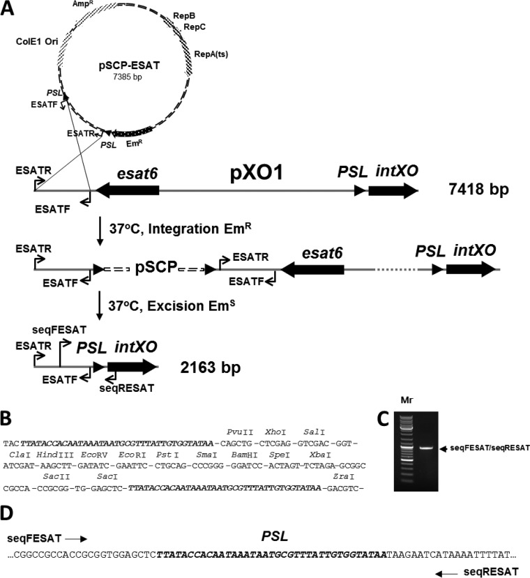
Creation and analysis of the deletion generated in pXO1K by insertion of an additional PSL site in the vicinity of the native PSL. (A) Scheme for insertion of an additional PSL site and consequent deletion of the DNA region flanked by the native and added PSL sites (details of the experiment are described in Results). (B) Sequence of the multiple-cloning site of pSCP flanked by two direct PSL sites (indicated in bold). The sequence is shown with 6-bp unique restriction site sequences (and several linkers) separated by dashes, with the sites identified above the sequence. (C) PCR verification of the deletion in Ames 35K Δesat6(pXO1). Primers used to verify the deletion are indicated at the right side of the gel and in panel A. The PCR fragment amplified with the primers is indicated by the arrow. Mr, GeneRuler DNA ladder mix for size determination (as in Fig. 1B). (D) The single PSL site remaining was found by sequencing of the PCR fragment with the indicated primers.
Plasmid construction for identification and analysis of the second pXO1 maintenance system.
Deleted variants of pXO1 were created in vivo (i.e., in pXO1K, within B. anthracis) to identify regions required for plasmid maintenance. In the Ames 35(pXO1K) strain (Table 2), the native pXO1 plasmid has the nonessential ORF GBAA_RS28540 (which lies between the two essential ORFs of the MR region) replaced by an Ω Kmr cassette (2). To produce the deletion designated L1 that covers ORFs GBAA_RS28725 to GBAA_RS29000 (Fig. 1A, pL1), we cloned two fragments, amplified with primers 46F/68R (left fragment) and 133F/137R (right fragment), into the pSC vector; both fragments, as well as other fragments, were used according to the Cre-loxP system protocol previously described. To produce the deletion L2 (Fig. 1A, pL1L2), two fragments, amplified with primers 190F/191R (left fragment) and 19F/19R (right fragment), were cloned into pSC in opposite orientations to change the orientation of the inserted loxP sequence. Three more deletions created with the Cre-loxP system included L3 (Fig. 1A, pL3), made using primers 46F/68R for the left fragment and 190F1/191R1 for the right fragment; L5 (Fig. 1A, pL5), made using primers 46F/68R for the left fragment and 172F/172R for the right fragment; and L6 (Fig. 1A, pL6), made using primers 46F/68R for the left fragment and 182F/184R for the right fragment. The Flp-FRT system was used twice. The first use was for deletion of the F1 region within pL1L2F1. To produce this deletion, fragments amplified with primers 26F/29R (left fragment) and 66F/67R (right fragment) were cloned into the pSCF vector. In the second case, the Flp-FRT system was used to delete the F2 interval from pL1L2. To produce this deletion, fragments were amplified with primers 26F/29R (left fragment) and 133F/137R (right fragment) and inserted into pSCF. All fragments cloned into pSCF were used according to the Flp-FRT system method described in the previous section. In both cases, the clockwise orientation of the FRT site was saved. All deletions created with the Cre-loxP and Flp-FRT systems are indicated in Fig. 1.
Direct cloning into the pMR plasmid was used to estimate the roles of gene clusters involved in the second pXO1 maintenance system. For this purpose, a fragment containing the tubZR cluster was amplified from pXO1 by use of Phusion high-fidelity DNA polymerase (New England BioLabs, Ipswich, MA) and the 67F/69R primer pair. The fragment was inserted between the BsrFI and PvuI sites of pMR to produce pZR. A fragment containing the IntXO recombinase gene along with the PSL sequence was amplified with the 183F/181R primer pair and inserted between the AfeI sites of pZR to produce pZR1. pZR1 was cut with BsrFI and PvuI, and the sticky ends were filled with corresponding deoxynucleotides by use of T4 DNA polymerase according to the New England BioLabs protocol and then ligated to produce pZR2.
Two additional plasmids were created to estimate IntXO-PSL recombinase system functionality. For the pSCP-ESAT plasmid, a fragment located downstream of esat6 (GBAA_RS29195) was amplified from pXO1 by use of Phusion high-fidelity DNA polymerase (New England BioLabs, Ipswich, MA) and the ESATF/ESATR primer pair. The fragment was inserted between the SpeI and XhoI sites of pSCP to produce pSCP-ESAT (see Fig. 4). The pSCP(Ω-km) plasmid was constructed by insertion of the Ω Kmr cassette flanked by two BamHI sites from pUC4-ΩKM2 into the BamHI site of pSCP.
Estimation of IntXO activity in vitro.
The IntXO protein was synthesized using a PURExpress in vitro protein synthesis kit (New England BioLabs). The NdeI-EcoRI fragment of the pIntPAS plasmid containing the IntXO recombinase gene was inserted into the DHFR control plasmid, replacing the DHFR gene. The resulting pIntCP plasmid was used as a template for protein expression. The PURExpress reaction mixture contained 750 ng of template with 10 μl solution A and 7.5 μl solution B (solutions provided with the kit). A murine RNase inhibitor (New England BioLabs) was added to each 30-μl reaction mixture to inhibit any RNases present in the template, as recommended. A negative-control reaction mixture devoid of template DNA and a positive-control reaction mixture with 250 ng of DHFR control plasmid DNA provided with the kit were also made in the same manner. The reaction mixtures were incubated at 37°C for 2.5 h, and reactions were stopped by placing the samples on ice. For SDS-PAGE analysis, 5-μl samples of the resulting protein mixtures were boiled at 95°C for 5 min in loading dye. The samples were analyzed on a Novex WedgeWell 10 to 20% Tris-glycine protein minigel (Life Technologies, Carlsbad, CA) with an unstained protein ladder (broad range, 10 to 250 kDa; New England BioLabs) and stained with Coomassie R250 staining solution. Reaction mixtures with and without the pIntCP template were treated with 0.5 μg/ml of RNase A (Qiagen, Germantown, MD) and used for estimation of IntXO activity in vitro. For this purpose, 1.5 μg of pSCP(Ω-km) was mixed with 2.5 μl of reaction mixture in 30 μl of reaction buffer (33 mM NaCl, 10 mM MgCl2, 50 mM Tris-HCl, pH 7.5). The mixtures were incubated at 37°C for 1 h, heated at 70°C for 10 min, and run in a 1% agarose gel in Tris-acetate buffer (Quality Biological, Gaithersburg, MD).
Segregational stability assay.
The stabilities of Kmr pXO1K derivative plasmids in Ames 35 (a pXO1+ pXO2− strain), as well as pMR and its Spr derivative plasmids in Ames 33 (a pXO1− pXO2− strain), were determined using previously described methods (2). Briefly, the B. anthracis strains containing plasmids were grown on LB agar with the corresponding antibiotic to obtain separated colonies. The separated colonies were spread on LB agar as a strip (approximately 3 × 20 mm). Twelve sequential passages on nonselective LB agar were performed from such strips, at 12-h intervals. After each passage, bacteria were diluted in phosphate-buffered saline (Quality Biological, Inc., Gaithersburg, MD) and titrated, and aliquots were plated on agar with and without antibiotic to determine the percentage of cells retaining the plasmid.
DNA sequencing and bioinformatic analyses.
Plasmid and chromosomal DNAs were sequenced using primers listed in Table 1. Primers for sequencing and PCR were synthesized by Integrated DNA Technologies, Inc., Coralville, IA. Sequences were determined using a primer walking strategy (Macrogen, Rockville, MD). Sequence data were assembled using the Vector NTI software (Life Technologies, Carlsbad, CA). The National Center for Biotechnology Information (NCBI) BLAST and FASTA programs (http://www.ncbi.nlm.nih.gov) as well as the Pfam database (http://pfam.sanger.ac.uk) were used for putative conserved-domain and homology searches in GenBank and nonredundant protein sequence databases. RepeatAround (15) software was used to locate direct and inverted repeats. The Mfold Web server for nucleic acid folding (http://mfold.rna.albany.edu/cgi-bin/mfold-3.4.cgi) was used for DNA palindrome folding and Gibbs free energy determination.
RESULTS
Deletions generated by tyrosine recombinases identify pXO1 regions constituting the second plasmid maintenance system.
The Cre-loxP and Flp-FRT systems were used to create large deletions of various regions of pXO1K (Fig. 1A), the pXO1 variant with a kanamycin resistance cassette inserted in the minireplicon region. All seven deletions shown were confirmed by PCR analysis with primers flanking each deleted region (Fig. 1B). Sequencing of the PCR products confirmed replacement of the deleted areas with either the loxP or FRT sequence (data not shown). The first deletion removed genes encoding ORFs 28725 to 29000. This region includes genes of the first maintenance system, i.e., amsP, minP, and sojP (2). However, the pL1 deletion plasmid was stable in host cells during 12 passages without selective pressure (Fig. 1C). This was clear evidence that another maintenance system is encoded on pXO1. Addition of a second deletion, L2, created plasmid pL1L2. The newly deleted region included ORF 29235, encoding a protein annotated as XerS because of its similarity to a tyrosine recombinase of Streptococcus suis that separates chromosomal dimers (16). However, the pL1L2 deletion plasmid remained stable (Fig. 1C), indicating that xerS and other genes in this region are not essential components of the second pXO1 maintenance system. The region retained in the pL1L2 plasmid includes two prominent features: a putative type III plasmid partitioning system (type III PPS; ORFs 28715 to 28720) and nearly all of the region designated a pathogenicity island (PAI; ORFs 28995 to 29190). The third deletion created (Fig. 1A) can be viewed as having the L1 deletion extended to ORF 29195 to make L3. The corresponding plasmid had very low stability (Fig. 1C), indicating that the deleted region contains at least one gene or sequence required for pXO1 stability. This deletion includes genes encoding the first maintenance system (MSI) and nearly all of the PAI region, indicating that the PAI contains an important part of the second pXO1 maintenance system. The pL1L2 plasmid described above was then further deleted in the F1 region, to make the fourth deleted plasmid shown in Fig. 1A, pL1L2F1, which retained the type III PPS and nearly all of the PAI. This variant had high plasmid stability, clearly indicating that the region (ORFs 28560 to 28710) between the MR and the type III PPS is not important for the second pXO1 maintenance system. The next deletion plasmid, pL2F2, differs from the one shown above it, pL1L2F1, only by the loss of ORFs RS28715 to RS28720 (tubZ and tubR). This plasmid had greatly decreased plasmid stability (Fig. 1D), consistent with the essential role of the type III PPS genes in the second pXO1 maintenance system. This finding was supported by the behavior of the L5 deletion plasmid, which retained the type III PPS genes and the 5′ end of the PAI, which includes the IntXO recombinase gene (Fig. 1A), whose annotation suggested its possible involvement in plasmid maintenance. The pL5 plasmid was stably maintained in the Ames 35 host strain (Fig. 1D).
The final deletion in Fig. 1A, L6, extended the previous deletion from ORFs 29135 to 29165, producing strain Ames 35K(pL6), which rapidly lost the pL6 plasmid, even though the type III PPS was present. The additionally deleted region contained ORFs 29140 to 29160 and the 5′ region of ORF 29165, the IntXO recombinase gene. This observation led us to conclude that features essential for the second pXO1 maintenance system include two noncontiguous regions: the tubZR genes of a type III PPS and ORF GBAA_RS29165, which encodes the putative integrase/recombinase that we named IntXO.
Creation of a small, stable recombinant plasmid.
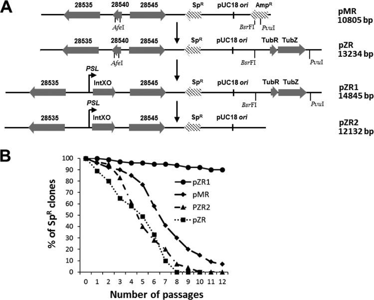
To further characterize the two noncontiguous pXO1 regions (the type III PPS genes and the IntXO recombinase gene) shown by the deletion analysis to be required for maintenance, we tested their ability to stabilize the minireplicon plasmid pMR. First, we cloned the type III PPS genes into pMR (Fig. 2A). A stability test of the resulting plasmid, pZR, in the Ames 33(pZR) strain demonstrated that these genes alone did not confer plasmid stability (Fig. 2B). Transformation of the Ames 33(pZR) strain with pSW4-intXO, containing the structural gene for IntXO behind the pagA promoter, did not enhance the stability of pZR in Ames 33(pZR, pSW4-intXO) (data not shown), indicating that some factor additional to the structural gene for IntXO is needed for pMR stabilization. Insertion into pZR of a PCR fragment encoding IntXO and upstream sequences amplified with the 183F/181R primer pair (as described in Materials and Methods) produced plasmid pZR1, which proved to be highly stable. Deletion of tubZR from pZR1 resulted in loss of stability of the resulting pZR2 plasmid in comparison with the parent pZR1 plasmid (Fig. 2B). These data showed that (i) 5′ and 3′ sequences of the IntXO recombinase gene are important for pZR1 stabilization and (ii) these surrounding 5′ and 3′ sequences should be in cis with the MR and type III PPS regions.
FIG 2.
Creation and stability analysis of plasmids containing selected components of the MSII system. (A) The pMR plasmid (1) was used as a vector for cloning of the type III PPS (producing the pZR plasmid) or both the type III PPS and the IntXO-PSL system (producing the pZR1 plasmid). The pZR1 plasmid was used to produce the pZR2 plasmid, containing only the IntXO-PSL system. (B) Percentages of spectinomycin-resistant (i.e., plasmid-containing) bacteria in cultures of B. anthracis Ames 33 containing either pMR or a pMR derivative (pZR, pZR1, or pZR2). Cultures were subcultured every 12 h on LB agar without spectinomycin and grown at 37°C. Bacteria from each subculture were diluted and plated on LB agar, with and without spectinomycin (150 μg/ml), to see the fractions retaining the plasmids. Maximum root mean square deviations in the percentage of spectinomycin-resistant colonies did not exceed 10%.
Bioinformatic analyses of the IntXO gene DNA region and the IntXO protein.
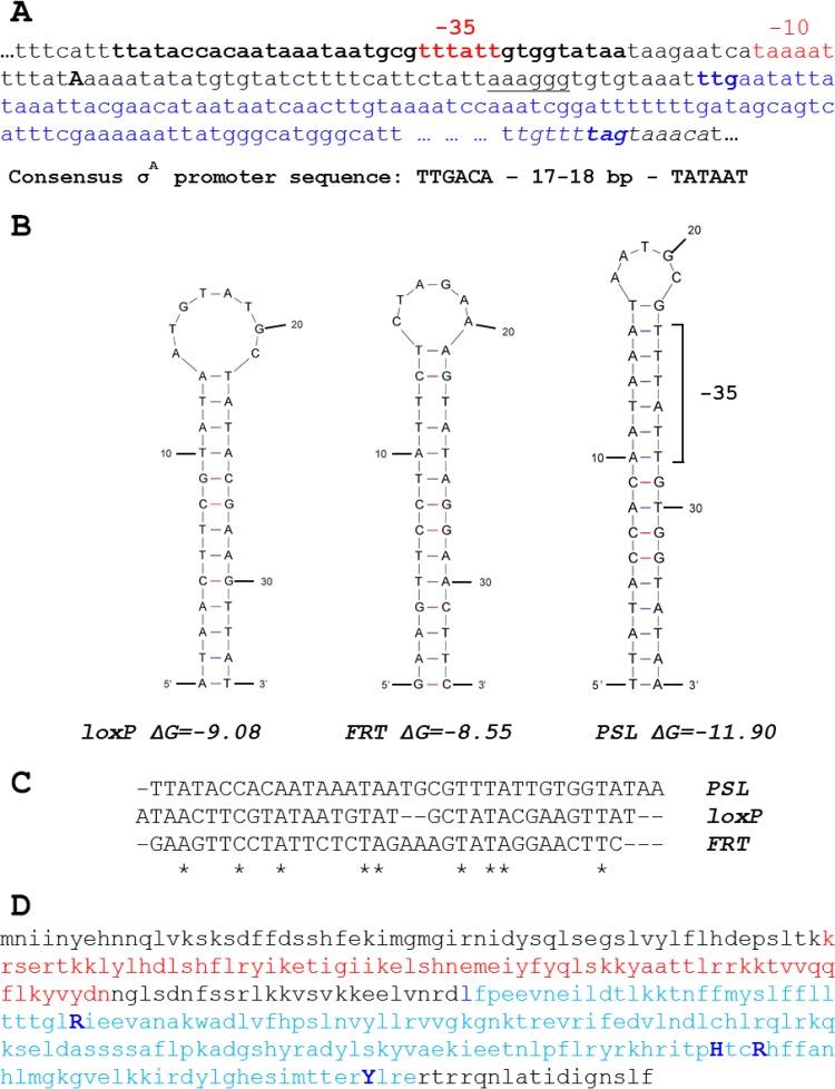
We noted a perfect stem-loop (PSL) of 37 bp in the 5′ untranslated region (5′ UTR) of ORF 29165 (the IntXO recombinase gene) and considered whether it may play a role (Fig. 3A). The PSL is located upstream of the transcriptional start site of the IntXO recombinase gene (17), and it contains a −35 region of a putative σA-dependent promoter. This conjoint location may be indicative of a complex pattern of IntXO gene regulation. The codon ttg, shown in bold blue, indicates the putative start translation codon (STC) for the GBAA_RS29165 ORF in the NCBI database (accession no. NC_007322.2). The underlined sequence aaaggg, located 9 nucleotides upstream of this codon, is the putative ribosome binding site (RBS). The short palindromic sequence partly overlapping the end of the IntXO gene ORF and the 3′ UTR of the IntXO gene mRNA may play a role as a transcriptional terminator for IntXO gene transcription (Fig. 3A).
FIG 3.
Nucleotide sequences of IntXO recombinase gene regulatory elements and amino acid sequence of IntXO tyrosine recombinase, with the recombinase catalytic site and domain structure also indicated. (A) IntXO recombinase gene 5′ and 3′ nucleotide sequences. The PSL sequence is indicated in bold lowercase letters. The −35 and −10 regions of the σA-like promoter are indicated in red. The transcriptional start site identified by RNA sequencing (17) is shown in uppercase letters. The structural part of the IntXO recombinase gene is indicated in blue (the potential translation start codon [ttg] and the stop codon [tag] are shown in bold). The putative ribosome binding site is underlined, and the potential transcriptional terminator sequence is denoted in italics. (B) Comparison of secondary structures and Gibbs free energies (http://mfold.rna.albany.edu/cgi-bin/mfold-3.4.cgi) for the loxP, FRT, and PSL target sites used by the Cre, Flp, and IntXO recombinases, respectively. The −35 region within PSL is indicated by a bracket. (C) Alignment of loxP, FRT, and PSL sequences. Similar nucleotides are indicated by stars. (D) The IntXO amino acid sequence contains the tyrosine recombinase catalytic site residues R-186, H-292, R-295, and Y-327 (indicated in dark blue) (see Fig. S1 in the supplemental material). The N-terminal SAM-like domain of phage integrase and the C-terminal catalytic domain of DNA breaking-rejoining enzymes are indicated in red and light blue, respectively. See Fig. S1 in the supplemental material for an alignment of all the tyrosine recombinases cited in this work.
The predicted DNA folding of the PSL in comparison with that of loxP (18) and the FRT sequence (19) is presented in Fig. 3B. The length of the stem region is longest for the PSL, while the Gibbs free energy is the smallest. This may facilitate interaction of the σA and IntXO proteins with the PSL. The sequence similarity among the loxP, FRT, and PSL sequences is very low (Fig. 3C), consistent with the expectation that the relevant enzymes have high specificity for their targets, as seen in our previous study where the Cre and Flp recombinases recognized only their respective targets and did not interfere with each other (2).
To clarify the role of GBAA_RS29165 in pXO1 maintenance, we analyzed the amino acid sequence of the 347-amino-acid (aa) hypothetical protein WP_001021546 (accession no. NC_007322.2) (Fig. 3D). Two conserved domains were found in the protein sequence: the N-terminal part of the sequence (aa 56 to 124), highlighted in red, has modest similarity to the family of phage integrase N-terminal SAM-like domains (pfam02899), while the C-terminal part of the sequence (aa 158 to 328), highlighted in blue, has high similarity to the superfamily of DNA-breaking enzymes (cd00397), which includes the Cre and FLP recombinases. Even higher homology was found for the entire protein (aa 30 to 330) and the conserved protein family designated “recombinase XerC” (TIGR02224). The XerC and XerD family enzymes are typically involved in separating both chromosomal and plasmid multimers during cell division. Thus, B. anthracis contains chromosomally encoded XerC and XerD enzymes which presumably contribute to chromosomal segregation. Information about the catalytic site of this family of enzymes comes from an analysis of structures of enzymes, such as the E. coli XerD enzyme (20). Alignment of the IntXO sequence with those of the chromosomally encoded E. coli and B. anthracis XerD recombinases and others is shown in Fig. S1 in the supplemental material. The conservation of all the active site residues is evidence for the conservation within this group (21). We named GBAA_RS29165 IntXO according to the Ames Ancestor plasmid pXO1 annotation for this protein as an “integrase” (NCBI accession no. NC_007322.2).
Confirmation of IntXO-PSL system functionality in vivo.
The ability of IntXO to recombine PSL palindromes was initially assessed within B. anthracis by creating a pXO1K target plasmid with two inserted PSL sequences as well as the endogenous one. Plasmid pSCP-ESAT (Fig. 4A), containing two PSL sites flanking a sequence matching the ESAT-like ORF 29195 gene, was electroporated into Ames 35 competent cells at 30°C to allow insertion into pXO1K by homologous recombination. After selection with erythromycin at 37°C, several additional passages were done without an antibiotic, and erythromycin-sensitive clones were then selected. The control primer pair seqFESAT/seqRESAT was used to verify the production of a 945-bp PCR product from the Ames 35K Δesat6 strain, consistent with deletion of the 5,255-bp region between the outer PSL sites (Fig. 4C). Sequencing of the PCR fragment confirmed the presence of a single remaining PSL site (Fig. 4D).
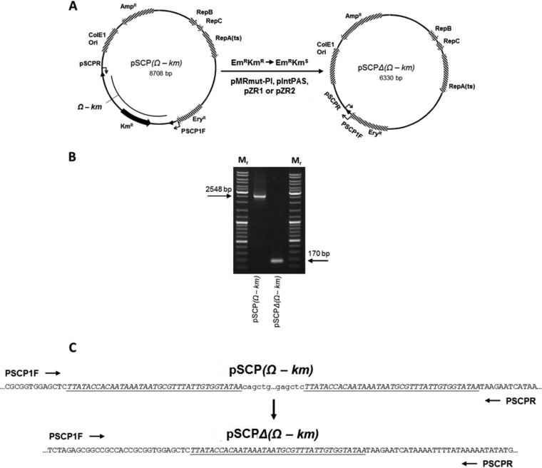
To unambiguously show that recombination at PSL sites is catalyzed by IntXO, we electroporated plasmid pSCP(Ω-km) into the pXO1-negative Ames 33 strain at 30°C with double selection (erythromycin plus kanamycin). The Ω Kmr gene in this plasmid is flanked by PSL sites, providing a target for IntXO. The Emr Kmr transformant was electroporated separately at 30°C with each of the IntXO-expressing plasmids, pMRmut-PI and pIntPAS, with double selection (erythromycin and spectinomycin). After only two passages at 37°C on plates containing medium with erythromycin, clones sensitive to both kanamycin and spectinomycin were selected (Fig. 5A). The control primer pair PSCP1F/PSCPR was used to verify the presence of the 170-bp fragment resulting from Ω Kmr cassette deletion (Fig. 5B). Sequencing of the PCR fragments obtained from pSCP(Ω-km) and pSCPΔ(Ω-km) confirmed the presence of 2 and 1 PSL sites, respectively (Fig. 5C). The IntXO protein produced by either the pZR1 or pZR2 plasmid also successfully deleted the cassette under the same conditions (data not shown). However, more passages were required to eliminate pZR2 from clones containing pSCPΔ(Ω-km), and we could not eliminate pZR1 during passages at 37°C. The data obtained show that PSL is an authentic site for the IntXO tyrosine recombinase.
FIG 5.
Deletion of a DNA fragment flanked by PSLs with the IntXO recombinase produced in a pXO1-negative strain. (A) Ames 33 containing pSCP(Ω-km) was transformed with pMRmut-PI at 30°C with double selection (erythromycin and spectinomycin). After two passages at 37°C on plates containing medium with erythromycin, clones sensitive to both kanamycin and spectinomycin were selected. (B) The control primer pair PSCP1F/PSCPR was used to verify the Ω Kmr cassette deletion. The sizes of the PCR fragments are shown above the arrows, identifying the fragments before and after the deletion. Mr, GeneRuler DNA ladder mix for size determination (as in Fig. 1B). (C) Sequences of both PCR fragments obtained from pSCP(Ω-km) and pSCPΔ(Ω-km). The directions of sequencing by use of the corresponding primers are indicated by arrows. The same result was obtained with the pIntPAS, pZR1, or pZR2 plasmid. See the explanation in the text.
Activity of recombinant IntXO in vitro.
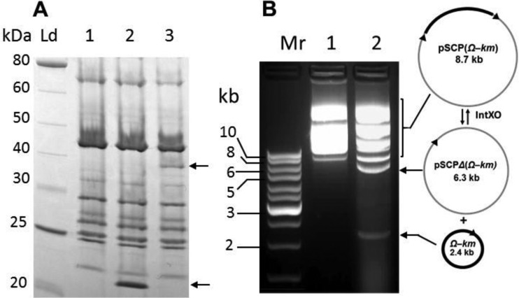
To assess whether IntXO can recombine PSL sites in the absence of additional substrates or cofactors, we produced the recombinant protein in vitro by using a coupled transcription-translation system. Reaction mixtures containing the pIntCP plasmid as an IntXO DNA template showed a new protein band of the expected size of 41 kDa (Fig. 6A, lane 3). Treatment of the PSL-containing substrate plasmid pSCP(Ω-km) with this recombinant IntXO protein generated two new DNA species, of approximately 5 and 2 kb (Fig. 6B, lane 2). These species were absent from the control reaction mixtures, which contained the proteins from transcription-translation system reaction mixtures lacking a DNA substrate (Fig. 6B, lane 1). The 5- and 2-kb species are the products expected from IntXO action on the PSL sites of the pSCP(Ω-km) circular form: the 5-kb species is the circular form of pSCPΔ(Ω-km) with a single PSL site, and the 2-kb species is the circular form of the Ω-km cassette with a single PSL site (Fig. 6B, diagrams). Also evident was the resolution of the large pSCP(Ω-km) plasmid multimers into smaller multimeric forms and the pSCP(Ω-km) monomer. These data show that recombinant IntXO is able to recombine PSL sites in the absence of cofactors, consistent with the activities of Cre and Flp tyrosine recombinases.
FIG 6.
Recombinant IntXO protein activity in vitro. (A) Profiles of proteins (5 μl in each lane) synthesized in vitro by use of a PURExpress kit (New England BioLabs). Lane 1, no template DNA; lane 2, DHFR control plasmid used as template DNA; lane 3, pIntCP plasmid used as template DNA. The bottom arrow indicates Escherichia coli dihydrofolate reductase; the top arrow indicates the IntXO protein. Ld, DNA ladder. (B) Determination of IntXO activity in vitro. The agarose gel image shows the pSCP(Ω-km) plasmid treated with proteins synthesized as described for panel A. Lane 1, DNA treated with the proteins from the reaction mixture without template DNA (lane 1 of panel A); lane 2, DNA treated with the proteins from the reaction mixture containing pIntPC template DNA (lane 3 of panel A; see details in Materials and Methods). The diagrams in panel B show the pSCP(Ω-km) substrate resolution, with the IntXO products designated by arrows. Arrowheads on the plasmids indicate the locations and directions of PSL targets.
Effect of pXO1 maintenance system deletion on stability of the plasmid in spores.
B. anthracis spores were prepared from strains lacking one or both of the two pXO1 maintenance systems by growth on NBY-Mn agar as described in Materials and Methods. The efficiency of sporulation was estimated by counting the CFU on LB agar before and after heating at 70°C for 30 min. The efficiency was approximately the same for all strains studied: 95% ± 5%. The heated spores were subsequently titrated on LB agar with and without kanamycin. We found that deletion of either MSI or MSII did not affect retention of pXO1K during sporulation. Thus, strains Ames 35K (having both MSI and MSII), Ames 35K Δ(29020–28520) (2) (with MSII deleted), and Ames 35K(pL1) (with MSI deleted) all demonstrated equal pXO1K retention levels: 97% ± 7%. Deletion of both maintenance systems in Ames 35K Δ(28560–29195) (2) led to a nearly complete loss of pXO1K during sporulation, with only 9% ± 5% of the population retaining the deleted plasmid.
DISCUSSION
The work presented here identifies a second maintenance system (MSII) for the B. anthracis plasmid pXO1 that functions independently of the first maintenance system (MSI), which we described previously (2). The locations of the components of each system on the pXO1 genome are shown in Fig. 1A. The second system consists of two noncontiguous regions on the pXO1 plasmid that work together to maintain mini-pXO1K plasmids created by deletion in vivo and recombinant pXO1-derived minireplicon plasmids containing the previously identified replication genes (1). The first region carries tubZ and tubR genes that are homologous to those of the maintenance system of the B. thuringiensis virulence plasmid pBtoxis (4). These genes along with a tubC site encode a type III PPS that provides a mechanism for pBtoxis movement and, ultimately, segregation (5, 22). However, the data for pXO1 presented here demonstrate that a classical type III PPS like that in pBtoxis is not sufficient for stability of either the pXO1K derivative plasmids present in strains Ames 35K(pL6) and Ames 35K(pL3) (Fig. 1) or the recombinant plasmid pZR, which combines pMR and type III PPS genes (Fig. 2). These data confirm our previous conclusion that these type III PPS components alone do not ensure the stability of pXO1 in B. anthracis (1). The second, complementary MSII region that we identified carries a tyrosine recombinase (IntXO) gene along with an upstream PSL sequence that serves as a target for the recombinase. The requirement for a recombinase is not surprising, because separation (or resolution) of plasmid multimers is required to maximize the number of individual plasmids available for segregation into daughter cells (11). Large plasmids usually contain a dedicated recombinase system consisting of a gene for a specific recombinase and a recombination site at which the recombinase acts. These site-specific recombinases resolve dimers to produce separate circular monomers suitable for partitioning during cell segregation. Two families of recombinases have been described for various plasmid species: active site tyrosine recombinases and active site serine recombinases (23).
The mutual action of plasmid maintenance functions was previously described, for example, by Bouet et al. (24). They showed that a SopB-sopC partition complex of F plasmid (similar to the TubR-tubC complex of the type III PPS) stimulates plasmid multimerization, which serves to decrease plasmid stability, while the ResD-resF site-specific recombination system (similar to IntXO-PSL from pXO1) resolves F plasmid multimers to monomers and guarantees retention of the plasmid in daughter cells. Thus, in the case of the F plasmid, the partition complex creates a requirement for dimer resolution by a recombinase. More information on the resolution of multimeric forms of circular plasmids and chromosomes can be found in recent reviews (25, 26).
The best-studied members of the DNA breaking-rejoining enzyme family, to which IntXO belongs, are the bacteriophage lambda integrase, the bacteriophage P1 Cre recombinase, the yeast Flp recombinase, and the bacterial XerDC recombinases. All these enzymes recognize specific DNA regions and catalyze three types of events: integration and excision, inversion, and dimer formation and resolution. Recombinase systems based on these enzymes are widely used in gene (and genome) modifications in both prokaryotes and eukaryotes. The newly identified B. anthracis pXO1 plasmid-encoded site-specific tyrosine recombinase IntXO (GBAA_RS29165), which recognizes the 37-bp DNA site PSL (perfect stem-loop) very specifically (Fig. 3), provides an additional tool for genetic manipulation. We created plasmids either producing the IntXO recombinase or carrying the PSL sites and showed that these are suitable for deleting and inserting DNA fragments into targeted DNA. The new IntXO-PSL system could be used for gene (genome) modifications in both prokaryotes and eukaryotes, in a manner similar to that of the Cre-loxP and other known recombinase systems. Modifications shown to be produced by the IntXO-PSL system include both deletions (Fig. 4 and 5) and insertions (27) of DNA. Thus, this system may have value for complex manipulations, including synthesis of designer chromosomes from scratch, as described by several groups (28–30). The ability of the IntXO protein to recognize and recombine PSL sites in vitro may also be useful for future work in the synthetic biology field.
Approximately three copies of pXO1 were found by sequence analysis to be present in various B. anthracis strains (31–34). This low copy number would not ensure plasmid maintenance if partitioning occurred passively and randomly, so it is not surprising that large plasmids have evolved and retained efficient partitioning systems. However, it is not clear why pXO1 contains two seemingly redundant systems. This may be an evolutionary “accident”—a consequence of the gene transfer events that presumably created pXO1 from several smaller precursor plasmids, thus creating the relatively “young” pathogen we recognize as B. anthracis. The ability of B. anthracis spores to remain dormant in soil for decades greatly limits the number of vegetative growth cycles that occur, thus decreasing the opportunity for selection of plasmid variants in which one of the systems has been lost. However, a more likely explanation for retention of two systems is that they are not truly redundant and that each is needed in different circumstances that are not easily identifiable by culture under laboratory conditions. The most experimentally accessible hypothesis of this type is that one of the systems plays a more dominant role in partitioning of pXO1 into the forespore during sporulation. However, we found that deletion of either MSI or MSII did not decrease the ability of the resulting pXO1 plasmids to penetrate into B. anthracis spores from mother cells (in comparison with native pXO1). On the other hand, deletion of both systems drastically decreased the penetration ability. It follows that each maintenance system can function during both vegetative cell division and sporulation. This apparent redundancy in function leaves open the question, discussed briefly above, of why the plasmid retains redundant maintenance systems. This question merits further study, which should be facilitated by use of the deletion plasmids described in this work.
Supplementary Material
ACKNOWLEDGMENT
This research was supported by the Intramural Research Program of the National Institute of Allergy and Infectious Diseases, NIH.
Footnotes
Supplemental material for this article may be found at http://dx.doi.org/10.1128/JB.01004-15.
REFERENCES
- 1.Pomerantsev AP, Camp A, Leppla SH. 2009. A new minimal replicon of Bacillus anthracis plasmid pXO1. J Bacteriol 191:5134–5146. doi: 10.1128/JB.00422-09. [DOI] [PMC free article] [PubMed] [Google Scholar]
- 2.Pomerantsev AP, Chang Z, Rappole C, Leppla SH. 2014. Identification of three noncontiguous regions on Bacillus anthracis plasmid pXO1 that are important for its maintenance. J Bacteriol 196:2921–2933. doi: 10.1128/JB.01747-14. [DOI] [PMC free article] [PubMed] [Google Scholar]
- 3.Lutkenhaus J. 2012. The ParA/MinD family puts things in their place. Trends Microbiol 20:411–418. doi: 10.1016/j.tim.2012.05.002. [DOI] [PMC free article] [PubMed] [Google Scholar]
- 4.Berry C, O'Neil S, Ben-Dov E, Jones AF, Murphy L, Quail MA, Holden MT, Harris D, Zaritsky A, Parkhill J. 2002. Complete sequence and organization of pBtoxis, the toxin-coding plasmid of Bacillus thuringiensis subsp. israelensis. Appl Environ Microbiol 68:5082–5095. doi: 10.1128/AEM.68.10.5082-5095.2002. [DOI] [PMC free article] [PubMed] [Google Scholar]
- 5.Larsen RA, Cusumano C, Fujioka A, Lim-Fong G, Patterson P, Pogliano J. 2007. Treadmilling of a prokaryotic tubulin-like protein, TubZ, required for plasmid stability in Bacillus thuringiensis. Genes Dev 21:1340–1352. doi: 10.1101/gad.1546107. [DOI] [PMC free article] [PubMed] [Google Scholar]
- 6.Aylett CH, Lowe J. 2012. Superstructure of the centromeric complex of TubZRC plasmid partitioning systems. Proc Natl Acad Sci U S A 109:16522–16527. doi: 10.1073/pnas.1210899109. [DOI] [PMC free article] [PubMed] [Google Scholar]
- 7.Hoshino S, Hayashi I. 2012. Filament formation of the FtsZ/tubulin-like protein TubZ from the Bacillus cereus pXO1 plasmid. J Biol Chem 287:32103–32112. doi: 10.1074/jbc.M112.373803. [DOI] [PMC free article] [PubMed] [Google Scholar]
- 8.Hoshino S, Maki T, Hayashi I. 2012. Crystallization and preliminary X-ray data analysis of the pXO1 plasmid-partitioning factor TubZ from Bacillus cereus. Acta Crystallogr Sect F Struct Biol Cryst Commun 68:1550–1553. doi: 10.1107/S1744309112045551. [DOI] [PMC free article] [PubMed] [Google Scholar]
- 9.Ge Y, Hu X, Zhao N, Shi T, Cai Q, Yuan Z. 2014. A new tubRZ operon involved in the maintenance of the Bacillus sphaericus mosquitocidal plasmid pBsph. Microbiology 160:1112–1124. doi: 10.1099/mic.0.075465-0. [DOI] [PubMed] [Google Scholar]
- 10.Ge Y, Zhao N, Hu X, Shi T, Cai Q, Yuan Z. 2014. A novel transcriptional activator, tubX, is required for the stability of Bacillus sphaericus mosquitocidal plasmid pBsph. J Bacteriol 196:4304–4314. doi: 10.1128/JB.01855-14. [DOI] [PMC free article] [PubMed] [Google Scholar]
- 11.Sengupta M, Austin S. 2011. Prevalence and significance of plasmid maintenance functions in the virulence plasmids of pathogenic bacteria. Infect Immun 79:2502–2509. doi: 10.1128/IAI.00127-11. [DOI] [PMC free article] [PubMed] [Google Scholar]
- 12.Gerdes K, Howard M, Szardenings F. 2010. Pushing and pulling in prokaryotic DNA segregation. Cell 141:927–942. doi: 10.1016/j.cell.2010.05.033. [DOI] [PubMed] [Google Scholar]
- 13.Sambrook J, Russell DW. 2001. Molecular cloning: a laboratory manual, 2nd ed Cold Spring Harbor Laboratory Press, Cold Spring Harbor, NY. [Google Scholar]
- 14.Thorne CB. 1993. Bacillus anthracis, p 113–124. In Sonenshein AB, Hoch JA, Losick R (ed), Bacillus subtilis and other Gram-positive bacteria: biochemistry, physiology, and molecular genetics. American Society for Microbiology, Washington, DC. [Google Scholar]
- 15.Goios A, Meirinhos J, Rocha R, Lopes R, Amorim A, Pereira L. 2006. RepeatAround: a software tool for finding and visualizing repeats in circular genomes and its application to a human mtDNA database. Mitochondrion 6:218–224. doi: 10.1016/j.mito.2006.06.001. [DOI] [PubMed] [Google Scholar]
- 16.Le Bourgeois P, Bugarel M, Campo N, Daveran-Mingot ML, Labonte J, Lanfranchi D, Lautier T, Pages C, Ritzenthaler P. 2007. The unconventional Xer recombination machinery of streptococci/lactococci. PLoS Genet 3:e117. doi: 10.1371/journal.pgen.0030117. [DOI] [PMC free article] [PubMed] [Google Scholar]
- 17.McKenzie AT, Pomerantsev AP, Sastalla I, Martens C, Ricklefs SM, Virtaneva K, Anzick S, Porcella SF, Leppla SH. 2014. Transcriptome analysis identifies Bacillus anthracis genes that respond to CO2 through an AtxA-dependent mechanism. BMC Genomics 15:229. doi: 10.1186/1471-2164-15-229. [DOI] [PMC free article] [PubMed] [Google Scholar]
- 18.Hoess RH, Ziese M, Sternberg N. 1982. P1 site-specific recombination: nucleotide sequence of the recombining sites. Proc Natl Acad Sci U S A 79:3398–3402. doi: 10.1073/pnas.79.11.3398. [DOI] [PMC free article] [PubMed] [Google Scholar]
- 19.Lacroix C, Giovannini D, Combe A, Bargieri DY, Spath S, Panchal D, Tawk L, Thiberge S, Carvalho TG, Barale JC, Bhanot P, Menard R. 2011. FLP/FRT-mediated conditional mutagenesis in pre-erythrocytic stages of Plasmodium berghei. Nat Protoc 6:1412–1428. doi: 10.1038/nprot.2011.363. [DOI] [PubMed] [Google Scholar]
- 20.Subramanya HS, Arciszewska LK, Baker RA, Bird LE, Sherratt DJ, Wigley DB. 1997. Crystal structure of the site-specific recombinase, XerD. EMBO J 16:5178–5187. doi: 10.1093/emboj/16.17.5178. [DOI] [PMC free article] [PubMed] [Google Scholar]
- 21.Guo F, Gopaul DN, Van Duyne GD. 1997. Structure of Cre recombinase complexed with DNA in a site-specific recombination synapse. Nature 389:40–46. doi: 10.1038/37925. [DOI] [PubMed] [Google Scholar]
- 22.Fink G, Lowe J. 2015. Reconstitution of a prokaryotic minus end-tracking system using TubRC centromeric complexes and tubulin-like protein TubZ filaments. Proc Natl Acad Sci U S A 112:E1845–E1850. doi: 10.1073/pnas.1423746112. [DOI] [PMC free article] [PubMed] [Google Scholar]
- 23.Stark WM, Boocock MR, Sherratt DJ. 1992. Catalysis by site-specific recombinases. Trends Genet 8:432–439. [PubMed] [Google Scholar]
- 24.Bouet JY, Bouvier M, Lane D. 2006. Concerted action of plasmid maintenance functions: partition complexes create a requirement for dimer resolution. Mol Microbiol 62:1447–1459. doi: 10.1111/j.1365-2958.2006.05454.x. [DOI] [PubMed] [Google Scholar]
- 25.Crozat E, Fournes F, Cornet F, Hallet B, Rousseau P. October 2014. Resolution of multimeric forms of circular plasmids and chromosomes. Microbiol Spectr doi: 10.1128/microbiolspec.PLAS-0025-2014. [DOI] [PubMed] [Google Scholar]
- 26.Midonet C, Barre FX. December 2014. Xer site-specific recombination: promoting vertical and horizontal transmission of genetic information. Microbiol Spectr doi: 10.1128/microbiolspec.MDNA3-0056-2014. [DOI] [PubMed] [Google Scholar]
- 27.Pomerantsev AP, Hepler NL, Chahoud M, Vrentas C, Leppla SH. 2015. Role of AtxA in toxin production by Bacillus anthracis as revealed by a new method for gene insertion, abstr 185 (B), p 66 Abstr 2015 ASM Biodefense Emerg Dis Res Meet. American Society for Microbiology, Washington, DC. [Google Scholar]
- 28.Hubbard EJ. 2014. FLP/FRT and Cre/lox recombination technology in C elegans. Methods 68:417–424. doi: 10.1016/j.ymeth.2014.05.007. [DOI] [PMC free article] [PubMed] [Google Scholar]
- 29.Annaluru N, Muller H, Mitchell LA, Ramalingam S, Stracquadanio G, Richardson SM, Dymond JS, Kuang Z, Scheifele LZ, Cooper EM, Cai Y, Zeller K, Agmon N, Han JS, Hadjithomas M, Tullman J, Caravelli K, Cirelli K, Guo Z, London V, Yeluru A, Murugan S, Kandavelou K, Agier N, Fischer G, Yang K, Martin JA, Bilgel M, Bohutski P, Boulier KM, Capaldo BJ, Chang J, Charoen K, Choi WJ, Deng P, Dicarlo JE, Doong J, Dunn J, Feinberg JI, Fernandez C, Floria CE, Gladowski D, Hadidi P, Ishizuka I, Jabbari J, Lau CY, Lee PA, Li S, Lin D, Linder ME, Ling J, Liu J, Liu J, London M, Ma H, Mao J, McDade JE, McMillan A, Moore AM, Oh WC, Ouyang Y, Patel R, Paul M, Paulsen LC, Qiu J, Rhee A, Rubashkin MG, Soh IY, Sotuyo NE, Srinivas V, Suarez A, Wong A, Wong R, Xie WR, Xu Y, Yu AT, Koszul R, Bader JS, Boeke JD, Chandrasegaran S. 2014. Total synthesis of a functional designer eukaryotic chromosome. Science 344:55–58. doi: 10.1126/science.1249252. [DOI] [PMC free article] [PubMed] [Google Scholar]
- 30.Cai Y, Agmon N, Choi WJ, Ubide A, Stracquadanio G, Caravelli K, Hao H, Bader JS, Boeke JD. 2015. Intrinsic biocontainment: multiplex genome safeguards combine transcriptional and recombinational control of essential yeast genes. Proc Natl Acad Sci U S A 112:1803–1808. doi: 10.1073/pnas.1424704112. [DOI] [PMC free article] [PubMed] [Google Scholar]
- 31.Read TD, Salzberg SL, Pop M, Shumway M, Umayam L, Jiang L, Holtzapple E, Busch JD, Smith KL, Schupp JM, Solomon D, Keim P, Fraser CM. 2002. Comparative genome sequencing for discovery of novel polymorphisms in Bacillus anthracis. Science 296:2028–2033. doi: 10.1126/science.1071837. [DOI] [PubMed] [Google Scholar]
- 32.Ravel J, Jiang L, Stanley ST, Wilson MR, Decker RS, Read TD, Worsham P, Keim PS, Salzberg SL, Fraser-Liggett CM, Rasko DA. 2009. The complete genome sequence of Bacillus anthracis Ames “Ancestor.” J Bacteriol 191:445–446. doi: 10.1128/JB.01347-08. [DOI] [PMC free article] [PubMed] [Google Scholar]
- 33.Ruckert C, Licht K, Kalinowski J, Espirito Santo C, Antwerpen M, Hanczaruk M, Reischl U, Holzmann T, Gessner A, Tiemann C, Grass G. 2012. Draft genome sequence of Bacillus anthracis UR-1, isolated from a German heroin user. J Bacteriol 194:5997–5998. doi: 10.1128/JB.01410-12. [DOI] [PMC free article] [PubMed] [Google Scholar]
- 34.Okinaka RT, Challacombe J, Drees K, Birdsell DN, Janke N, Naumann A, Seymour M, Hornstra H, Schupp J, Sahl J, Foster JT, Pearson T, Turnbull P, Keim P. 2014. Genome sequence of Bacillus anthracis STI, a Sterne-like Georgian/Soviet vaccine strain. Genome Announc 2:e00853-14. doi: 10.1128/genomeA.00853-14. [DOI] [PMC free article] [PubMed] [Google Scholar]
- 35.Pomerantsev AP, Kalnin KV, Osorio M, Leppla SH. 2003. Phosphatidylcholine-specific phospholipase C and sphingomyelinase activities in bacteria of the Bacillus cereus group. Infect Immun 71:6591–6606. doi: 10.1128/IAI.71.11.6591-6606.2003. [DOI] [PMC free article] [PubMed] [Google Scholar]
- 36.Oliva MA, Martin-Galiano AJ, Sakaguchi Y, Andreu JM. 2012. Tubulin homolog TubZ in a phage-encoded partition system. Proc Natl Acad Sci U S A 109:7711–7716. doi: 10.1073/pnas.1121546109. [DOI] [PMC free article] [PubMed] [Google Scholar]
Associated Data
This section collects any data citations, data availability statements, or supplementary materials included in this article.


