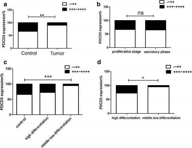
Fig. 4.

Statistical analysis of PDCD5 expression in 51 endometrioid endometrial carcinoma specimens and 53 control endometrium. a The staining index of PDCD5 in endometrial carcinoma specimens was significantly lower than that in control endometrium (P < 0.01); b No significant differences in PDCD5 expression were observed between proliferative phase and secretory phase of control endometrium (P > 0.05); c The staining index of PDCD5 in middle-low differentiation of endometrial carcinoma was significantly lower than that in control endometrium (P < 0.001), but there were no obvious differences between high differentiation of endometrial carcinoma and control endometrium; d The staining index of PDCD5 in high differentiation of endometrial carcinoma was significantly higher than that in middle-low differentiation of endometrial carcinoma (P < 0.05). *P < 0.05; **P < 0.01; ***P < 0.001
