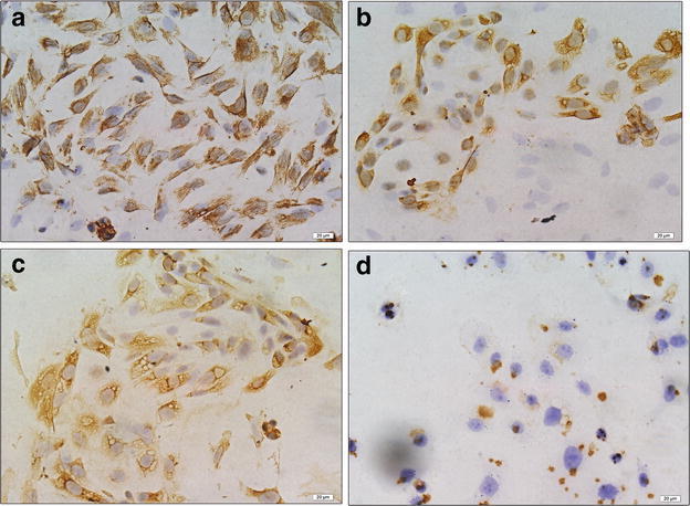
Fig. 5.

Immunocytochemical staining for vimentin, cytokeratin and PDCD5 protein in endometrial stromal cells, glandular epithelial cells and KLE cells (magnification, ×400). a The immunostaining of vimentin in endometrial stromal cells; b the immunostaining of cytokeratin in endometrial glandular epithelial cells; c the immunostaining of PDCD5 protein in endometrial glandular epithelial cells; d the immunostaining of PDCD5 protein in KLE cells
