Figure 2.
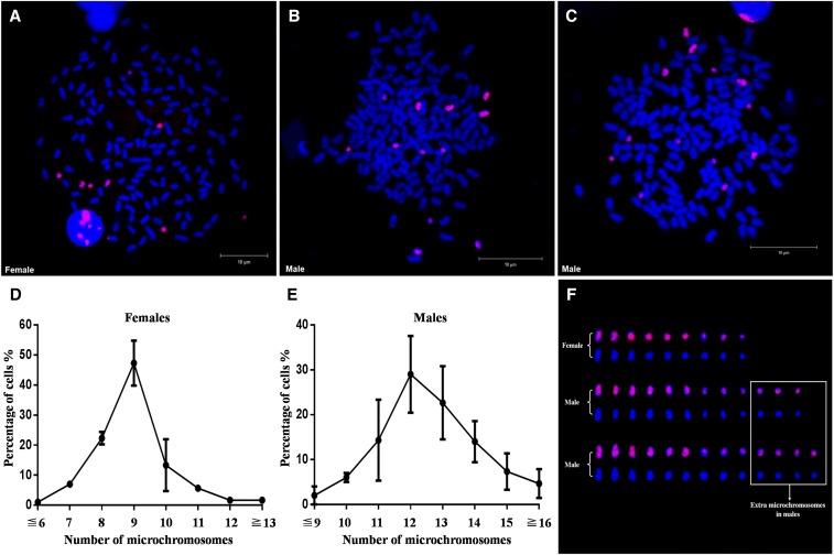
Discovery of extra microchromosomes in males via FISH analysis. (A) FISH analysis in metaphase of female 1. (B) FISH analysis in metaphase of male 1. (C) FISH analysis in metaphase of male 3. (D) Statistical data of microchromosome number in females. (E) Statistical data of microchromosome number in males. The number of microchromosomes is shown on the x-axis, and the percentage of total cells is shown on the y-axis. Three females and three males in the offspring were used in statistical analysis, and a total of 100 metaphases were counted for each tested individual. (F) Morphological comparisons of microchromosomes in (A–C). Extra microchromosomes in males are indicated in the white box. The probe was labeled with Biotin, and red fluorescence was produced. All metaphase chromosomes were counterstained with DAPI and appeared blue.

