Abstract
Objective(s):
The energy resolution of a cadmium-zinc-telluride (CZT) solid-state semiconductor detector is about 5%, and is superior to the resolution of the conventional Anger type detector which is 10%. Also, the window width of the high-energy part and of the low-energy part of a photo peak window can be changed separately. In this study, we used a semiconductor detector and examined the effects of changing energy window widths for 99mTc and 123I simultaneous SPECT.
Methods:
The energy “centerline” for 99mTc was set at 140.5 keV and that for 123I at 159.0 keV. For 99mTc, the “low-energy-window width” was set to values that varied from 3% to 10% of 140.5 keV and the “high-energy-window width” were independently set to values that varied from 3% to 6% of 140.5 keV. For 123I, the “low energy-window-width” varied from 3% to 6% of 159.0 keV and the high-energy-window width from 3% to 10% of 159 keV. In this study we imaged the cardiac phantom, using single or dual radionuclide, changing energy window width, and comparing SPECT counts as well as crosstalk ratio.
Results:
The contamination to the 123I window from 99mTc (the crosstalk) was only 1% or less with cutoffs of 4% at lower part and 6% at upper part of 159KeV. On the other hand, the crosstalk from 123I photons into the 99mTc window mostly exceeded 20%. Therefore, in order to suppress the rate of contamination to 20% or less, 99mTc window cutoffs were set at 3% in upper part and 7% at lower part of 140.5 KeV. The semiconductor detector improves separation accuracy of the acquisition inherently at dual radionuclide imaging. In, this phantom study we simulated dual radionuclide simultaneous SPECT by 99mTc-tetrofosmin and 123I-BMIPP.
Conclusion:
We suggest that dual radionuclide simultaneous SPECT of 99mTc and 123I using a CZT semiconductor detector is possible employing the recommended windows.
Keywords: Breast cancer, Myocardial perfusion, Radiotherapy, SPECT
Introduction
In nuclear cardiology, the mismatch of benzenepentadecanoicacid, 4-(iodo-123I)-b-methyl-(123I-BMIPP) myocardial fatty-acid metabolism single photon emission CT (SPECT) compared to technetium Tc-99m 1,2-bis (bis(2-ethoxyethyl) phosphino) ethane (99mTc-tetrofosmin) myocardial perfusion gated SPECT is a good predictor of myocardial viability (1-3).
For practical reasons as well as to increase accuracy and to improve patient comfort and convenience, one-time simultaneous acquisition is desirable. But the energy resolution of an Anger camera is only about 10%, so separation of the counts from the 140.5 keV photons of 99mTc and those from the 159.0 keV photons of 123I is difficult. Up to now, the method for performing dual radioisotopes simultaneous acquisition usually relied on separating the energy windows as much as possible by narrowing one or more of the energy windows. The window width employed was 15% or 20% (4, 5), and a symmetric window was the only choice possible (6-8).
On the other hand, it is reported that the energy resolution of a semiconductor SPECT system is 5% (9). And an asymmetric window setting is a possible choice. This study investigates dual radionuclide simultaneous SPECT employing a semiconductor detector and various asymmetric window choices.
Methods
The SPECT system used was Discovery NM 530c (GE Healthcare, Milwaukee, WI, USA) equipped with 19 pinhole collimators (9), employed list-mode raw data acquisition over 5 minutes. The matrix size was 70 × 70, and the image reconstruction voxel size was 4.0 × 4.0 × 4.0 mm. The data processor was the Xeleris (GE Healthcare, Milwaukee, WI, USA).
In this study, reconstruction was based on an implementation of a 3-D iterative Bayesian reconstruction algorithm. A Butterworth filter (order 7, cutoff frequency = 0.37 cycles/cm) was used as a post-filter (10).
Crosstalk measurement
For crosstalk measurement using the cardiac phantom without defect, initially the crosstalk into various-sized windows was determined for both 99mTc and 123I. The energy “centerline” for 99mTc was set at 140.5 keV and that for 123I at 159.0 keV. For 99mTc, the part of the window from the “centerline” down to a low-energy cutoff (the low-energy-window width) was set to values that varied from 3%-10% of the 99mTc photopeak energy and the part of the window from the centerline up to a high-energy cutoff (the high-energy-window width) was independently set to values that varied from 3%-6% of the 99mTc photopeak energy. On the other hand, the window-width variations for 123I covered a larger range on the high energy side and a smaller range on the low energy side: the low energy-window-width varied from 3%-6% of 159.0 keV and the high-energy-window width from 3% to 10% of 159.0 keV.
After reduction of the counts of 123I within energy window of the 99mTc, the presence of down-scattered 123I counts subtracted by the dual energy window (DEW) method (11). The energy window width for scatter correction is 120 keV±5%.
In one initial study, we used the cardiac phantom (HL type, Kyoto-kagaku, Kyoto, Japan). The rate of crosstalk and the concentration linearity were analyzed with the data obtained from the cardiac phantom. The myocardium was set to the center of effective field of view. The acquisition position is shown in Figure 1.
Figure 1.
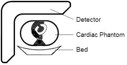
Geometric arrangement of the c cardiac phantom study
The radionuclide injected to the phantom was based on previous human studies that injected 1.8% (12) of 259 MBq of 99mTc-Tetrofosmin and 5.4% (13) of 111 MBq of 123I-BMIPP was accumulated in the myocardium. Therefore, the injection rate was set to 45.0 kBq/ml, nearly same. Single nuclide and dual simultaneous energy spectrum is shown in Figure 2.
Figure 2.
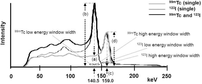
The energy spectrum of 99mTc and 123I by the Discoverly NM530c. Intensity was made similar to the clinical study
The measurement itself involved using either the cardiac phantom of 99mTc, or the cardiac phantom of 123I. The count rate results were appropriately normalized for the activity levels. After that normalization, it was possible to compute the ratio of the count rate of 123I photons in the window centered on the 99mTc photopeak, defined as 123I (140KeV), divided by the count rate of 99mTc photons in the window centered on its own photopeak, defined as 99mTc (140KeV). The ratio then can be represented by 123I (140KeV) / 99mTc (140KeV); (equal to a/b in Figure 2). This ratio was found with different settings of the 99mTc windows (low energy part and high energy part). This was the iodine to technetium-window crosstalk. It was also possible to compute a similar crosstalk ratio in the opposite direction, the technetium in the iodine window crosstalk. The ratio then can be represented by 99mTc (159KeV) / 123I (159KeV); (equal to c/d in Figure 2) count ratio. The count per pixel is the average.
The linearity of the concentration
Another experiment was performed to check the linearity of image results when the concentration of each radionuclide was varied. The cardiac phantom used contained only a single radionuclide or a mixture of both (dual) radionuclides. The concentration of the cardiac phantom with only 99mTc or only 123I was 20, 40, 60, 80 or 100% of 45.0 kBq/ml. A mixture of both (dual) radionuclides, the concentration of 99mTc + 123I were 20%+80%, 40%+60%, 60%+40% and 80%+20%, respectively.
Selection of the energy window width
The following points were considered in coming to a recommendation for the window settings for 99mTc, (1) The iodine to technetium crosstalk should be 20% or less, (2) not too many potential true counts should be lost, (3) we do not want the high-energy cutoff for the technetium window to overlap the low-energy cutoff of the iodine window. On the other hand, for 123I the following points were considered; (1) Although the ratio for technetium-to-iodine-window crosstalk was almost constant; not too many potential true counts should be lost, (2) stability of counts was observed for a high-energy-window width greater that 7%; (3) We do not want the low-energy cutoff for the iodine window to overlap the high-energy cutoff of the technetium window.
The cardiac phantom study
The cardiac phantom study placed 1.5 cm, 3.0 cm and left anterior descending defect into the anterior, and compared the detectability of that defect under various conditions. Only 99mTc (single), only 123I (single), or a mixture of both (dual) radionuclides was injected into the phantom. 99mTc and 123I of 45.0 kBq/ml, the same volumes were injected into the myocardium. And the same volumes of 10.0 kBq/ml (14) were injected into the lung, the LV cavity, the mediastinum and the liver (Figure 3.upper left). This static image (schema) was acquired by an Anger type gamma camera (Infinia; GE Healthcare, Milwaukee, WI, USA).
Figure 3.
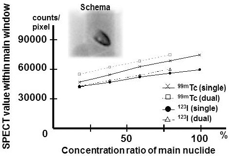
Schema is the image of the cardiac phantom with injection radionuclide. This static image was acquired by an Anger type gamma camera. The count ratio of single radionuclide and a mixture of both radionuclides to which concentration was changed. X-axis: Concentration of 99mTc or 123I in a single radionuclide (100%) and a mixture of both radionuclides (20, 40, 60 and 80%). Y-axis: The count of the point source per pixel
The count for the anterior view within the 99mTc window averaged 600 counts/pixel and that within the 123I window averaged 400 counts/pixel. The acquisition count was similar to the clinical study. Single radionuclide was decided that the high-energy-window width should be 5% and the low-energy-window width should be 5%. This condition is conventional symmetrical window width.
Human study
A 54-year-old man with hypertrophic cardiomyopathy participated in this study. Informed consent was obtained after a detailed explanation of the purpose of the study and scanning procedures. This patient was injected with 111 MBq of 123I-BMIPP at rest and SPECT imaging was performed 20 minutes after injection. After completing 123I-BMIPP SPECT, 295 MBq of 99mTc-tetrofosmin was administered to obtain simultaneous 123I-BMIPP and 99mTc-tetrofosmin SPECT. As in the myocardial phantom studies, the protocols given above were employed.
Results
Energy spectrum
The energy spectra for single radionuclide acquisitions and for a dual radionuclide simultaneous acquisition are shown in Figure 2. Although it is the same activity, energetic differs.
Result of crosstalk measurements
The technetium in the iodine window crosstalk leads to a ratio for 99mTc (159) divided by 123I (159) that is 1% or smaller (Figure 4.upper right). However, the iodine in the technetium window crosstalk leads to a ratio for 123I (140) divided by 99mTc (140) that is above 20% for most of the choices for the technetium windows (Figure 4. upper left).
Figure 4.
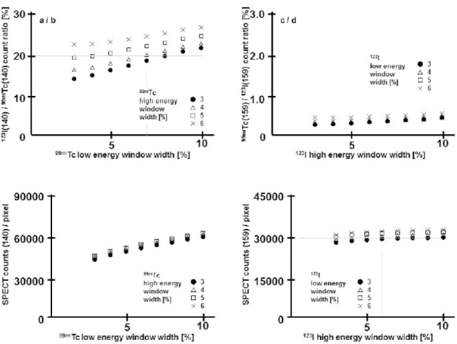
In crosstalk measurement using cardiac phantom, the crosstalk into various-sized windows was determined for both 99mTc and 123I.
Based on these initial results, it was decided that the high-energy-window width should be 3% and the low-energy-window width should be 7% for 99mTc. Also it was decided that the high-energy-window width should be 6% and the low-energy-window width should be 4% for 123I.
-
a.
The rate in which the counts of 123I crosstalk to the window of 99mTc(140)
-
b.
The rate in which the counts of 99mTc crosstalk to the window of 123I(159)
-
c.
The count of the 99mTc(140) window including the count of 123I(140) crosstalk to which the 99mTc window width was changed
-
d.
The count of the 123I(159) window including the count of 99mTc(159) crosstalk to which the 123Tc window width was changed
In concentration change study, the count of each single radionuclide was compared with the count of a mixture of both radionuclides. The count was measured every 20% and it had good linearity (Figure 3). The increase in crosstalk was remarkable by an increase in 123I (140) / 99mTc (140) concentration.
The cardiac phantom study
Bull’s eye map of a cardiac phantom without defect was compared with three pattern of defects of the cardiac phantom regarding distribution of the tracer.
For distribution of the tracer we used the contrast ratio divided 17 segments. Bull’s eye map were produced for single 99mTc in the phantom, a 99mTc image from a mixed radionuclide phantom (dual 99mTc image), a 123I image from a mixed radionuclide phantom (dual 123I image), and an image with single 123I in the phantom (Figure 5). Bull’s eye map of single 99mTc was similar to dual 99mTc (7-3). Also Bull’s eye map for single 123I was similar to dual 123I (4-6). Without defect and with defect size of 1.5 cm had similar 99mTc single image (white and black line oval).
Figure 5.
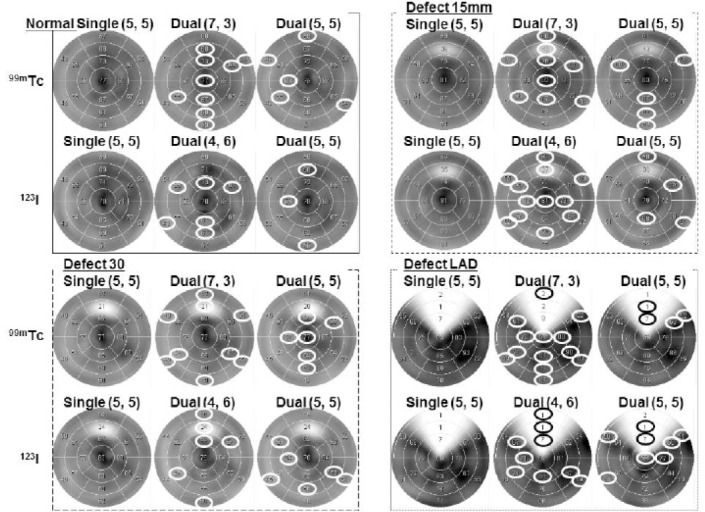
Bull’s eye map for the cardiac phantom with anterior defect in single- and dual-radionuclide. The defect is 1.5 cm (upper right), 3.0 cm (lower left) and left anterior descending (lower right). Upper left image was without defect
Human study
The energy window width that image reconfiguration used was: 99mTc photo peak 140.5 keV, high-energy-window width 3%, low-energy-window width 7%, and 123I photo peak 159.0keV, high-energy-window width 6% and low-energy- window width 4%.
An example of a mismatch between perfusion and 123I-BMIPP
Images from our 54-year-old patient with hypertrophic cardiomyopathy using dual isotope SPECT images with 99mTc-tetrofosmin and 123I-BMIPP are displayed in Figure 6 in short-axis views. 123I-BMIPP uptake was moderately to severely reduced from anterolateral to apical and inferior region, while 99mTc-tetrofosmin uptake is slightly decreased or almost normal in basal anterior area and apex. Invasive coronary angiography was normal (not shown).
Figure 6.
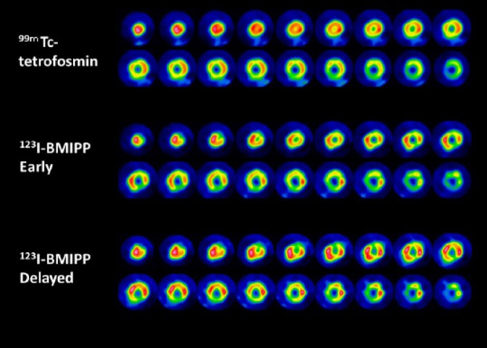
99mTc-tetrofosmin and 123I-BMIPP are displayed (top: 99mTc-tetrofosmin, middle: 123I-BMIPP in early phase, and bottom: 123I-BMIPP in delayed phase)
Discussion
Dual radionuclide simultaneous acquisition of 99mTc and 123I uses the technique of separating an energy window as much as possible. Usually energy window width must be symmetrical in the high- energy-window and low- energy-window parts. When energy window width is 15% or 20% (4, 5), energy window width overlaps.
Therefore, acquisition energy peak which 99mTc should move to low energy side, and 123I should move to high energy side, to prevent overlap. However using that technique, acquisition count decreases remarkably and quality of image deteriorated. As energy resolution was not optimal, perfect separation was difficult.
As for the energy window width of Discovery NM530c system, it can be changed symmetrically and freely. It is not necessary to shift a photo peak and an energy window can be separated. Therefore, it is possible to acquire as many photons as possible in an efficient manner. Since sensitivity is better with a semiconductor detector than with an Anger camera, one can acquire sufficient counts even if an energy window was narrow.
We calculated in consideration of energy resolution and the rate of crosstalk according to the phantom study the suitable window width. 99mTc energy window were photo peak 140.5 keV, high-energy-window width 3%, low-energy-window width 7%, and 123I energy window were photo peak 159.0 keV, high-energy-window width 6% and low-energy- window width 4%. The change of energy window width (4, 5) does not largely influence the single nuclide image.
Additionally in the linearity study of the concentration, a mixture of both radionuclides of the same concentration increased the count rate about 20% compared with the count of 99mTc only single radionuclide. The Compton scattering of 123I is included in the crosstalk to a main energy window of the 99mTc, about this, it considers the DEW subtraction method (11).
Uptake rates differ remarkably by the radionuclide. The uptake rate of 99mTc-tetrofosmin is 1.8% (12) and 123I-BMIPP is about 5.1% (13). The dose (accumulation) to 123I-BMIPP is nearly equal to 99mTc-tetrofosmin. However, the energy spectrum of the energetics of 123I is only 50% or less by the intensity of 99mTc. For this reason, we have to make window width change according to the activity. It can be imagined that it is satisfactory even if it changes a small percent for the window width for this result.
In this study we showed that dual radionuclide separated well according to the presented technique. We started to use this technique for many clinical studies.
Conclusion
A semiconductor detector is better in energy resolution and sensitivity compared to the conventional Anger type detector. Therefore, energy window width could be narrowed and it was possible for dual radionuclide simultaneous SPECT by 99mTc and 123I.
References
- 1.Dobbeleir AA, Hambys ASE, Franken PR. Influence of methodology on the presence and extent of mismatching between 99mTc-MIBI and 123I-BMIPP in myocardial viability studies. J Nucl Med. 1999;40:707–14. [PubMed] [Google Scholar]
- 2.Tamaki N, Tadamura E, Kawamoto M Magata, Yonekura Y, Fujibayashi Y, et al. Decreased uptake of iodinated branched fatty acid analog indicates metabolic alterations in ischemic myocardium. J Nucl Med. 1995;36:1974–80. [PubMed] [Google Scholar]
- 3.Kumita S, Mizumura S, Kijima T, Machida M, Kumazaki T, Tetsuou Y, et al. ECG-gated dual isotope myocardial SPECT with 99mTc-MIBI and 123I-BMIPP in patiens with ischemic heart disease. Kaku Igaku. 1995;32:547–55. [PubMed] [Google Scholar]
- 4.Standards publication NU-1–1994. Washington, DC: NEMA; 1994. National Electric Manufacturers Association (NEMA). Performance Measurements of Scintillation Cameras. [Google Scholar]
- 5.USA, Rosslyn: National Electrical Manufacturers Association; 2001. NEMA Standard Publication NU 1-001, Performance Measurements of Scintillation Cameras. [Google Scholar]
- 6.Mizumura S, Kumita S, Kumazaki T. A study of the simultaneous acquisition of dual energy SPECT with 99mTc and 123I: Evaluation of optimal window setting with myocardial phantom. Kaku Igaku. 1995;32(2):183–90. [PubMed] [Google Scholar]
- 7.Hirata M, Monzen H, Suzuki T, Ogasawara M, Nakanishi A, Sumi N, et al. Evaluation of a new protocol for two-isotope 123I-BMIPP/99mTc-TF single photon emission computed tomography (SPECT) to detect myocardial damage within one hour. Jpn J Med Phys. 2009;29:3–11. [PubMed] [Google Scholar]
- 8.Inoue T. Basic study of dual radionuclide data acquisition with Tc-99m and I-123 to establish quantitative brain SPECT. Ehime Medical Journal. 1993;12:228–37. [Google Scholar]
- 9.Bocher M, Blevis IM, Tsukerman L, Shrem Y, Kovalski G, Volokh L. A fast cardiac gamma camera with dynamic SPECT capabilities: design, system validation and future potential. Eur J Nucl Med. 2010;37:1887–902. doi: 10.1007/s00259-010-1488-z. [DOI] [PMC free article] [PubMed] [Google Scholar]
- 10.Takahashi Y, Miyagawa M, Nishiyama Y, Ishimura H, Mochizuki T. Performance of a semiconductor SPECT system: comparison with a conventional Anger-type SPECT instrument. Ann Nucl Med. 2013;27:11–6. doi: 10.1007/s12149-012-0653-9. [DOI] [PMC free article] [PubMed] [Google Scholar]
- 11.Jaszcak RJ, Greer KL. Improved SPECT quantification using compensation for scattered photons. J Nucl Med. 1984;25:893–900. [PubMed] [Google Scholar]
- 12.Kubo A, Nakamura K, Hashimoto J, Sammiya T, Iwanaga S, Hashimoto S, et al. Phase I clinical trial of a new myocardial imaging agent, 99mTc-PPN1011. Kaku Igaku. 1992;29(10):1165–76. [PubMed] [Google Scholar]
- 13.Torizuka K, Yonekura Y, Nishimura T, Tamaki N, Uehara T, Ikekubo K, et al. A Phase 1 study of beta-methyl-p-(123I)-iodophenyl-pentadecanoic acid (123I-BMIPP) Kaku Igaku. 1991;28(7):681–90. [PubMed] [Google Scholar]
- 14.Hatakeyama R. Heart phantom with liver object. A new textbook of nuclear medicine technology. 2001;1:243–246. in Japanese. [Google Scholar]


