Abstract
The incidence of melanoma among the Asian population is lower compared to that among the Western European population. These populations differed in their most common histopathologic subtypes, acral lentiginous melanoma being the most common in the Asian population. Although the dermoscopic features of the melanomas on the acral skin have been thoroughly investigated in the Asian population, studies concerning the dermoscopic patterns of melanomas on the non-acral skin have been scarce. The aim of this study was to investigate the dermoscopic patterns of melanomas on the trunk and extremities in the Asian population. To achieve this, we evaluated the dermoscopic patterns of 22 primary melanomas diagnosed at two university hospitals in Korea. In addition, 100 benign melanocytic lesions were included as the control group for comparative analysis. A P value less than 0.05 was regarded as statistically significant. Melanoma-associated dermoscopic features such as asymmetry (odds ratio [OR], 30.00), multicolor pattern (OR, 30.12), blotches (OR, 13.50), blue white veils (OR, 15.75), atypical pigment networks (OR, 9.71), irregular peripheral streaks (OR, 6.30), atypical vascular patterns (OR, 11.50), ulcers (OR, 15.83), atypical dots/globules (OR, 3.15), shiny white lines (OR, 5.88), and regression structures (OR, 7.06) were more commonly observed in patients with melanomas than in patients of the control group. The mean dermoscopic scores obtained on the 7-point checklist, revised 7-point checklist, 3-point checklist, ABCD rule, and CASH algorithm were 5.36, 3.41, 2.05, 6.89, and 9.68, respectively, in the primary melanomas, and 1.33, 0.93, 0.46, 2.45, and 3.60, respectively, in the control group (all, P < 0.001). The present study showed that melanoma-related dermoscopic patterns were common in Asian patients. Dermoscopy is a reliable diagnostic tool for the melanomas of the trunk and extremities in the Asian populations.
Introduction
Malignant melanoma (MM) is among the most aggressive and treatment-resistant human cancers [1]. It has been an increasingly important public health problem worldwide [2]. The incidence of melanoma has been steadily increasing 4–6% annually in the USA [3]. Although the incidence of melanoma in the Asian population is lower than that in the Western European population, melanoma is the most common cause of cancer-related mortality among Asian patients with skin cancers [4]. Because of the low incidence rates and low public awareness associated with MM, its diagnosis is often delayed, resulting in more advanced stages of disease at presentation among the individuals of non-European descent [5]. The early detection of melanoma is crucial for a favorable prognosis because prognosis is directly associated with the invasion depth of the melanoma.
Dermoscopy improves the diagnostic accuracy rates for melanomas by 49% compared to the clinical diagnosis with the naked eye [6]. Although the dermoscopic patterns of melanomas have been widely reported in the Western European population, data on the dermoscopic patterns of the melanoma of the trunk in the Asian population are scarce. This is attributed to the fact that the most common histological subtype is acral lentiginous melanoma, constituting roughly 45–66% of the cases compared to the 2–3% of all cases in patients from Western European descent [7–9]. Therefore, previous dermoscopic studies on the melanomas in the Asian population were mainly focused on the acral melanomas [10–12]. Because dermoscopic patterns of the melanomas on the non-glabrous skin are different from acral melanomas, a study investigating the dermoscopic features of the melanomas on the trunk in the Asian population is necessary. Although the dermoscopic principles and the fundamental patterns of melanomas are the same regardless of the race, the differences between the skin and demographic characteristics of the Asian patients and those from the Western European descent necessitated this study.
Materials and Methods
The patients who were diagnosed with MM at the Seoul National University Hospital, Seoul, and Pusan National University Hospital, Busan, between 2007 and 2014 were enrolled. The inclusion criteria were as follows: 1) histopathologically confirmed melanomas, 2) anatomic location of the trunk and limb with the acral, facial, and other special areas excluded, 3) availability of high quality clinical and dermoscopic photography. Dermoscopic photographs were acquired using a dermatoscope (DermLite II Pro HR or DL3 equipment) attached to a digital camera. The images were acquired under cross-polarized light without any liquid medium. For the comparative analysis, we included benign nevi on the trunk and the extremities (arm and leg) for the control group.
We evaluated the images according to the melanoma-related patterns that were previously described in the literature [13–16]. In addition, we calculated the scores for each lesion using dermoscopic algorithms such as the 7-point checklist, revised 7-point checklist, 3-point checklist, ABCD rule, and the color, architecture, symmetry, and homogeneity (CASH) algorithm [17–21]. The evaluation was performed by two dermatologists experienced in dermoscopy without knowledge of the final diagnosis (JHM and WIK). All statistical analyses were performed using the IBM SPSS version 21. We used simple cross-tabulations and the Pearson chi-square or Fisher exact test for the comparison of the proportions. Student t-test was used to analyze the continuous variables. Odds ratios (ORs) with the corresponding 95% confidence intervals (CIs) were calculated. Inter-observer agreement was examined using the Cohen kappa. All given p values were 2-tailed, and p values ≤0.05 (95% confidence) were considered statistically significant.
The present study was approved by the institutional review board of the Seoul National University Hospital (IRB No.: H-1510-120-714), and it was conducted in compliance with the principles of the Declaration of Helsinki. The need for informed consent for participation in this study was waived because the data was analyzed anonymously. The clinical and dermoscopic photographs were obtained with the patients’ consent during skin examination, and they agreed that the images could be used in research if their identity would not be disclosed.
Results
During the study period, 276 patients were diagnosed with MM. MM occurred on the acral area (n = 180, 65.2%), head and neck (n = 31, 11.2%), and the trunk and the extremities (n = 65, 23.6%). Among these cases, only those that were anatomically located in the trunk and the limbs were enrolled; the acral, facial, and other special areas were excluded. In addition, we excluded patients with unavailable or poor clinical and dermoscopic photographs. Finally, 22 MM cases were analyzed in the present study. The mean age of the patients with MM was 59.4 years (range, 32–84 years), and 63.6% (n = 14) of the patients were women. Nineteen cases were invasive melanomas (86.4%), and three cases were melanoma in situ. Twelve cases were superficial spreading melanomas, and 10 cases were nodular type. The mean Breslow’s thickness was 3.4 ± 4.0 mm.
The control group consisted of 100 benign nevi including 86 banal melanocytic nevi, 6 congenital nevi, 5 intradermal nevi, 2 Spitz nevi, and 1 blue nevus. Thirty-seven cases were histopathologically examined. We included 63 cases of non-biopsied banal nevi to avoid a selection bias because only difficult or suspicious nevi were excised, and majority of the typical lesions were managed with observation. The diagnosis of banal nevi was made based on typical clinical and dermoscopic findings.
In primary melanomas, the following melanoma-associated dermoscopic patterns were detected: asymmetry (90.9%), blotches (81.8%), multicolor patterns (81.8%), blue-white veils (63.6%), atypical pigment networks (52.5%), irregular peripheral streaks (54.5%), atypical vascular patterns (50.0%), ulcers (45.5%), atypical dots/globules (40.9%), white shiny lines (27.3%), brown peripheral structureless areas (22.7%), and regression structures (22.7%). The comparison of the dermoscopic features between the primary melanoma group and the control group is demonstrated in Table 1.
Table 1. The frequency distributions of dermoscopic patterns between melanomas and benign nevi showing significant associations.
| Dermoscopic patterns | Melanoma (N = 22), n (%) | Benign nevus (N = 100), n (%) | OR | 95% CI | P value |
|---|---|---|---|---|---|
| Asymmetry | 20 (90.9) | 25 (25.0) | 30.00 | 6.55–137.50 | <0.001 |
| Multicolor pattern | 18 (81.8) | 13 (13.0) | 30.12 | 8.80–103.05 | <0.001 |
| Irregular blotches | 18 (81.8) | 25 (25.0) | 13.50 | 4.17–43.68 | <0.001 |
| Blue-white veil | 14 (63.6) | 10 (10.0) | 15.75 | 5.31–46.70 | <0.001 |
| Atypical pigment network | 12 (54.5) | 11 (11.0) | 9.71 | 3.41–27.67 | <0.001 |
| Irregular peripheral streaks | 12 (54.5) | 16 (16.0) | 6.30 | 2.33–17.04 | <0.001 |
| Atypical vascular patterns | 11 (50.0) | 8 (8.0) | 11.50 | 3.81–34.71 | <0.001 |
| Ulcers | 10 (45.5) | 5 (5.0) | 15.83 | 4.63–54.17 | <0.001 |
| Atypical dots/globules | 9 (40.9) | 18 (18.0) | 3.15 | 1.17–8.50 | 0. 026 |
| Shiny white lines | 6 (27.3) | 6 (6.0) | 5.88 | 1.68–20.50 | 0.008 |
| Brown peripheral structureless area | 5 (22.7) | 12 (12.0) | 2.15 | 0.67–6.72 | 0.189 |
| Regression structure | 5 (22.7) | 4 (4.0) | 7.06 | 1.72–28.98 | 0.010 |
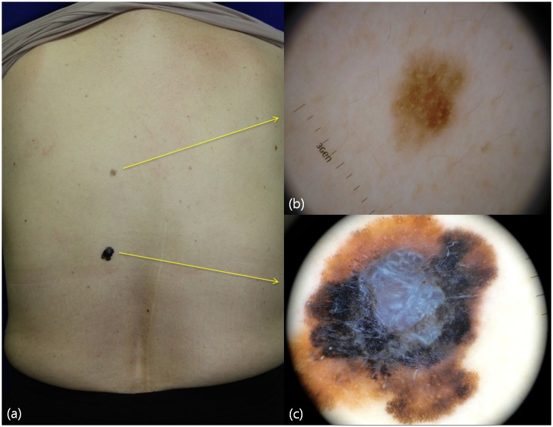
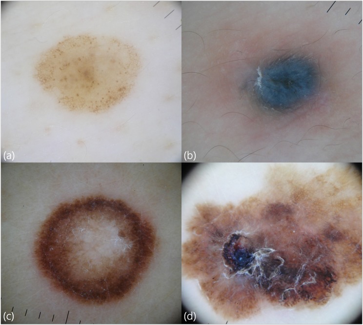
In melanomas, the more commonly observed dermoscopic patterns were the following: asymmetry (OR, 30.00; 95% CI, 6.55–137.50; P < 0.001); multicolor pattern (OR, 30.12; 95% CI, 8.80–103.05; P < 0.001); blotches (OR, 13.50; 95% CI, 4.17–43.68; P < 0.001); blue white veils (OR, 15.75; 95% CI, 5.31–46.70; P < 0.001); atypical pigment networks (OR, 9.71; 95% CI, 3.41–27.67; P < 0.001); irregular peripheral streaks (OR, 6.30; 95% CI, 2.33–17.04; P < 0.001); atypical vascular patterns (OR, 11.50; 95% CI, 3.81–34.71.76; P < 0.001); ulcers (OR, 15.83; 95% CI, 4.63–54.17; P < 0.001); atypical dots/globules (OR, 3.15; 95% CI, 1.17–8.50; P = 0.026); shiny white lines (OR, 5.88; 95% CI, 1.68–20.50; P = 0.008); and regression structures (OR, 7.06; 95% CI, 1.72–28.98; P = 0.010). The brown peripheral structureless areas did not differ significantly from the benign lesions (OR, 2.15; 95% CI, 0.67–6.72; P = 0.189) (Figs 1 and 2).
Fig 1. Clinical image and dermoscopic features of a malignant melanoma and benign nevus in a 60-year-old woman.
Compared to the clinical picture (a), dermoscopic evaluation provides detailed clues for the diagnosis of melanomas (b), dermoscopy of benign nevus showing a reticular pattern (c). Dermoscopy of malignant melanoma exhibits melanoma-associated patterns such as asymmetry, atypical pigment networks, blue-white veil, irregular blotches, irregular dots, peripheral streaks, shiny white lines, and multicolor patterns.
Fig 2. Dermoscopic features of a melanocytic nevus with a globular and reticular pattern (a), a blue nevus (b), a spitz nevus (c), and a malignant melanoma (d).
Compared to benign nevi, a melanoma exhibits more patterns such as asymmetry, irregular blotches, atypical pigment networks, irregular dots, peripheral streaks, multicolor patterns, and ulcer.
The mean scores on the 7-point checklist, revised 7-point checklist, 3-point checklist, ABCD rule, and CASH algorithm were 5.36 ± 1.79, 3.41 ± 1.33, 2.05 ± 0.90, 6.89 ± 2.10, and 9.68 ± 2.57 in primary melanomas, and 1.33 ± 1.72, 0.93 ± 1.13, 0.46 ± 0.76, 2.45 ± 2.03, and 3.60 ± 2.54 in the control group, respectively. The score differences between the primary melanomas and the control group were statistically significant for all algorithms (all, P < 0.001). The sensitivity, specificity, positive predictive value, and negative predictive value for each algorithm were as follows: 95.5%, 79.0%, 50.0%, 98.8% for the 7-point checklist; 100%, 48.0%, 29.7%, 100% for the revised 7-point checklist; 90.9%, 68.0%, 38.5%, 97.1% for the 3-point checklist; 81.8%, 83.0%, 51.4%, 95.4% for the ABCD rule; and 81.8%, 88.0%, 60.0%, 95.7% for the CASH algorithm.
Cohen kappa ranged from 0.554 to 0.804, showing moderate to excellent agreement between the 2 observers for all variables (asymmetry, multicolor pattern, blotches, blue-white veils, atypical pigment networks, irregular peripheral streaks, atypical vascular patterns, ulcers, atypical dots/globules, shiny white lines, brown peripheral structureless areas, and regression structures).
Discussion
Dermoscopy is a rapid, noninvasive magnifying tool that allows clinicians to visualize morphologic structures that are not discernible to the naked eye [22]. It improves diagnostic sensitivity for melanomas (90%) compared to that achieved with the naked eye (74%) [23]. Although data on the dermoscopic patterns of melanomas have been widely reported in the Western European population, the data on the dermoscopic patterns of malignant melanomas on the trunk have been scarce in the Asian population. To our knowledge, this is the first original study to report the dermoscopic features of malignant melanomas on the trunk and the limbs, excluding the acral skin, in the Asian population. In melanomas, the commonly observed dermoscopic patterns were asymmetry, blotches, multicolor patterns, blue-white veils, atypical pigment networks, irregular peripheral streaks, atypical vascular patterns, ulcers, atypical dots/globules, white shiny lines, brown peripheral structureless areas, and regression structures.
Asymmetry was determined by visually dividing the lesion into two perpendicular axes to evaluate them in terms of structures or colors. Asymmetry in 2 axes was described as a criterion found in 94–96% of MMs [24]. In our study, asymmetry was the most common finding among the primary melanomas (90.9%). Melanomas frequently show more colors, while benign nevi usually have only one or two colors. Therefore, the assessment of the number of colors (light brown, dark brown, gray, black, red, and white) in a lesion could help discriminate between the benign nevi from melanomas. In this study, multicolor patterns (more than four colors) were more commonly observed (81.8%). Invasion might be associated with an increasing variety of colors [25]. The high detection rate of a multicolor pattern could be attributed to the fact that the majority of the cases were invasive melanomas.
Because the melanocytes in melanomas grow haphazardly in the vertical and horizontal planes, the pigmentary structures are common. In the present study, melanomas showed irregular blotches, blue-white veils, atypical pigment networks, irregular peripheral streaks, atypical dots/globules, and brown structureless areas in 81.8% 63.6%, 54.5%, 54.5%, 40.9%, and 22.7% of the primary melanomas, respectively. All structures showed statistical significance except for the brown structureless areas. This could be attributed to the fact that the majority of cases in the present study were invasive melanomas as brown structureless areas are usually found in thin melanomas [26].
Atypical vascular structures in melanomas are associated with tumor-induced angiogenesis [16]. Therefore, the presence of vessels increases the possibility of melanoma development. In the present study, 50% of the melanomas had atypical vascular structures. The presence of shiny white lines, also called shiny white streaks or chrysalis structures, increased the possibility of malignant tumors [27, 28]. In this study, shiny white lines were observed in 27.3% of the melanomas. Regression structures are white scar-like areas with peppering overlying flat or thin portions of a lesion [16]. They were found in 22.7% of the melanomas. An ulcer is a non-specific finding. However, it can increase the index of a malignant tumor, especially when it occurs without apparent trauma. In our data, only 5% of benign nevi showed ulcers, while 45.5% of melanomas had ulcers.
Different dermoscopic algorithmic methods for melanocytic lesions were developed to discriminate melanomas from benign nevi. In the present study, the mean scores of the 7-point checklist, revised 7-point checklist, 3-point checklist, ABCD rule, and the CASH algorithm in melanomas were 5.36, 3.41, 2.05, 6.89, and 9.68, respectively. These algorithms had 81.8–100% sensitivity in discriminating melanomas from benign nevi with modest to high specificities (48–88%). Recently, Argenziano et al. revised their 7-point checklist [29]. They lowered the threshold to increase the sensitivity for optimizing melanoma screening, which warranted excision only if one of the seven criteria was observed [29]. In the present study, the sensitivity of the 7-point checklist was the highest (100%); however, its specificity was the lowest (48.0%).
There were some limitations to the present study. First, only a small number of melanomas were included and the majority of the cases were in advanced stages. Therefore, further studies with larger samples and the inclusion of thin melanomas in the Asian population are necessary. Second, we did not include data from the non-Asian group. Therefore, we could not compare the dermoscopic characteristics of the melanoma in the Asian and the Western European groups. Future studies should be conducted to compare the features of the melanomas.
In conclusion, we showed that melanoma-related dermoscopic patterns are common among Asian patients. Consequently, dermoscopy is a reliable tool in diagnosing melanomas of the trunk and the extremities in the Asian population. Compared to Western European population, the use of dermoscopy in Asia is not widespread, and melanomas are diagnosed at rather advanced stages. In this regard, the daily use of dermoscopy might facilitate the detection of melanomas at an early stage. We hope that our report will encourage the popularization of dermoscopy in Asian countries.
Data Availability
All relevant data are within the paper.
Funding Statement
The authors received no specific funding for this work.
References
- 1.Lo JA, Fisher DE. The melanoma revolution: from UV carcinogenesis to a new era in therapeutics. Science. 346(6212):945–9. Epub 2014/11/22. 346/6212/945 [pii] 10.1126/science.1253735 . [DOI] [PMC free article] [PubMed] [Google Scholar]
- 2.Rigel DS, Russak J, Friedman R. The evolution of melanoma diagnosis: 25 years beyond the ABCDs. CA Cancer J Clin. 2010;60(5):301–16. Epub 2010/07/31. caac.20074 [pii] 10.3322/caac.20074 . [DOI] [PubMed] [Google Scholar]
- 3.Rigel DS. Trends in dermatology: melanoma incidence. Arch Dermatol. 2010;146(3):318 Epub 2010/03/17. 146/3/318 [pii] 10.1001/archdermatol.2009.379 . [DOI] [PubMed] [Google Scholar]
- 4.Sng J, Koh D, Siong WC, Choo TB. Skin cancer trends among Asians living in Singapore from 1968 to 2006. J Am Acad Dermatol. 2009;61(3):426–32. Epub 2009/07/25. S0190-9622(09)00386-7 [pii] 10.1016/j.jaad.2009.03.031 . [DOI] [PubMed] [Google Scholar]
- 5.Cormier JN, Xing Y, Ding M, Lee JE, Mansfield PF, Gershenwald JE, et al. Ethnic differences among patients with cutaneous melanoma. Arch Intern Med. 2006;166(17):1907–14. Epub 2006/09/27. 166/17/1907 [pii] 10.1001/archinte.166.17.1907 . [DOI] [PubMed] [Google Scholar]
- 6.Kittler H, Pehamberger H, Wolff K, Binder M. Diagnostic accuracy of dermoscopy. Lancet Oncology. 2002;3(3):159–65. 10.1016/S1470-2045(02)00679-4 ISI:000174890300020. [DOI] [PubMed] [Google Scholar]
- 7.Jang HS, Kim JH, Park KH, Lee JS, Bae JM, Oh BH, et al. Comparison of Melanoma Subtypes among Korean Patients by Morphologic Features and Ultraviolet Exposure. Annals of Dermatology. 2014;26(4):485–90. 10.5021/ad.2014.26.4.485 ISI:000341073000009. [DOI] [PMC free article] [PubMed] [Google Scholar]
- 8.Bradford PT, Goldstein AM, McMaster ML, Tucker MA. Acral Lentiginous Melanoma Incidence and Survival Patterns in the United States, 1986–2005. Archives of Dermatology. 2009;145(4):427–34. ISI:000265411100007. 10.1001/archdermatol.2008.609 [DOI] [PMC free article] [PubMed] [Google Scholar]
- 9.Lee HY, Chay WY, Tang MBY, Chio MTW, Tan SH. Melanoma: Differences between Asian and Caucasian Patients. Annals Academy of Medicine Singapore. 2012;41(1):17–20. ISI:000301038500004. [PubMed] [Google Scholar]
- 10.Saida T, Miyazaki A, Oguchi S, Ishihara Y, Yamazaki Y, Murase S, et al. Significance of dermoscopic patterns in detecting malignant melanoma on acral volar skin: results of a multicenter study in Japan. Arch Dermatol. 2004;140(10):1233–8. Epub 2004/10/20. 140/10/1233 [pii] 10.1001/archderm.140.10.1233 . [DOI] [PubMed] [Google Scholar]
- 11.Saida T, Koga H, Uhara H. Key points in dermoscopic differentiation between early acral melanoma and acral nevus. J Dermatol. 2011;38(1):25–34. Epub 2010/12/24. 10.1111/j.1346-8138.2010.01174.x . [DOI] [PubMed] [Google Scholar]
- 12.Mun JH, Kim GW, Jwa SW, Song M, Kim HS, Ko HC, et al. Dermoscopy of subungual haemorrhage: its usefulness in differential diagnosis from nail-unit melanoma. Br J Dermatol. 2013;168(6):1224–9. Epub 2013/01/11. 10.1111/bjd.12209 . [DOI] [PubMed] [Google Scholar]
- 13.Argenziano G, Soyer HP, Chimenti S, Talamini R, Corona R, Sera F, et al. Dermoscopy of pigmented skin lesions: Results of a consensus meeting via the Internet. Journal of the American Academy of Dermatology. 2003;48(5):679–93. 10.1067/mjd.2003.281 ISI:000182977200004. [DOI] [PubMed] [Google Scholar]
- 14.Neila J, Soyer HP. Key points in dermoscopy for diagnosis of melanomas, including difficult to diagnose melanomas, on the trunk and extremities. Journal of Dermatology. 2011;38(1):3–9. 10.1111/j.1346-8138.2010.01131.x ISI:000285760100003. [DOI] [PubMed] [Google Scholar]
- 15.Haliasos HC, Zalaudek I, Malvehy J, Lanschuetzer C, Hinter H, Hofmann-Wellenhof R, et al. Dermoscopy of Benign and Malignant Neoplasms in the Pediatric Population. Seminars in Cutaneous Medicine and Surgery. 2010;29(4):218–31. 10.1016/j.sder.2010.10.003 ISI:000287382200004. [DOI] [PubMed] [Google Scholar]
- 16.Haliasos EC, Kerner M, Jaimes N, Zalaudek I, Malvehy J, Hofmann-Wellenhof R, et al. Dermoscopy for the Pediatric Dermatologist Part III: Dermoscopy of Melanocytic Lesions. Pediatric Dermatology. 2013;30(3):281–93. 10.1111/pde.12041 ISI:000318360200014. [DOI] [PubMed] [Google Scholar]
- 17.Nachbar F, Stolz W, Merkle T, Cognetta AB, Vogt T, Landthaler M, et al. The ABCD rule of dermatoscopy. High prospective value in the diagnosis of doubtful melanocytic skin lesions. J Am Acad Dermatol. 1994;30(4):551–9. Epub 1994/04/01. . [DOI] [PubMed] [Google Scholar]
- 18.Argenziano G, Fabbrocini G, Carli P, De Giorgi V, Sammarco E, Delfino M. Epiluminescence microscopy for the diagnosis of doubtful melanocytic skin lesions. Comparison of the ABCD rule of dermatoscopy and a new 7-point checklist based on pattern analysis. Arch Dermatol. 1998;134(12):1563–70. Epub 1999/01/06. . [DOI] [PubMed] [Google Scholar]
- 19.Zalaudek I, Argenziano G, Soyer HP, Corona R, Sera F, Blum A, et al. Three-point checklist of dermoscopy: an open internet study. Br J Dermatol. 2006;154(3):431–7. Epub 2006/02/01. BJD6983 [pii] 10.1111/j.1365-2133.2005.06983.x . [DOI] [PubMed] [Google Scholar]
- 20.Henning JS, Dusza SW, Wang SQ, Marghoob AA, Rabinovitz HS, Polsky D, et al. The CASH (color, architecture, symmetry, and homogeneity) algorithm for dermoscopy. J Am Acad Dermatol. 2007;56(1):45–52. Epub 2006/12/28. S0190-9622(06)02527-8 [pii] 10.1016/j.jaad.2006.09.003 . [DOI] [PubMed] [Google Scholar]
- 21.Argenziano G, Catricala C, Ardigo M, Buccini P, De Simone P, Eibenschutz L, et al. Seven-point checklist of dermoscopy revisited. Br J Dermatol. 2011;164(4):785–90. Epub 2010/12/24. 10.1111/j.1365-2133.2010.10194.x . [DOI] [PubMed] [Google Scholar]
- 22.Mun JH, Park SM, Kim TW, Kim BS, Ko HC, Kim MB. Importance of keen observation for the diagnosis of epidermal cysts: dermoscopy can be a useful adjuvant tool. J Am Acad Dermatol. 2014;71(4):e138–40. Epub 2014/09/16. S0190-9622(14)01411-X [pii] 10.1016/j.jaad.2014.04.054 . [DOI] [PubMed] [Google Scholar]
- 23.Vestergaard ME, Macaskill P, Holt PE, Menzies SW. Dermoscopy compared with naked eye examination for the diagnosis of primary melanoma: a meta-analysis of studies performed in a clinical setting. Br J Dermatol. 2008;159(3):669–76. Epub 2008/07/12. BJD8713 [pii] 10.1111/j.1365-2133.2008.08713.x . [DOI] [PubMed] [Google Scholar]
- 24.da Silva VPM, Ikino JK, Sens MM, Nunes DH, Di Giunta G. Dermoscopic features of thin melanomas: a comparative study of melanoma in situ and invasive melanomas smaller than or equal to 1mm. Anais Brasileiros De Dermatologia. 2013;88(5):712–7. 10.1590/abd1806-4841.20132017 ISI:000328202900002. [DOI] [PMC free article] [PubMed] [Google Scholar]
- 25.Emiroglu N, Cengiz FP, Hofmann-Wellenhof R. Dermoscopic and clinical features of trunk melanomas. Postepy Dermatologii I Alergologii. 2014;31(6):362–7. 10.5114/pdia.2014.47119 ISI:000346042500004. [DOI] [PMC free article] [PubMed] [Google Scholar]
- 26.Annessi G, Bono R, Sampogna F, Faraggiana T, Abeni D. Sensitivity, specificity, and diagnostic accuracy of three dermoscopic algorithmic methods in the diagnosis of doubtful melanocytic lesions. Journal of the American Academy of Dermatology. 2007;56(5):759–67. 10.1016/j.jaad.2007.01.014 ISI:000246041400005. [DOI] [PubMed] [Google Scholar]
- 27.Balagula Y, Braun RP, Rabinovitz HS, Dusza SW, Scope A, Liebman TN, et al. The significance of crystalline/chrysalis structures in the diagnosis of melanocytic and nonmelanocytic lesions. Journal of the American Academy of Dermatology. 2012;67(2):194.e1–8. ARTN 194.e1 10.1016/j.jaad.2011.04.039 ISI:000306368600013. [DOI] [PubMed] [Google Scholar]
- 28.Shitara D, Ishioka P, Alonso-Pinedo Y, Palacios-Bejarano L, Carrera C, Malvehy J, et al. Shiny White Streaks: A Sign of Malignancy at Dermoscopy of Pigmented Skin Lesions. Acta Dermato-Venereologica. 2014;94(2):132–7. 10.2340/00015555-1683 ISI:000332820000001. [DOI] [PubMed] [Google Scholar]
- 29.Argenziano G, Catricala C, Ardigo M, Buccini P, De Simone P, Eibenschutz L, et al. Seven-point checklist of dermoscopy revisited. British Journal of Dermatology. 2011;164(4):785–90. 10.1111/j.1365-2133.2010.10194.x ISI:000289150600015. [DOI] [PubMed] [Google Scholar]
Associated Data
This section collects any data citations, data availability statements, or supplementary materials included in this article.
Data Availability Statement
All relevant data are within the paper.


