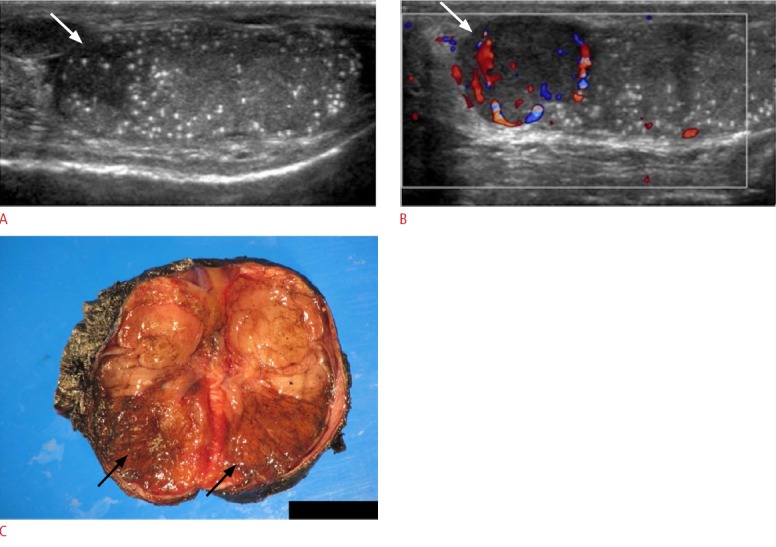
Fig. 18. Microcalcifications associated with a testicular mass in a 32-year-old male with a non-tender left scrotal mass.

A, B. Ultrasonogram reveals microcalcifications throughout the heterogeneously echogenic testicle (A) and presence of a hypoechoic vascular lesion (arrow in A and B) arising from the superior aspect of the left testicle (B). The left testicle was subsequently resected and pathology evaluation identified the mass as a seminoma. C. Macroscopic pathology specimen shows the entire left testicle cut in half. A solid, firm greyish-colored mass measuring 2.0×1.8×1.8 cm (arrows) is located at the superior pole of the testicle abutting the epididymis and tunica albuginea.
