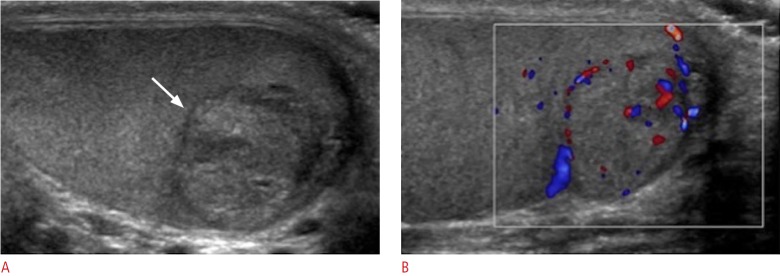
Fig. 20. A 26-year-old male with a painless right testicular mass.
A, B. Longitudinal gray-scale ultrasonogram of the right testicle (A) shows a heterogeneous mass (arrow), with internal vascular flow on color Doppler imaging (B). The patient subsequently underwent right orchiectomy. Histologically the mass was found to represent a seminoma.

