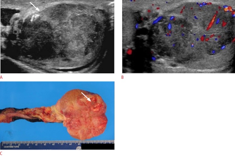
Fig. 22. A 33-year-old male with several months of left testicular pain and swelling.

A. Gray-scale ultrasonogram of the left testicle demonstrates an enlarged and heterogeneous testicle (arrow) with nodular, irregular contour. B. Color Doppler evaluation shows diffusely increased vascular flow throughout the testicle. The patient underwent left orchiectomy and the pathology revealed a mixed germ cell tumor with 80% seminoma and 20% embryonal carcinoma component. C. Macroscopic pathology specimen of the left testicular mass reveals a well-circumscribed, homogeneous, brown-gray tumor (arrow).
