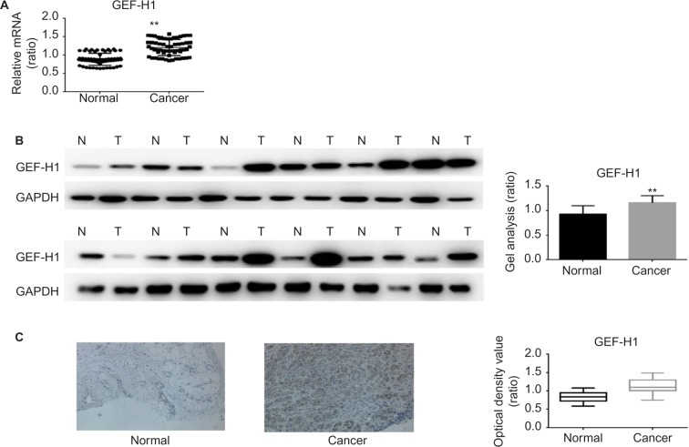
Figure 1.
mRNA and protein expressions of GEF-H1 in adjacent tissues and melanoma tissues.
Notes: (A) GEF-H1 mRNA expression in 60 samples of melanoma tissues and normal tissues was detected, respectively, by real-time RT-PCR. Results normalized to those of GAPDH. The expression of GEF-H1 was higher in the melanoma tissues. Data are shown as mean ± SEM. **P< 0.01 versus adjacent tissues group. (B) The protein level of GEF-H1 expression in melanoma tissues and normal tissues was detected by Western blot. The expression of GEF-H1 was higher in the melanoma tissues. Data are shown as mean ± SEM. **P<0.01 versus normal tissues group. (C) Representative immunohistochemical staining for GEF-H1 expression in melanoma. Left (normal): low expression of GEF-H1 in adjacent tissue. Right (cancer): high expression of GEF-H1 in melanoma tissues. Original magnification ×200.
Abbreviations: GEF-H1, guanine nucleotide exchange factor H1; mRNA, messenger RNA; RT-PCR, reverse transcription polymerase chain reaction; SEM, standard error of the mean.

