Abstract
Willed-movement training has been demonstrated to be a promising approach to increase motor performance and neural plasticity in ischemic rats. However, little is known regarding the molecular signals that are involved in neural plasticity following willed-movement training. To investigate the potential signals related to neural plasticity following willed-movement training, littermate rats were randomly assigned into three groups: middle cerebral artery occlusion, environmental modification, and willed-movement training. The infarct volume was measured 18 d after occlusion of the right middle cerebral artery. Reverse transcription-polymerase chain reaction (PCR) and immunofluorescence staining were used to detect the changes in the signal transducer and activator of transcription 3 (STAT3) mRNA and protein, respectively. A chromatin immunoprecipitation was used to investigate whether STAT3 bound to plasticity-related genes, such as brain-derived neurotrophic factor (BDNF), synaptophysin, and protein interacting with C kinase 1 (PICK1). In this study, we demonstrated that STAT3 mRNA and protein were markedly increased following 15-d willed-movement training in the ischemic hemispheres of the treated rats. STAT3 bound to BDNF, PICK1, and synaptophysin promoters in the neocortical cells of rats. These data suggest that the increased STAT3 levels after willed-movement training might play critical roles in the neural plasticity by directly regulating plasticity-related genes.
Keywords: Motor training, Signal transducer and activator of transcription 3 (STAT3), Brain-derived neurotrophic factor (BDNF), Protein interacting with C kinase 1 (PICK1), Neural plasticity
1. Introduction
Motor training is known to be able to enhance the neuronal structural and functional synaptic plasticity in the motor cortex. Willed-movement (WM) training is a type of motor training, in which the individual makes an effort to accomplish motor tasks (Tang et al., 2005; 2007). WM therapy for rats can improve neurobehavioral performance, upregulate the messenger RNAs (mRNAs) for the α-amino-3-hydroxy-5-methyl-4-isoxazolepropionic acid (AMPA)-type glutamate receptor subunits GluR1 and GluR4 (Tang et al., 2007), and increase the levels of protein interacting with C kinase 1 (PICK1) (Tang et al., 2013) and synaptophysin (data not shown) proteins in the ischemia hemispheres at their subacute stage. Additionally, WM training has been clinically demonstrated to be a promising approach to increase the motor recovery of stroke subjects with cognitive function deficits (Tang et al., 2005). These studies suggested that WM training might play a role in synaptic plasticity. The activity-dependent synaptic changes are a function of the precise synaptic transmission (Bliss and Collingridge, 1993; Huang et al., 2015). Neuronal transmission networks are involved in the molecular signaling that translates activities into long-lasting changes (Gao et al., 2010).
Signal transducer and activator of transcription 3 (STAT3) has been identified as a transcription factor involved in the maintenance of early embryonic neocortical development (Yoshimatsu et al., 2006; Oatley et al., 2010) and neuronal survival (Yadav et al., 2005). STAT3 signaling mediates axon elongation (Selvaraj et al., 2012; Quarta et al., 2014) and plays a pivotal role in early neural circuit formation (Bouret et al., 2012). These studies suggest that STAT3 is involved in neural plasticity. However, studies have not tested the change of STAT3 expression levels following motor training, nor whether STAT3 is involved in regulating plasticity-related genes. In the present study, we investigated the possible role of STAT3 on neural plasticity in WM-trained rats which had suffered focal cerebral ischemia.
2. Materials and methods
2.1. Design
Ninety-eight adult male Sprague Dawley rats weighing 200 to 250 g were used in this study. All animals were housed at (23±2) °C room temperature with a 12-h light/dark cycle. A right middle cerebral artery occlusion (MCAO) was performed as in our previous study (Tang et al., 2007). Only 36 survived rats with a neurological deficit score of 2 or 3 two hours after recirculation were recruited in this study.
Littermate rats were randomly assigned to three groups before ischemic surgery: rats that received only right MCAO, rats that additionally received environmental modification (EM), and rats that additionally received WM therapy. The housing, feeding, and training protocols of the rats in the EM and WM groups were the same as that in a previous study (Tang et al., 2013). The rats were sacrificed 18 d after the surgery. The climbing frequency and neurological and neurobehavioral examinations were assessed according to our previous studies (Tang et al., 2013).
2.2. Measurement of infarct volume
The infarct volume was measured using a 20 mg/ml 2,3,5-triphenyl tetrazolium chloride (TTC) staining protocol 18 d after MCAO. The rats were deeply anesthetized with sodium pentobarbital (60 mg/kg) and decapitated. The brain was then removed and coronally sliced into five 2-mm thick coronal sections. The slices were immersed in the TTC solution for 30 min and then fixed with 10% paraformaldehyde. The infarct volumes were calculated by measuring the infarct areas on each of the slices, multiplying these values by the slice thicknesses, and adding the infarct areas of all slices together. To eliminate the effect of brain edema, the corrected infarct volume was calculated using the following formula: intact contralateral hemisphere volume−(ipsilateral hemisphere volume−measured infarct volume) (Schabitz et al., 1999).
2.3. Reverse transcription-polymerase chain reaction (RT-PCR)
The tissue samples of the ischemic penumbra (IP) were dissected according to the method descripted by Ashwal et al. (1998). Briefly, a longitudinal cut approximately 2 mm from the midline in the right hemisphere was made. Then a transverse diagonal cut was made to separate the penumbra from the core. The RNA extraction was performed as in our previous study (Tang et al., 2007). Briefly, on Day 18 after surgery, rats were sacrificed and total RNA was extracted from the IP. Then the RNA was pelleted by centrifugation, washed and redissolved in 20 μl of diethylpyrocarbonate (DEPC)-treated water. The concentration and purity of RNA were confirmed and total RNA quality was assessed.
The RevertAid™ First Strand cDNA Synthesis Kit (Thermo, Cat. No. K1622) was utilized to synthesize complementary DNA (cDNA) according to the kit instructions. The 2-μg RNA was added to the cDNA synthesis reaction and one-twentieth volume of the final cDNA product (1 μl per reaction) was added to PCR reactions.
Oligonucleotide primers were designed on a computer (Primer 5 software) and synthesized by BGI (Shenzhen, China). The primers were designed for STAT3 cDNA as follows: sense, 5'-AAAGGACATC AGTGGCAAGA-3' and antisense, 5'-ACATCGGC AGGTCAATGGTA-3' with a length of 305 bp. As a control, the primers for glyceraldehyde-3-phosphate dehydrogenase (GAPDH) mRNA were designed from the published sequences as follows: sense, 5'-ACCA CAGTCCATGCCATCAC-3' and antisense, 5'-TCC ACCACCCTGTTGCTGTA-3' with a length of 452 bp (Niture and Jaiswal, 2012). Specificity of the primers was determined by a BLAST (basic local alignment search tool) search. After preparation of the first-strand cDNA, the reaction solution was mixed with PCR reagents to make a 20-μl reaction solution containing 10 μl of 2× pre-Taq PCR master mix, 1 μl of each set of primer, 1 μl of template cDNA, and 8 μl of double-distilled water (ddH2O). The reverse transcription reactions were carried on among the internal standard GAPDH and STAT3 in each cDNA production but in different separate tubes. PCR was performed in a T100 thermal cycler (Bio-Rad) by initial denaturation at 95 °C for 3 min, followed by 22 cycles of PCR amplification: denaturation at 95 °C for 30 s, annealing of primers STAT3 at 72 °C or GAPDH at 58 °C for 30 s, and extension at 72 °C for 1 min. PCR was completed for a final extension of 72 °C for 10 min. Negative controls for PCR were performed using templates derived from reverse transcription reactions lacking either reverse transcriptase or total RNA. Finally, PCR products were run on a 1.2% (12 mg/ml) agarose gel containing 0.2 μg/ml ethidium bromide, visualized under ultraviolet (UV) light (Bio-Rad), and analyzed with Quantity One 1-D analysis software (Bio-Rad).
2.4. Immunofluorescence staining
Immunofluorescence staining was performed on frozen sections of rat brain samples fixed by perfusion with 40 mg/ml fresh paraformaldehyde in 0.1 mol/L phosphate buffer solution (pH 7.4, 4 °C) (Tang et al., 2013). The coronal sections (30 μm, −0.3 to 1.3 mm vs. bregma (Liu et al., 2006)) of the brain were then cut on a Leica CM1900 cryostat (Leica Microsystems, Wetzlar, Germany). A free-floating immunofluorescence method similar to that previously described (Tang et al., 2013) was utilized to detect STAT3. After the brain slices were incubated with a mouse anti-STAT3 antibody (Ab119352, 1:400; Abcam, Cambridge, MA, USA) for 48 h at 4 °C, the antibody was detected with an Alexa-594-labeled donkey anti-mouse antibody (A21203, 1:200; Invitrogen, Paisley, PA, USA) for 2 h at 37 °C. The stained slices were observed on an inverted fluorescence microscope (Eclipse T1, Nikon, Melville, NY, USA). Three bregma sections (1.2, 0.48, −0.24 mm) were selected and observed at 40×10 magnification for each rat. The regions with STAT3-positive cells expressed in the polymorphic layers of the cortical area and its adjacent corpus callosum, and the neural nuclei below the putamen in each section were scanned. The expression of STAT3 was evaluated by counting the number of STAT3-positive cells in each scanning field.
2.5. Cell culture
Mixed cultures of cortical neurons and glial cells were prepared from the cerebral cortices of embryonic Sprague Dawley rats at early embryonic stage (E18) (Doyle et al., 2010) with minor modifications. Briefly, the dissected cortical tissue was dissociated with 1.25 mg/ml trypsin for 20 min at 37 °C. The cells were then plated in poly-D-lysine-coated 6-well clusters at a density of 7.5×10⁵ cells per well and cultured in Neurobasal-A medium supplemented with 20 mg/ml B27 (Gibco-Life Technologies) and 0.5 mmol/L GlutaMAX™-I. Half the volume of the culture media was replenished 3 times per week and incubated at 37 °C with 5% CO2.
2.6. Immunocytochemistry
Neurons at Day 7 were fixed in 40 mg/ml paraformaldehyde for 20 min, permeabilized for 10 min in 2 mg/ml Triton X-100, and blocked for 30 min in 5% normal donkey serum. The cells were then incubated with a mouse anti-NeuN (diluted 1:100; Millipore, Billerica, MA, USA) monoclonal antibody overnight at 4 °C. The cells were then treated with Alexa-594-labeled donkey anti-mouse antibody (diluted 1:200; Invitrogen) for 2 h at 37 °C. After removing the secondary antibody, the nuclei were stained with Hoechst 33342 (diluted 1:2000; Invitrogen), and the cells were visualized using a Nikon Eclipse TE2000-S inverted microscope (Nikon Instruments, Melville, NY, USA).
2.7. Chromatin immunoprecipitation
A chromatin immunoprecipitation (ChIP) assay was performed using the ChIP kit from Upstate (Billerica, MA, USA) according to the manufacturer’s protocol. Briefly, neocortical cells on Day 7 in vitro were used in ChIP experiences, and cells were cross-linked with 10 mg/ml formaldehyde for 10 min at 37 °C. The cells were then homogenized in lysis buffer. The lysates were sonicated at 152 W for 6 min (cycles of 5 s “on” and 9 s “off”, SCIENTZ-IID, Scientz, China) on wet ice. After pre-clearing the chromatin with protein G agarose, the sonicated chromatin (100 μl) was immunoprecipitated with 10 μg of STAT3 mouse antibody (Cell Signaling Technology Inc., Beverly, MA, USA), 1.0 μg of positive control (anti-RNA polymerase, Upstate), or 1.0 μg of negative control (normal mouse IgG, Upstate) in duplicate. Bound chromatin was eluted and was reverse cross-linked overnight at 65 °C. The ChIPed DNA was purified using spin columns and eluted with elution buffer. The purified DNA was used for quantitative PCR (qPCR).
2.8. PCR of immunoprecipitated DNA
Immunoprecipitated DNA was subjected to PCR analyses in a 20-μl reaction solution containing 2 μl DNA template, 1 μl of each set of primer, 7 μl H2O, and 10 μl GoTaq® GreenMaster Mix (Promega, USA). The PCR reaction program consisted of an initial denaturation at 94 °C for 5 min, and 32 repeated cycles as follows: a heat denaturation at 94 °C for 30 s, annealing of STAT3-synaptophysin at 55 °C for 30 s, STAT3-brain-derived neurotrophic factor (BDNF) at 46 °C for 30 s, STAT3-PICK1 at 58 °C for 30 s, and GAPDH at 58 °C for 30 s, with extension at 72 °C for 30 s. PCR was completed for a final extension at 72 °C for 2 min. The PCR fragments were analyzed by electrophoresis on a 4% (40 mg/ml) agarose gel containing 0.2 mg/ml ethidium bromide and visualized under a UV light (Molecular Imager® ChemiDoc™ XRS+System, Bio-Rad, USA). The control primers were designed for the rats’ GAPDH gene in immunoprecipitated DNA, including a forward primer 5'-ACCACAGTCCATGCCATCAC-3' and a reverse primer 5'-TCCACCACCCTGTTGCTGTA-3' with a length of 452 bp (Niture and Jaiswal, 2012). The primers were designed according to the putative STAT3 binding sites at BDNF, PICK1, and synaptophysin promoters using the TFSEARCH server (http://cbrc3.cbrc.jp/research/db/TFSEARCH.html). The binding sites, the exact position of the primer design for each gene, and sequences of these primers are given in Table 1. The specificity of the primers was confirmed by a BLAST search.
Table 1.
STAT3 binding sites and primers used for quantitative PCR for ChIP
| Gene | GenBank No. | Binding site | Primer position | Primer sequence | Size (bp) |
| BDNF | NC_005102.3 | TTCCAAGAA | 5874‒6067 | Forward: 5'-GCCTGGCAACTCTAA-3' | 194 |
| Reverse: 5'-TCCTAATGCGTTCTATG-3' | |||||
| PICK1 | NC_005106.3 | TTCCCTGAA | 2613‒2909 | Forward: 5'-CTGGCTGTGGGACGGACT-3' | 287 |
| Reverse: 5'-AAGAGCACAGGGCTTCAGG-3' | |||||
| Syn | NC_005120.3 | TTCCTCTAA | 1335‒1509 | Forward: 5'-AACTGAGCGGTCCTCCTAC-3' | 175 |
| Reverse: 5'-TCGCACCCTGTCTGTCTT-3' |
2.9. Statistical analysis
All data were analyzed using the SPSS 19.0 statistical package. Data were presented as the mean±standard deviation (SD). One-way analysis of variance (ANOVA) was used for the expression data, followed by the least significant difference (LSD) test for post-hoc comparisons. Statistically significant differences were established at P<0.05. The effect size was computed using partial eta squared (η 2) value.
3. Results
3.1. Infarct volumes
The mean infarct volumes were (68.62±10.07), (67.95±10.81), and (66.88±14.86) mm3 for the MCAO, EM, and WM rats, respectively. Significant differences were not found among the three groups (Fig. 1; df=2, F=0.03, η 2=0.00, P>0.05).
Fig. 1.
TTC staining of the rats 18 d after surgery
Representative TTC staining of the MCAO (a), EM (b), and WM (c) rats, respectively. (d) Histogram demonstrates the infarct volumes of MCAO, EM, and WM rats 18 d after surgery. Data are expressed as mean±SD of 6 rats each group
3.2. RT-PCR of STAT3 mRNA
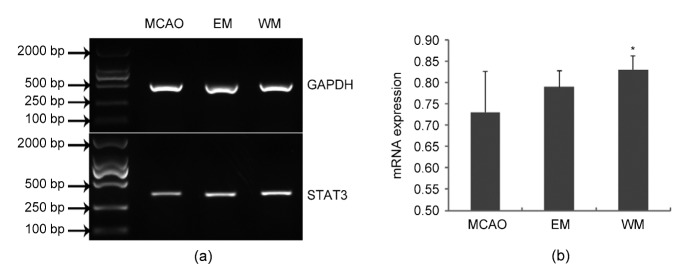
At 18 d after perfusion, the STAT3 mRNAs of the three groups were significantly different from each other in the IP regions (Fig. 2; df=2, F=4.58, η 2=0.34, P<0.05). Further LSD indicated that STAT3 mRNA of WM rats in IP region (0.83±0.03) was upregulated at an average level of 13.70% compared with MCAO rats (0.73±0.09), P<0.01. No significant differences were found in STAT3 mRNA expressional levels between MCAO and EM rats (0.79±0.04), nor between EM and WM in IP regions (Fig. 2b).
Fig. 2.
Expression of STAT3 and GAPDH detected by RT-PCR
(a) Representative RT-PCR of mRNAs of STAT3 (305 bp) and GAPDH (452 bp) in the IP region of ischemic rats from MCAO, EM, and WM groups. The intensity of band for STAT3 in the WM rats is significantly higher than that in the MCAO rats. (b) Histogram demonstrates the intensity of STAT3 mRNAs in the IP region of ischemic rats for MCAO, EM, and WM rats. Data are expressed as mean±SD of 6 rats each group. * P<0.01, vs. MCAO group
3.3. Immunofluorescence labeling of STAT3
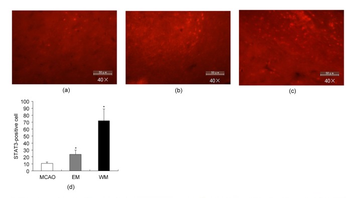
The immunofluorescence signals for antibodies to STAT3 were mainly localized in the cytoplasm and the nucleus at the polymorphic layers of cortical area and its adjacent corpus callosum, neural nuclei below the putamen. The STAT3-positive cells of the three groups were significantly different from each other in the ischemic hemisphere (Fig. 3; df=2, F=107.87, η 2=0.86, P<0.001). The WM rats showed a more than 7-fold increase and the EM rats showed a more than 2-fold increase in STAT3-positive cells compared with the MCAO rats (MCAO 10.42±2.39, EM 23.67±6.08, and WM 71.75±17.46 in 40×10 microscope, P<0.01).
Fig. 3.
Immunofluorescence signals of STAT3 at the polymorphic layers of the cortical area and its adjacent corpus callosum in the ischemic hemisphere for MCAO, EM, and WM rats
Representative immunofluorescence staining of STAT3 for MCAO (a), EM (b), and WM (c) rats, respectively. (d) Histogram demonstrates the number of STAT3-positive cells in the ischemic hemisphere for MCAO, EM, and WM rats. Data are expressed as mean±SD of 6 rats each group. * P<0.01, vs. MCAO group
3.4. Cortical neurons and glial cells in the culture
NeuN has been recognized as an excellent marker for neurons in the central and peripheral nervous systems (Mullen et al., 1992). In our study, the Hoechst and NeuN staining indicated that most of the culture cells were neurons (86.7%) and a few cells were glial cells (13.3%), as shown in Fig. 4.
Fig. 4.
Cortical neurons and glial cells on Day 7
(a) Cortical neurons and glial cells. (b) Hoechst staining nuclei (blue) of the cortical neurons and glial cells. (c) NeuN (red) cells with cytoplasm and nuclear staining in the neurons (Note: for interpretation of the references to color in this figure legend, the reader is referred to the web version of this article)
3.5. STAT3 binding to the BDNF, PICK1, and synaptophysin promoters
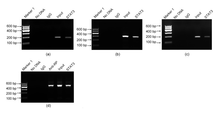
ChIP-PCR analyses were performed in neocortical cells to validate that STAT3 mediates the BDNF, PICK1, and synaptophysin genes. DNA was immunoprecipitated with anti-STAT3, normal mouse IgG, or anti-RNA polymerase using primers designed from the BDNF, PICK1, and synaptophysin promoter fragments. The BDNF, PICK1, and synaptophysin promoter sequences were amplified specifically from the STAT3 samples (Fig. 5), which indicated the binding of STAT3 to these plasticity-related genes in neocortical cells.
Fig. 5.
Binding of STAT3 to the BDNF, PICK1, and synaptophysin promoters
The ChIP of synchronized neocortical cells was performed using an antibody to STAT3, a negative control antibody (IgG), or a positive control anti-RNA polymerase (anti-RP). A sample without DNA was tested as an additional control for DNA contamination, and an input sample was also detected. Immunoprecipitated DNA analyzed by PCR using primers that amplify the BDNF (a), PICK1 (b), synaptophysin (c) promoters and GAPDH gene (d), respectively
4. Discussion
This study indicated that the levels of STAT3 mRNA and protein markedly increased in the ischemic hemisphere following 15 d of WM training. Studies have demonstrated that STAT3 was elevated from approximately 0.5 h to several days after ischemia. The increased STAT3 levels were thought to be involved in the process of cerebral ischemia and reperfusion injury in the acute stage of ischemia (Suzuki et al., 2001; Lei et al., 2011; Shulga and Pastorino, 2012). However, the increase in STAT3 in the present study was found after 18 d of ischemic reperfusion. Additionally, although the infarct volumes and tissue loss were similar in all the rats in the three groups, a significant increase of STAT3 expressional level persisted in WM rats compared with MCAO or/and EM rats. Therefore, the enhancement of STAT3 protein was not involved in ischemia and reperfusion injury for WM rats.
Motor training can increase synaptic transmission and enhance neural plasticity (Jones et al., 1999; Maldonado et al., 2008; Tang et al., 2013). Our previous data (Tang et al., 2007; 2013) indicated that WM training for rats could improve neurobehavioral performance compared with EM and MCAO rats. STAT3 activation is associated with early embryonic and neocortical development (Yoshimatsu et al., 2006; Oatley et al., 2010), increasing cell survival (Yadav et al., 2005; Dziennis and Alkayed, 2008), regulating neuronal differentiation (Chang et al., 2014), and mediating axon elongation (Liu and Snider, 2001; Selvaraj et al., 2012) and early neural circuit formation (Bouret et al., 2012). Therefore, we speculated that the motor activity in the WM rats induced the increase of STAT3 protein that might be related to neural plasticity in the subacute stage of focal ischemia.
Our ChIP analysis confirmed that STAT3 bound to BDNF, synaptophysin, and PICK1 in neocortical cells. BDNF (Nakai et al., 2014), synaptophysin (Liang et al., 2007; Gordon et al., 2011; Kwon and Chapman, 2011), and PICK1 (Antoniou et al., 2014; Xu et al., 2014) have been proven to be the key proteins in the regulation of synaptic plasticity. Numerous studies indicated that motor exercise increased the expressions of BDNF (Gómez-Pinilla et al., 2002; Ploughman et al., 2009; MacLellan et al., 2011; Wilhelm et al., 2012), PICK1 (Volk et al., 2010), and synaptophysin (Seo et al., 2010), which promoted neuroplasticity and was beneficial for recovery after a stroke (MacLellan et al., 2011). Our previous studies have proven that the expressional levels of PICK1 (Tang et al., 2013) and synaptophysin proteins were elevated after WM training in the IP regions of ischemic rats. The STAT3-mediated activations of BDNF, synaptophysin, and PICK1 may therefore play a role in synaptic plasticity in WM training for focal ischemic rats. In WM training, STAT3 may affect synaptic vesicle efficiency via its synaptophysin-activating function (Liang et al., 2007; Gordon et al., 2011; Kwon and Chapman, 2011). Additionally, STAT3 may facilitate nerve regeneration, neuronal development, and synaptic transmission by a BDNF mechanism (Gómez-Pinilla et al., 2002; Ng et al., 2006; Ploughman et al., 2009; MacLellan et al., 2011; Waterhouse et al., 2012; Wilhelm et al., 2012). Furthermore, STAT3 may indirectly regulate receptors, transporters, and ion channel trafficking by activating PICK1 (Citri et al., 2010; Hu et al., 2010). Future studies could be designed to clarify which of these mechanisms are responsible for the neuroplasticity regulated by STAT3 after WM training.
Our findings agree with a study by Ng et al. (2006), in which the inhibition of STAT3 markedly attenuated BDNF-induced axon outgrowth in cultured hippocampal neurons. This finding suggested that BDNF signaling is regulated by the STAT3 pathway during axon elongation (Dominguez et al., 2010). However, as far as we know, no studies have directly shown that STAT3 can bind to BDNF, synaptophysin, and PICK1 promoters using ChIP assays.
In summary, our study discovered that STAT3 increases after WM training and is an important transcription factor for regulating plasticity-related genes, such as BDNF, synaptophysin, and PICK1. These results indicate a novel gene regulatory mechanism mediated by STAT3 that is involved in neural plasticity. Furthermore, gene interference or knockout of STAT3 in WM rats is recommended to investigate whether these signaling pathways functionally regulate the neural plasticity following WM training.
Footnotes
Project supported by the National Natural Science Foundation of China (Nos. 30973167, 81472160, and 81173595), the China Postdoctoral Science Foundation (Nos. 2011M501301 and 2012T50711), and the China-Japan Friendship Hospital Youth Science and Technology Excellence Project (No. 2014-QNYC-A-04)
Compliance with ethics guidelines: Qing-ping TANG, Qin SHEN, Li-xiang WU, Xiang-ling FENG, Hui LIU, Bei WU, Xiao-song HUANG, Gai-qing WANG, Zhong-hao LI, and Zun-jing LIU declare that they have no conflict of interest.
All institutional and national guidelines for the care and use of laboratory animals were followed.
References
- 1.Antoniou A, Baptista M, Carney N, et al. PICK1 links Argonaute 2 to endosomes in neuronal dendrites and regulates miRNA activity. EMBO Rep. 2014;15(5):548–556. doi: 10.1002/embr.201337631. (Available from: http://dx.doi.org/10.1002/embr.201337631) [DOI] [PMC free article] [PubMed] [Google Scholar]
- 2.Ashwal S, Tone B, Tian HR, et al. Core and penumbral nitric oxide synthase activity during cerebral ischemia and reperfusion. Stroke. 1998;29(5):1037–1046. [PubMed] [Google Scholar]
- 3.Bliss TV, Collingridge GL. A synaptic model of memory: long-term potentiation in the hippocampus. Nature. 1993;361(6407):31–39. doi: 10.1038/361031a0. (Available from: http://dx.doi.org/10.1038/361031a0) [DOI] [PubMed] [Google Scholar]
- 4.Bouret SG, Bates SH, Chen S, et al. Distinct roles for specific leptin receptor signals in the development of hypothalamic feeding circuits. J Neurosci. 2012;32(4):1244–1252. doi: 10.1523/JNEUROSCI.2277-11.2012. (Available from: http://dx.doi.org/10.1523/jneurosci.2277-11.2012) [DOI] [PMC free article] [PubMed] [Google Scholar]
- 5.Chang YJ, Chen KW, Chen CJ, et al. SH2B1β interacts with STAT3 and enhances fibroblast growth factor 1-induced gene expression during neuronal differentiation. Mol Cell Biol. 2014;34(6):1003–1019. doi: 10.1128/MCB.00940-13. (Available from: http://dx.doi.org/10.1128/MCB.00940-13) [DOI] [PMC free article] [PubMed] [Google Scholar]
- 6.Citri A, Bhattacharyya S, Ma C, et al. Calcium binding to PICK1 is essential for the intracellular retention of AMPA receptors underlying long-term depression. J Neurosci. 2010;30(49):16437–16452. doi: 10.1523/JNEUROSCI.4478-10.2010. (Available from: http://dx.doi.org/10.1523/jneurosci.4478-10.2010) [DOI] [PMC free article] [PubMed] [Google Scholar]
- 7.Dominguez E, Mauborgne A, Mallet J, et al. SOCS3-mediated blockade of JAK/STAT3 signaling pathway reveals its major contribution to spinal cord neuroinflammation and mechanical allodynia after peripheral nerve injury. J Neurosci. 2010;30(16):5754–5766. doi: 10.1523/JNEUROSCI.5007-09.2010. (Available from: http://dx.doi.org/10.1523/jneurosci.5007-09.2010) [DOI] [PMC free article] [PubMed] [Google Scholar]
- 8.Doyle S, Pyndiah S, de Gois S, et al. Excitation-transcription coupling via calcium/calmodulin-dependent protein kinase/ERK1/2 signaling mediates the coordinate induction of VGLUT2 and Narp triggered by a prolonged increase in glutamatergic synaptic activity. J Biol Chem. 2010;285(19):14366–14376. doi: 10.1074/jbc.M109.080069. (Available from: http://dx.doi.org/10.1074/jbc.M109.080069) [DOI] [PMC free article] [PubMed] [Google Scholar]
- 9.Dziennis S, Alkayed NJ. Role of signal transducer and activator of transcription 3 in neuronal survival and regeneration. Rev Neurosci. 2008;19(4-5):341–361. doi: 10.1515/revneuro.2008.19.4-5.341. [DOI] [PMC free article] [PubMed] [Google Scholar]
- 10.Gao J, Wang WY, Mao YW, et al. A novel pathway regulates memory and plasticity via SIRT1 and miR-134. Nature. 2010;466(7310):1105–1109. doi: 10.1038/nature09271. (Available from: http://dx.doi.org/10.1038/nature09271) [DOI] [PMC free article] [PubMed] [Google Scholar]
- 11.Gómez-Pinilla F, Ying Z, Roy RR, et al. Voluntary exercise induces a BDNF-mediated mechanism that promotes neuroplasticity. J Neurophysiol. 2002;88(5):2187–2195. doi: 10.1152/jn.00152.2002. (Available from: http://dx.doi.org/10.1152/jn.00152.2002) [DOI] [PubMed] [Google Scholar]
- 12.Gordon SL, Leube RE, Cousin MA. Synaptophysin is required for synaptobrevin retrieval during synaptic vesicle endocytosis. J Neurosci. 2011;31(39):14032–14036. doi: 10.1523/JNEUROSCI.3162-11.2011. (Available from: http://dx.doi.org/10.1523/jneurosci.3162-11.2011) [DOI] [PMC free article] [PubMed] [Google Scholar]
- 13.Hu ZL, Huang C, Fu H, et al. Disruption of PICK1 attenuates the function of ASICs and PKC regulation of ASICs. Am J Physiol Cell Physiol. 2010;299(6):C1355–C1362. doi: 10.1152/ajpcell.00569.2009. (Available from: http://dx.doi.org/10.1152/ajpcell.00569.2009) [DOI] [PubMed] [Google Scholar]
- 14.Huang J, Du FL, Yao Y, et al. Numerical magnitude processing in abacus-trained children with superior mathematical ability: an EEG study. J Zhejiang Univ-Sci B (Biomed & Biotechnol) 2015;16(8):661–671. doi: 10.1631/jzus.B1400287. (Available from: http://dx.doi.org/10.1631/jzus.B1400287) [DOI] [PMC free article] [PubMed] [Google Scholar]
- 15.Jones TA, Chu CJ, Grande LA, et al. Motor skills training enhances lesion-induced structural plasticity in the motor cortex of adult rats. J Neurosci. 1999;19(22):10153–10163. doi: 10.1523/JNEUROSCI.19-22-10153.1999. [DOI] [PMC free article] [PubMed] [Google Scholar]
- 16.Kwon SE, Chapman ER. Synaptophysin regulates the kinetics of synaptic vesicle endocytosis in central neurons. Neuron. 2011;70(5):847–854. doi: 10.1016/j.neuron.2011.04.001. (Available from: http://dx.doi.org/10.1016/j.neuron.2011.04.001) [DOI] [PMC free article] [PubMed] [Google Scholar]
- 17.Lei C, Deng J, Wang B, et al. Reactive oxygen species scavenger inhibits STAT3 activation after transient focal cerebral ischemia-reperfusion injury in rats. Anesth Analg. 2011;113(1):153–159. doi: 10.1213/ANE.0b013e31821a9fbe. (Available from: http://dx.doi.org/10.1213/ANE.0b013e31821a9fbe) [DOI] [PubMed] [Google Scholar]
- 18.Liang YJ, Wu DF, Yang LQ, et al. Interaction of the μ-opioid receptor with synaptophysin influences receptor trafficking and signaling. Mol Pharmacol. 2007;71(1):123–131. doi: 10.1124/mol.106.026062. (Available from: http://dx.doi.org/10.1124/mol.106.026062) [DOI] [PubMed] [Google Scholar]
- 19.Liu H, Honmou O, Harada K, et al. Neuroprotection by PlGF gene-modified human mesenchymal stem cells after cerebral ischaemia. Brain. 2006;129(Pt 10):2734–2745. doi: 10.1093/brain/awl207. (Available from: http://dx.doi.org/10.1093/brain/awl207) [DOI] [PMC free article] [PubMed] [Google Scholar]
- 20.Liu RY, Snider WD. Different signaling pathways mediate regenerative versus developmental sensory axon growth. J Neurosci. 2001;21(17):RC164. doi: 10.1523/JNEUROSCI.21-17-j0003.2001. [DOI] [PMC free article] [PubMed] [Google Scholar]
- 21.MacLellan CL, Keough MB, Granter-Button S, et al. A critical threshold of rehabilitation involving brain-derived neurotrophic factor is required for poststroke recovery. Neurorehabil Neural Repair. 2011;25(8):740–748. doi: 10.1177/1545968311407517. (Available from: http://dx.doi.org/10.1177/1545968311407517) [DOI] [PubMed] [Google Scholar]
- 22.Maldonado MA, Allred RP, Felthauser EL, et al. Motor skill training, but not voluntary exercise, improves skilled reaching after unilateral ischemic lesions of the sensorimotor cortex in rats. Neurorehabil Neural Repair. 2008;22(3):250–261. doi: 10.1177/1545968307308551. (Available from: http://dx.doi.org/10.1177/1545968307308551) [DOI] [PMC free article] [PubMed] [Google Scholar]
- 23.Mullen RJ, Buck CR, Smith AM. NeuN, a neuronal specific nuclear protein in vertebrates. Development. 1992;116(1):201–211. doi: 10.1242/dev.116.1.201. [DOI] [PubMed] [Google Scholar]
- 24.Nakai T, Nagai T, Tanaka M, et al. Girdin phosphorylation is crucial for synaptic plasticity and memory: a potential role in the interaction of BDNF/TrkB/Akt signaling with NMDA receptor. J Neurosci. 2014;34(45):14995–15008. doi: 10.1523/JNEUROSCI.2228-14.2014. (Available from: http://dx.doi.org/10.1523/JNEUROSCI.2228-14.2014) [DOI] [PMC free article] [PubMed] [Google Scholar]
- 25.Ng YP, Cheung ZH, Ip NY. STAT3 as a downstream mediator of Trk signaling and functions. J Biol Chem. 2006;281(23):15636–15644. doi: 10.1074/jbc.M601863200. (Available from: http://dx.doi.org/10.1074/jbc.M601863200) [DOI] [PubMed] [Google Scholar]
- 26.Niture SK, Jaiswal AK. Nrf2 protein up-regulates antiapoptotic protein Bcl-2 and prevents cellular apoptosis. J Biol Chem. 2012;287(13):9873–9886. doi: 10.1074/jbc.M111.312694. (Available from: http://dx.doi.org/10.1074/jbc.M111.312694) [DOI] [PMC free article] [PubMed] [Google Scholar]
- 27.Oatley JM, Kaucher AV, Avarbock MR, et al. Regulation of mouse spermatogonial stem cell differentiation by STAT3 signaling. Biol Reprod. 2010;83(3):427–433. doi: 10.1095/biolreprod.109.083352. (Available from: http://dx.doi.org/10.1095/biolreprod.109.083352) [DOI] [PMC free article] [PubMed] [Google Scholar]
- 28.Ploughman M, Windle V, MacLellan CL, et al. Brain-derived neurotrophic factor contributes to recovery of skilled reaching after focal ischemia in rats. Stroke. 2009;40(4):1490–1495. doi: 10.1161/STROKEAHA.108.531806. (Available from: http://dx.doi.org/10.1161/strokeaha.108.531806) [DOI] [PubMed] [Google Scholar]
- 29.Quarta S, Baeumer BE, Scherbakov N, et al. Peripheral nerve regeneration and NGF-dependent neurite outgrowth of adult sensory neurons converge on STAT3 phosphorylation downstream of neuropoietic cytokine receptor gp130. J Neurosci. 2014;34(39):13222–13233. doi: 10.1523/JNEUROSCI.1209-13.2014. (Available from: http://dx.doi.org/10.1523/jneurosci.1209-13.2014) [DOI] [PMC free article] [PubMed] [Google Scholar]
- 30.Schabitz WR, Li F, Irie K, et al. Synergistic effects of a combination of low-dose basic fibroblast growth factor and citicoline after temporary experimental focal ischemia. Stroke. 1999;30(2):427–431. doi: 10.1161/01.str.30.2.427. [DOI] [PubMed] [Google Scholar]
- 31.Selvaraj BT, Frank N, Bender FL, et al. Local axonal function of STAT3 rescues axon degeneration in the pmn model of motoneuron disease. J Cell Biol. 2012;199(3):437–451. doi: 10.1083/jcb.201203109. (Available from: http://dx.doi.org/10.1083/jcb.201203109) [DOI] [PMC free article] [PubMed] [Google Scholar]
- 32.Seo HG, Kim DY, Park HW, et al. Early motor balance and coordination training increased synaptophysin in subcortical regions of the ischemic rat brain. J Korean Med Sci. 2010;25(11):1638–1645. doi: 10.3346/jkms.2010.25.11.1638. (Available from: http://dx.doi.org/10.3346/jkms.2010.25.11.1638) [DOI] [PMC free article] [PubMed] [Google Scholar]
- 33.Shulga N, Pastorino JG. GRIM-19-mediated translocation of STAT3 to mitochondria is necessary for TNF-induced necroptosis. J Cell Sci. 2012;125(Pt 12):2995–3003. doi: 10.1242/jcs.103093. (Available from: http://dx.doi.org/10.1242/jcs.103093) [DOI] [PMC free article] [PubMed] [Google Scholar] [Retracted]
- 34.Suzuki S, Tanaka K, Nogawa S, et al. Phosphorylation of signal transducer and activator of transcription-3 (Stat3) after focal cerebral ischemia in rats. Exp Neurol. 2001;170(1):63–71. doi: 10.1006/exnr.2001.7701. (Available from: http://dx.doi.org/10.1006/exnr.2001.7701) [DOI] [PubMed] [Google Scholar]
- 35.Tang QP, Yang QD, Wu YH, et al. Effects of problem-oriented willed-movement therapy on motor abilities for people with poststroke cognitive deficits. Phys Ther. 2005;85(10):1020–1033. [PubMed] [Google Scholar]
- 36.Tang QP, Yang QD, Hu ZY, et al. The effects of willed movement therapy on AMPA receptor properties for adult rat following focal cerebral ischemia. Behav Brain Res. 2007;181(2):254–261. doi: 10.1016/j.bbr.2007.04.013. (Available from: http://dx.doi.org/10.1016/j.bbr.2007.04.013) [DOI] [PubMed] [Google Scholar]
- 37.Tang QP, Tan LH, Yang XS, et al. Willed-movement training reduces motor deficits and induces a PICK1-dependent LTD in rats subjected to focal cerebral ischemia. Behav Brain Res. 2013;256:481–487. doi: 10.1016/j.bbr.2013.08.039. (Available from: http://dx.doi.org/10.1016/j.bbr.2013.08.039) [DOI] [PubMed] [Google Scholar]
- 38.Volk L, Kim CH, Takamiya K, et al. Developmental regulation of protein interacting with C kinase 1 (PICK1) function in hippocampal synaptic plasticity and learning. PNAS. 2010;107(50):21784–21789. doi: 10.1073/pnas.1016103107. (Available from: http://dx.doi.org/10.1073/pnas.1016103107) [DOI] [PMC free article] [PubMed] [Google Scholar]
- 39.Waterhouse EG, An JJ, Orefice LL, et al. BDNF promotes differentiation and maturation of adult-born neurons through GABAergic transmission. J Neurosci. 2012;32(41):14318–14330. doi: 10.1523/JNEUROSCI.0709-12.2012. (Available from: http://dx.doi.org/10.1523/jneurosci.0709-12.2012) [DOI] [PMC free article] [PubMed] [Google Scholar]
- 40.Wilhelm JC, Xu M, Cucoranu D, et al. Cooperative roles of BDNF expression in neurons and Schwann cells are modulated by exercise to facilitate nerve regeneration. J Neurosci. 2012;32(14):5002–5009. doi: 10.1523/JNEUROSCI.1411-11.2012. (Available from: http://dx.doi.org/10.1523/jneurosci.1411-11.2012) [DOI] [PMC free article] [PubMed] [Google Scholar]
- 41.Xu J, Kam C, Luo JH, et al. PICK1 mediates synaptic recruitment of AMPA receptors at neurexin-induced postsynaptic sites. J Neurosci. 2014;34(46):15415–15424. doi: 10.1523/JNEUROSCI.0296-14.2014. (Available from: http://dx.doi.org/10.1523/JNEUROSCI.0296-14.2014) [DOI] [PMC free article] [PubMed] [Google Scholar]
- 42.Yadav A, Kalita A, Dhillon S, et al. JAK/STAT3 pathway is involved in survival of neurons in response to insulin-like growth factor and negatively regulated by suppressor of cytokine signaling-3. J Biol Chem. 2005;280(36):31830–31840. doi: 10.1074/jbc.M501316200. (Available from: http://dx.doi.org/10.1074/jbc.M501316200) [DOI] [PubMed] [Google Scholar]
- 43.Yoshimatsu T, Kawaguchi D, Oishi K, et al. Non-cell-autonomous action of STAT3 in maintenance of neural precursor cells in the mouse neocortex. Development. 2006;133(13):2553–2563. doi: 10.1242/dev.02419. (Available from: http://dx.doi.org/10.1242/dev.02419) [DOI] [PubMed] [Google Scholar]


