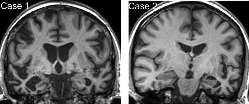
Figure 1.

Brain MRI profiles in patients with semantic dementia and food aversion.
Representative coronal T1-weighted MR sections through the anterior temporal lobes are presented for each of the patients described; the left hemisphere is shown on the right for both sections. In each case, there is relatively focal, asymmetric atrophy of the anterior temporal lobes, most marked medially and inferiorly (predominantly right-sided though bilateral in Case 1, predominantly left-sided in Case 2).
