

Abstract
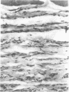
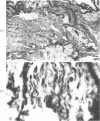
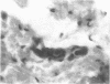
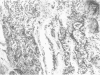
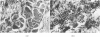
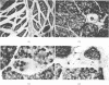
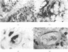
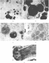
Pathological findings are reported on 34 specimens from 16 cases of arthrogryposis multiplex congenita (AMC), including initial observations on paraffin sections from 28 muscles, and subsequent observations on six additional specimens from three of these cases studied both histologically and histochemically. Thirteen of the 34 specimens (from 11 cases) were histologically normal, probably on account of an unaffected muscle being sampled. The most constant pathological feature in the remaining specimens was a disorganization of the muscle fibres and fascicles by severe fibrosis; only three specimens (from two cases) did not show this. Very thin faintly striated muscle fibres embedded in this matrix were encountered in 10 specimens from nine cases. An attempt at grouping of these atrophic or ill-developed fibres was noticed in four specimens; but this may not be denervation atrophy. Two specimens (from two cases) showed `myopathic' features. Repeat biopsy after two to three years was carried out on two affected muscles each from three patients. Case 3 showed well preserved but uniformly small fibres. Case 4 showed extremely few and scattered small rounded fibres. Case 14 showed pronounced variation of fibre size in both, with both atrophic and hypertrophied fibres. Normal nerves and spindles were seen in all these six specimens irrespective of the state of the muscle, and excessive fibro-fatty tissue in cases 4 and 14. Histochemical examination for oxidative enzymes, ATPase, and phosphorylase in these six specimens revealed a normal checkerboard pattern and ratio of type I and type II fibres, in case 3 only. The muscles of case 4 showed a preponderance of type I fibres. One specimen from case 14, showed the same fibres reacting for both oxidative enzymes and phosphorylase, suggesting a lack of development of fibres. The intrafusal fibres were mainly of type I in all. Two possible pathogenetic mechanisms operating in early embryonic life, which may lead to the characteristic changes of AMC, are discussed: (1) a defect in the development of the muscle whereby the full recruitment of myoblasts from the mesenchyme of the limb-bud does not take place and muscles do not form adequately; (2) a lack of innervation of the muscles on account of arrested growth of anterior horn cells. The combined operation of both these mechanisms is also considered. Fibrous tissue replaces the muscle tissue that is lacking, and contractures and deformities ensue. The evidence gathered on our material, such as the very thin smooth muscle fibres, the large numbers of well-formed nerves and spindles especially in the repeat biopsies, and the above-mentioned histochemical feature, would appear to favour the hypothesis of ill-developed muscles in the production of AMC in the majority; in the rest denervation playing either a major or concurrent role.
Full text
PDF


Images in this article

Selected References
These references are in PubMed. This may not be the complete list of references from this article.
- BANKER B. Q., VICTOR M., ADAMS R. D. Arthrogryposis multiplex due to congenital muscular dystrophy. Brain. 1957 Sep;80(3):319–334. doi: 10.1093/brain/80.3.319. [DOI] [PubMed] [Google Scholar]
- Bharucha E. P., Pandya S. S., Dastur D. K. Arthrogryposis multiplex congenita. I. Clinical and electromyographic aspects. J Neurol Neurosurg Psychiatry. 1972 Aug;35(4):425–434. doi: 10.1136/jnnp.35.4.425. [DOI] [PMC free article] [PubMed] [Google Scholar]
- DRACHMAN D. B., COULOMBRE A. J. Experimental clubfoot and arthrogryposis multiplex congenita. Lancet. 1962 Sep 15;2(7255):523–526. doi: 10.1016/s0140-6736(62)90399-9. [DOI] [PubMed] [Google Scholar]
- Dubowitz V. Enzyme histochemistry of skeletal muscle. 3. Neurogenic muscular atrophies. J Neurol Neurosurg Psychiatry. 1966 Feb;29(1):23–28. doi: 10.1136/jnnp.29.1.23. [DOI] [PMC free article] [PubMed] [Google Scholar]
- Fenichel G. M. Cerebral influence on muscle fiber typing. The effect of fetal immobilization. Arch Neurol. 1969 Jun;20(6):644–649. doi: 10.1001/archneur.1969.00480120090008. [DOI] [PubMed] [Google Scholar]
- HILLMAN J. W., JOHNSON J. T. H. Arthrogryposis multiplex congenita in twins. J Bone Joint Surg Am. 1952 Jan;34-A(1):211–214. [PubMed] [Google Scholar]
- HUGHES B. P. A method for the estimation of serum creatine kinase and its use in comparing creatine kinase and aldolase activity in normal and pathological sera. Clin Chim Acta. 1962 Sep;7:597–603. doi: 10.1016/0009-8981(62)90137-7. [DOI] [PubMed] [Google Scholar]
- Howard R. A Case of Congenital Defect of the Muscular System (Dystrophia muscularis congenita) and its Association with Congenital Talipes equino-varus. Proc R Soc Med. 1908;1(PATHOL):157–166. doi: 10.1177/003591570800101014. [DOI] [PMC free article] [PubMed] [Google Scholar]
- KITE J. H. Arthrogryposis multiplex congenita; review of fifty-four cases. South Med J. 1955 Nov;48(11):1141–1146. [PubMed] [Google Scholar]
- PADYKULA H. A., HERMAN E. The specificity of the histochemical method for adenosine triphosphatase. J Histochem Cytochem. 1955 May;3(3):170–195. doi: 10.1177/3.3.170. [DOI] [PubMed] [Google Scholar]
- PEARSON C. M., FOWLER W. G., Jr Hereditary non-progressive muscular dystrophy inducing arthrogryposis syndrome. Brain. 1963 Mar;86:75–88. doi: 10.1093/brain/86.1.75. [DOI] [PubMed] [Google Scholar]
- Patel A. N., Razzak Z. A., Dastur D. K. Disuse atrophy of human skeletal muscles. Arch Neurol. 1969 Apr;20(4):413–421. doi: 10.1001/archneur.1969.00480100089013. [DOI] [PubMed] [Google Scholar]
- SWINYARD C. A., MAYER V. Multiple congenital contractures. Public health considerations of arthrogryposis multiplex congenita. JAMA. 1963 Jan 5;183:23–27. [PubMed] [Google Scholar]