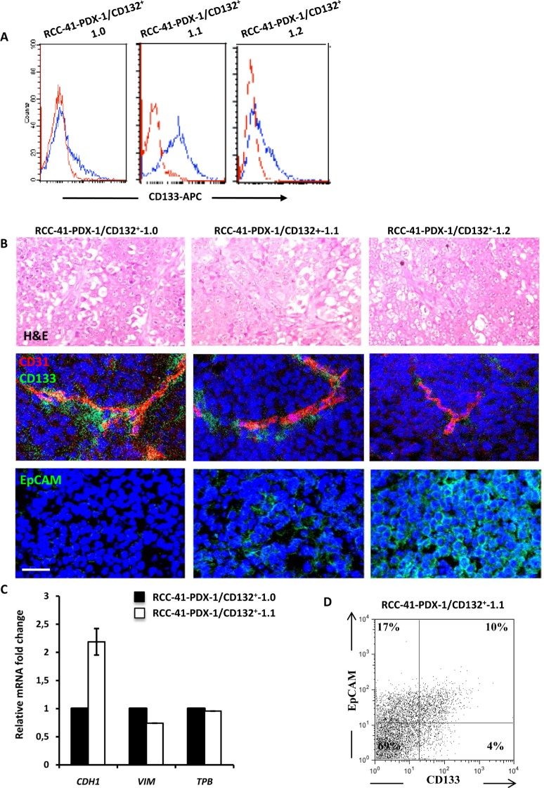
Figure 5. Tumorigenic potential of human renal RCC-41-PDX-1/CD132+ in SCID mice.
(A) Flow cytometry analysis of CD133 expression in RCC-41-PDX-1/CD132+ cell suspensions recovered from serial xenografts in SCID mice. Red outline histograms correspond to analyzed markers, and blue outline histograms correspond to cells incubated with the isotype-matched control antibody. (B) Vessel staining (CD31) and serial tumor formation by RCC-41-PDX-1/CD132+ cells subcutaneously injected in SCID mice. Top row: Representative hematoxylin and eosin staining showing the morphological appearance of an undifferentiated renal carcinoma with rare clear cells. Middle row: Representative immunohistochemistry of serial tumor sections showing positivity for human CD31 using a mAb that did not cross-react with the mouse CD31 (tumor vessels) and human CD133. CD133+ cells show a typical peri-vascular distribution. Bottom row: Representative immunohistochemistry of serial tumor sections showing positivity for human EpCAM. Original view × 40 (top) and × 60 (middle and bottom). Data are representative of 3 experiments with similar results. (C) Quantitative PCR analysis of E-cadherin (CDH1) and Vimentin (VIM), expression in RCC-41-PDX-1/CD132+ cells. Data were normalized using TATA binding gene mRNA (TBP), as endogenous control. (D) Flow cytometry analysis of CD133 and EpCAM expression in RCC-41/PDX-1/CD132+-1.1 cell suspension recovered from serial xenografts in SCID mice.

