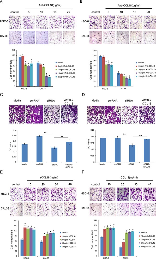
Figure 4. CCL18 enhances OSCC cell migration and invasion.

(A and B) The migration and invasion abilities of HSC-6 and CAL33 cells treated with the indicated concentrations of anti-CCL18 were evaluated by transwell migration and invasion assays. Representative pictures and the mean number of cells that migrated (A) or invaded (B) to the lower surface of three independent experiments (± SEM) are shown. (*P < 0.05 vs. control) (C and D) The migration and invasion abilities of HSC-6 cells were measured by transwell assay after the following treatments: transfection with 20 nM scrRNA, transfection with 20 nM siCCL18, or transfection with 20 nM siCCL18 and treatment with 20 ng/ml exogenous rCCL18 (siCCL18 + rCCL18). Representative pictures are shown and the mean OD values (± SEM) of migratory (C) or invasive (D) cells were measured at 590 nm. (*P < 0.05, **P < 0.01). (E and F) The migration and invasion abilities of HSC-6 and CAL33 cells treated with the indicated concentrations of exogenous rCCL18 were evaluated. Representative pictures and the mean number of migrated (E) or invaded (F) cells of three independent experiments (± SEM) are shown. (*P < 0.05 vs. control).
