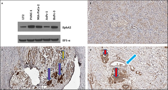
Figure 2. Expression pattern of EphA2 in cancer cells and pancreatic tissues.

(A) Western blotting of normal pancreatic fibroblasts (LT2) and pancreatic cancer cells (Panc-1, MIA-PaCa-2, AsPc-1, BxPc-3). Immunostaining of (B) normal pancreatic cancer tissue, (C) chronic pancreatitis tissue, and (D) pancreatic ductal adenocarcinoma tissue. EphA2 antibody appears to stain the surviving acinar tissue of chronic pancreatitis tissue (panel C), indicated by the orange arrow. In addition, some unknown tissue with islets characteristic morphology with high intensity within stroma of CP tissue is stained, as indicated by violet arrows (panel C). In the pancreatic ductal adenocarcinoma tissue, the EphA2 antibody stains cancer glands (red arrow, panel D) but not normal appearing tissues and ducts. Fibroblast cells also stains slightly. Normal ducts indicated by blue arrow.
