INTRODUCTION
Robotic ventral hernia repair is considered a new approach in the minimally invasive arena. There is a paucity of literature surrounding the safety and feasibility of robotic ventral hernia repair. The objective of our study is to evaluate the safety and feasibility of robotic ventral hernia repair and evaluate early outcomes. Numerous studies have demonstrated that laparoscopic ventral hernia has a low rate of conversion to open, shorter hospital stay as compared to open, moderate complication rates, and a low risk of recurrence. Despite these findings, the adoption of this technique across the US is not the dominant approach. The da Vinci robot (Si, Sie, Xi Intuitive Surgical, Sunnyvale, CA, USA) as an enabling tool, offers numerous advantages overall including 7° of freedom, three-dimensional (3D) imaging, and superior ergonomics that enable precise suturing and dissection at difficult angles. In this study, we are sharing our own case series and literature review.
METHODS
This is a retrospective single center, single surgeon review of prospectively collected data between 2012 and 2015 through institutional internal review board (IRB) approval. We performed a total of 106 consecutive robotic ventral hernia repairs. We used the robot (Si daVinci™, Intuitive) for hernia defect closure in selected cases using 0 monofilament Stratafix barbed sutures and underlay mesh placement which was fixed using running monofilament barbed 2-0 Stratafix absorbable barbed sutures, thereby avoiding the insertion of transfascial sutures through 8.5 mm reusable trocars. We used synthetic composite meshes with 3–5 cm overlap coverage in addition to primary closure of the fascial defect. We did not use a mesh deployment device. The study was designed to describe the technique and evaluate the initial results of robotic ventral hernia repair by reviewing the total operative time, console time, conversion, early recurrence, and initial outcomes including associated morbidity and mortality.
RESULTS
We reviewed one hundred six consecutive robotic ventral hernia repairs at our institution, of which sixty were Primary Ventral Hernia Repairs, forty-five were Incisional Hernia Repairs, and one was a Posterior Component Separation. Three cases of diastasis recti repair were performed in conjunction with hernia repair. A total of seven patients did not undergo primary fascial closure due to Swiss cheese like abdominal defects. None were converted to open or conventional laparoscopy only (two cases were performed as a combined sandwich technique for a flank incisional hernia and a chevron incisional hernia repair). Drains were employed for the chevron incisional hernia repair but not for the flank hernia repair. We utilized Symbotex™ (Medtronic, CT) Composite Synthetic mesh in eighty-five cases [sizes including 12 cm (n = 29), size 9 cm (n = 28), size 10 × 15 cm (n = 7), size 15 cm (n = 10), size 10 × 20 cm (n = 1), size 15 × 20 cm (n = 3), size 20 × 20 cm (n = 1), size 20 × 25 cm (n = 4), size 30 × 30 cm (n = 1), size 20 × 35 cm (n = 1), size 25 × 35 cm (n = 1)] (Table I). We used Proceed™ mesh (Ethicon) in three cases; size 20 × 20 cm (n = 1), size 20 × 10 cm (n = 2) (Table I). Phasix™ (Bard) mesh was used one time during the sandwich technique (35 × 20 cm) (Table I). Five cases were considered emergency cases and one hundred one were considered elective cases. We closed hernia defects in selected cases using O-monofilament Stratafix absorbable barbed sutures. We placed the synthetic mesh in an underlay fashion and secured the mesh to the abdominal wall using running barbed 2-0 monofilament barbed Stratafix absorbable sutures, except in the initial three cases where tackers were employed (initial surgeon preference until proficiency was achieved with robotic suturing). There was no need for the use of percutaneous trans-fascial sutures, mesh deployment devices, or human assistant through additional trocars. Fifty-five women were in the study compared to sixty-one men. The mean age was 54 years (27–84 years) with a mean BMI of 33 (22–48). The mean operative time was 85.7 minutes (35–335 minutes). The mean console operative time was 61 (20–300 minutes). The mean estimated blood loss was 7 mL (2–50 mL). The mean length of stay was 0.20 days (0–5 days). Median follow-up was at 6 months (1–24 months). The fascial defect sizes (in largest dimension) ranged from 2 to 25 cm with a mean defect size of 4.3 cm. Post-operative morbidities (Table II) were 6% (n = 7); one surgical-site infection that required operative Incision and Drainage on post-operative day 5 without the need for mesh explantation, one Ileus which resolved with conservative management, one rectus sheath hematoma on post operative day 42 related to therapeutic anticoagulation requiring admission and conservative management, one Small Bowel Obstruction requiring exploration and enterolysis (no adhesions noted to the mesh or barbed sutures). One patient experienced persistent nonspecific pain at 6-month follow up. One patient had a symptomatic seroma that required drainage at the office after flank incisional hernia repair (underlay and overlay meshes were placed). We had two patients develop incisional hernia recurrences (1.8%); 1 patient (BMI 48) had symptomatic recurrence at 6 month follow up from an incisional hernia due to Cesarian operation and the other patient had a recurrence requiring recurrent subcostal incisional hernia repair at 5 month follow up (The defects were not closed primarily in both patients who developed recurrences. The mesh was not fixed to Cooper's ligament in the Cesarian hernia repair, which was a technical error).
Table I.
Mesh type and quantity used.
| Symbotex | |||||||||||
| Mesh size (cm) | 9 × 9 | 12 × 12 | 15 × 15 | 10 × 15 | 10 × 20 | 15 × 20 | 20 × 20 | 20 × 25 | 30 × 30 | 20 × 35 | 25 × 35 |
| Quantity (n) | 28 | 29 | 10 | 7 | 1 | 3 | 1 | 4 | 1 | 1 | 1 |
| Phasix | |||||||||||
| Mesh size (cm) | 35 × 20 | ||||||||||
| Quantity (n) | 1 | ||||||||||
| Proceed | |||||||||||
| Mesh size (cm) | 20 × 20 | 20 × 10 | |||||||||
| Quantity (n) | 1 | 2 | |||||||||
Table II.
Post-operative complications.
| Complication | Surgical site infection | Ileus | Rectus sheath hematoma | SBO | Chronic abdominal pain | Symptomatic seroma | Hernia recurrence |
|---|---|---|---|---|---|---|---|
| Number of patients (n) | 1 | 1 | 1 | 1 | 1 | 1 | 2 |
DISCUSSION
The introduction of laparoscopy to the field of General Surgery in the 1980's marked the start of minimally invasive surgery in this field. Since then, numerous advances have been made in laparoscopy including the advent of robotic assisted laparoscopic surgery. LeBlanc et al. first described the use of laparoscopy for ventral hernia repair in the early 1990's.9 This group used PTFE mesh patches, which were secured to the anterior abdominal wall using staples. Wider acceptance of this method of repair brought new advances including meshes that were easier to handle in the abdomen and novel securing methods including tacking, transfascial suture fixation, and a combination of both techniques. This method showed clear benefits in these patients including fewer surgical site infections and decreased hospital stay compared to those who underwent the open technique.3 Yet, these advances led to potential problems, namely increased post-operative pain with the use of transfascial suture fixation4 and the potential for intestinal fistulae formation and adhesions from the tacks. Also, the laparoscopic technique still had recurrence rates as high as 9%.6
The advent of robotic assisted laparoscopic surgery introduced numerous advantages to the minimally invasive arena compared to standard laparoscopy including 7° of freedom, three-dimensional (3D) imaging, and superior ergonomics that enable precise suturing and dissection for mesh placement at difficult angles. (Kudsi, 2015) Given these potential benefits, robotic assisted laparoscopy has been applied to ventral hernia repairs. Schluender et al. described the first robotic ventral hernia repair in a porcine model using central and circumferential suture fixation,11 revealing a relative ease in intra-corporeal suturing as well as unparalleled precision in suture placement as compared to standard laparoscopy. This technology was subsequently applied to humans revealing that this technique was not only technically feasible, but also provided excellent visualization and precision of suture placement.1 Recent publications have shown that primary closure of the fascial defect provides added benefits in laparoscopic ventral hernia repair.10 Standard laparoscopy provides a very difficult channel for primary closure of the fascial defect and transfascial closure has been known to lead to significant postoperative pain. A recent study comparing robotic assisted laparoscopic ventral hernia repair with closure of fascial defect to laparoscopic ventral hernia repair without fascial defect closure showed decreased recurrence rates and complications in the robotic assisted patient group,5 revealing a tangible benefit to the use of robotic technology for ventral hernia repair. The robotic technology also allows ease in performing more challenging types of repairs, such as the pre-peritoneal placement technique with overlying peritoneal closure, which could potentially eliminate the need for dual sided mesh.
Our study demonstrates that application of robotic technology to ventral hernia repair is safe and feasible. Though our study has short follow-up of only 6 months, we report excellent early outcomes with a total of two recurrences in our patient population at 6-month follow up. In both patients who developed recurrences, the fascia was not primarily closed, which, in retrospect, was a technical error. We believe that this may have been a significant factor in the development of hernia recurrence in these patients but our study is not powered to analyze the role that this omission played on the outcome of recurrence. We reveal that robotic assisted ventral hernia repair can be effectively performed and is a viable option for patients when performed by a surgeon with expertise in this platform. Comparing console times of two similar cases of elective non-incarcerated 4 cm defect repairs from the beginning of the study period to the end of the study period reveals a console time of 65 minutes in the 2nd case in our series compared to 30 minutes in the 106th case. This clearly demonstrates that with more experience, operative time decreases, thereby cutting costs. The average number of robotic instruments that were used in our series was three. Ensuring that opening instruments that were routinely used rather than opening several unnecessary robotic instruments is another way to attempt to drive down cost with robotic ventral hernia repair.
Robotic assisted laparoscopic ventral hernia repair seems to be a promising approach given the numerous added benefits using this novel technology. Yet, no randomized controlled trials have been performed comparing open, laparoscopic, and robotic assisted laparoscopic ventral hernia repair. A known deterrent for the use of robotic surgery is the notable cost associated with this technology, with a recent study revealing that the per procedure cost is increased by up to $1,500.00 with the use of the robot.2 In this current era of significant healthcare costs, comparative trials must evaluate the costs of robotic surgery in ventral hernia repair and whether we are justified in using this novel and expensive technology.
Many surgeons have adopted the robotic technology for ventral hernia repair across the country in the past several years. In this current era in the United States, a significant number of Ventral Hernia Repairs are done via the open approach. Robotics may play a role in attracting these surgeons to a minimally invasive approach where standard laparoscopy has failed. We believe that robotic surgery has the potential to raise the bar in minimally invasive general surgery in experienced hands and enable less experienced minimally invasive surgeons to adapt and expand the arena of minimally invasive surgery. There is a strong need for collaboration amongst surgeons, institutions and industry in order to publish high level data to best serve our patients. Cost, experience and outcomes will all likely improve over time as it has with prior technological advances in General Surgery.
CONCLUSION
Robotic ventral hernia is considered a new approach in minimally invasive ventral hernia repair associated with a short a hospital stay, low rate of complications, and a low rate of conversion to open surgery. Technically, the use of robotic technology may facilitate handling and mesh deployment without the need of tackers, trans-fascial sutures or mesh deployment devices. Our study demonstrates that robotic ventral hernia repair is a safe procedure with excellent short-term outcomes. Further studies are planned for the future including long-term data follow-up. Although these early results are promising, multi center randomized controlled trials and long-term follow up are needed.
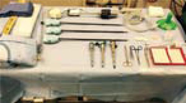
Figure 1.
Robotic scrub technician table, it demonstrates the efficiency and specific instruments needed for robotic ventral hernia repair. (Robotic needle driver, robotic monopolar scissors, robotic bipolar Maryland grasper).
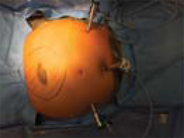
Figure 2.
Port placements for robotic ventral hernia repair. Epigastric hernia is marked with the expected 5 cm overlap. Note ports placed with adequate distance from the expected mesh placement. Three 8.5 mm ports were used (camera trocar placed infra-umbilical, each working trocar placed at each anterior axillary line).
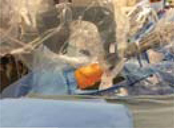
Figure 3.
Docking Si DaVinci system by driving the cart over patient's head (Cephalad is to the reader's left side). We prefer to place the patient in 15–30 degree Trendelenburg with the table flexed at the patient's hip to avoid any arm collision.
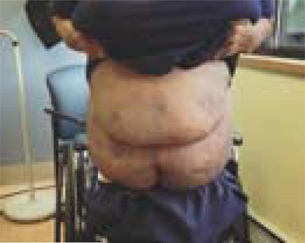
Figure 4.
Two weeks post robotic ventral hernia repair demonstrating the advantage of minimal invasive surgery in morbidly obese patients.
Figure 5.
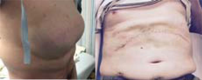
Left photo of hernia before surgery and right photo at two week follow up after incisional hernia repair. He underwent combined robotic enterolysis, defect closure, underlay mesh placement followed by open plication of the fascia and placement of overlay mesh as well.
Footnotes
Conflict of Interest
Dr. Kudsi received consulting fees for speaking/teaching robotic surgery from Intuitive. The remaining authors have no conflicts of interest.
REFERENCES
- 1.Allison N, Tieu K, Snyder B. Technical feasibility of robot-assisted ventral hernia repair. World J. Surg. 2012;36(2):447–452. doi: 10.1007/s00268-011-1389-8. [DOI] [PubMed] [Google Scholar]
- 2.Barbash GI, Glied SA. New technology and health care costs—The case of robot-assisted surgery. N. Engl. J. Med. 2010;363(8):701–4. doi: 10.1056/NEJMp1006602. [DOI] [PubMed] [Google Scholar]
- 3.Beldi G, Ipaktchi R, Wagner M. Laparoscopic ventral hernia repair is safe and cost effective. Surg Endosc. 2006;20(1):92–95. doi: 10.1007/s00464-005-0442-9. [DOI] [PubMed] [Google Scholar]
- 4.Beldi G, Wagner M, Bruegger L, Kurmann A. Mesh shrinkage and pain in laparoscopic ventral hernia repair: A randomized clinical trial comparing suture versus tack mesh fixation. Surg. Endosc. 2011;25(3):749–755. doi: 10.1007/s00464-010-1246-0. [DOI] [PubMed] [Google Scholar]
- 5.Gonzalez AM, Romero RJ, Seetharamaiah R. Laparoscopic ventral hernia repair with primary closure versus no primary closure of the defect: Potential benefits of the robotic technology. Int. J. Med. Robot. 2015;11(2):120–5. doi: 10.1002/rcs.1605. [DOI] [PubMed] [Google Scholar]
- 6.Heniford BT, Park A, Ramshaw BJ. Laparoscopic ventral and incisional hernia repair in 407 patients. J. Am. Coll. Surg. 2000;190(6):645–650. doi: 10.1016/s1072-7515(00)00280-5. [DOI] [PubMed] [Google Scholar]
- 7.Heniford BT, Park A, Ramshaw BJ. Laparoscopic repair of ventral hernias: Nine years’ experience with 850 consecutive hernias. Ann. Surg. 2003;238(3):391–400. doi: 10.1097/01.sla.0000086662.49499.ab. [DOI] [PMC free article] [PubMed] [Google Scholar]
- 8.Kudsi OY, Mabardy A, Paluvoi N. Robotic approaches to ventral and inguinal hernias. Operative Endoscopic and Minimally Invasive Surgery. In Press. [Google Scholar]
- 9.Leblanc KA, Booth WV. Laparoscopic repair of incisional abdominal hernias using expanded polytetrafluoroethylene: Preliminary findings. Surg. Laparosc. Endosc. 1993;3(1):39–41. [PubMed] [Google Scholar]
- 10.Rea R, Falco P, Izzo D. Laparocopic ventral hernia repair with primary transparietal closure of the hernial defect. BMC Surg. 2012;12(Suppl 1):S33. doi: 10.1186/1471-2482-12-S1-S33. [DOI] [PMC free article] [PubMed] [Google Scholar]
- 11.Schluender S, Conrad J, Divino CM. Robot-assisted laparoscopic repair of ventral hernia with intracorporeal suturing An experimental study. Surg. Endosc. 2003;17(9):1391–1395. doi: 10.1007/s00464-002-8795-9. [DOI] [PubMed] [Google Scholar]


