Abstract
Please cite this paper as: Peltola et al. (2011) Pandemic influenza A (H1N1) virus in households with young children. Influenza and Other Respiratory Viruses 6(3), e21–e24.
Abstract
Background Influenza viruses may cause a severe infection in infants and young children. The transmission patterns of pandemic 2009 influenza A (H1N1) within households with young children are poorly characterized.
Methods Household members of six children younger than 1·5 years with documented 2009 influenza A (H1N1) infection were studied by daily symptom diaries and serial parent‐collected nasal swab samples for detection of influenza A by reverse transcription polymerase chain reaction (RT‐PCR) assay.
Results Laboratory‐confirmed, symptomatic influenza was documented in 11 of 15 household contacts of young children with pandemic influenza (73%; 95% CI, 48–99). In five contact cases symptoms started earlier, in three cases on the same day, and in three cases after the onset of symptoms in the youngest child. The first case with influenza A (H1N1) within the household was an elder sibling in two households, father in two households, the youngest child in one household, and the youngest child at the same time with a sibling in one household. The median copy number of influenza virus was higher in children than in adults (4·2 × 107 versus 4·9 × 104, P = 0·02).
Conclusions This study demonstrates the feasibility of nasal swab sampling by parents in investigation of household transmission of influenza. The results support influenza vaccination of all household contacts of young children.
Keywords: Household transmission, infant, influenza A (H1N1) virus, pandemic influenza
Introduction
Young children are a risk group for severe infection and hospitalization with seasonal influenza. 1 , 2 As the 2009 influenza pandemic caused by a novel A (H1N1) virus spread throughout the world, this virus was soon documented to attack children more severely than seasonal influenza viruses. 3 , 4 Twice as many pediatric hospitalizations and 5–10 times more deaths than from seasonal influenza were reported from Argentina, and infants were identified as having the highest risk of hospitalization or death. 5
Protection of young children against influenza is problematic. Inactivated vaccines are not approved for children younger than 6 months, and data on their efficacy against seasonal or pandemic influenza in children under 2 years of age are limited. 6 , 7 , 8 Use of oseltamivir for the treatment of influenza in children under the age of 1 year was approved during the 2009 pandemic as an emergency action in the absence of proper documentation of safety and effectiveness in this age group.
The sources from where young children acquire pandemic influenza infection should be identified to improve prevention strategies. To find out the routes of transmission of the 2009 pandemic influenza in families with young children, we embedded a household follow‐up by parent‐collected nasal swab samples in our ongoing prospective study of respiratory infections in a child cohort.
Methods
Study population and design
Children born in the Hospital District of Southwest Finland during a 2‐year period starting from April 2008 were recruited in a prospective cohort study of respiratory infections before or soon after birth (the STEPS study). At the time of the first wave of the 2009 influenza A (H1N1) pandemic in Finland in October–November 2009, we screened 23 febrile children younger than 1.5 years visiting the cohort study clinic by a rapid antigen test (Coris, Gembloux, Belgium, or ArcDia, Turku, Finland) and a reverse transcription polymerase chain reaction (RT‐PCR) assay for influenza in nasal swab samples. Six children positive for influenza A, and all persons living in the same household with them were enrolled in this study, except one adult contact who did not participate. Participants or parents of participating children gave their written informed consent. The study protocol was approved by the Ethics Committee of the Hospital District of Southwest Finland.
Information on underlying illnesses, influenza vaccinations, and symptoms of a respiratory infection during the preceding 14 days was recorded. Nasal swab samples for RT‐PCR were taken from all participants by use of flocked swabs (Copan, Brescia, Italy). Parents were instructed to obtain nasal swabs at home from themselves and their children on days 3, 6, and 14 of the follow‐up and to send the swabs in dry sterile tubes to the laboratory by mail. Symptoms were recorded in a diary for 14 days.
Quantitative RT‐PCR for influenza A (H1N1) virus
Swabs were vortexed with 800 μl of phosphate‐buffered saline and 200‐μl aliquots of the suspensions were subjected to automated nucleic acid extraction with viral nucleic acid small volume kit on MagNA Pure 96 instrument (Roche, Penzberg, Germany) using an elution volume of 50 μl. An 8‐μl aliquot of each extract was reverse‐transcribed using RevertAid H minus first‐strand cDNA synthesis reagents (Fermentas, St. Leon‐Rot, Germany) with random hexamer primers. Of the 20‐μl RT reaction, 2 μl was used in a 20‐μl PCR using ABsolute Fast QPCR mix (Thermo Scientific, Waltham, MA, USA) with specific primers and probe described by Health Protection Agency, 9 UK. A pCR‐TOPO plasmid (Invitrogen, Carlsbad, CA, USA) constructed to contain nucleotides 773–1674 of the hemagglutinin gene of a Finnish 2009 influenza A (H1N1) isolate was used as the standard for copy number calculations.
Statistical analysis
In household contacts, 95% confidence intervals (CI) were calculated for confirmed influenza. Categorical variables in children and adults with influenza were compared using the Fisher test. Virus copy numbers had unequal variances and were compared using the Mann–Whitney test. Statistical analyses were performed using spss for Windows, version 16·0 (SPSS, Chicago, IL, USA).
Results
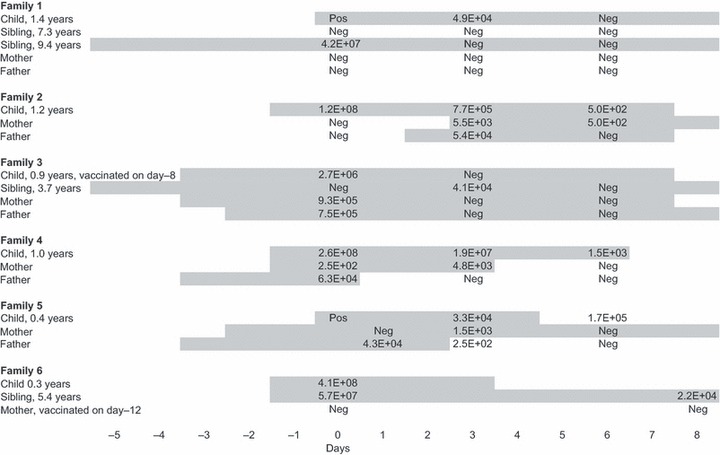
Six families were followed in this study. Symptoms of documented influenza and virus copy numbers in the families are shown in Figure 1. The mean age of the youngest children in the families was 0.9 years (range, 0·3–1·4 years). None of the children had an underlying risk condition. One child attended a day‐care center (Family 2). One child had received monovalent, adjuvanted pandemic influenza vaccine 5 days before the onset of symptoms of influenza (Family 3). Household members (n = 15) included four siblings (age, 5–9 years) and 11 parents. One adult with asthma had received pandemic influenza vaccine (Family 6). Other household members had no underlying risk conditions.
Figure 1.

Transmission patterns of pandemic 2009 influenza A (H1N1) in families with young children. Shaded areas show the duration of respiratory symptoms (cough, runny nose, or sore throat with or without fever) during laboratory‐confirmed influenza. Viral copy numbers are shown. Prospective follow‐up was started on day 0. Families were followed until day 14, but symptoms until day 8 are shown here for readability of the figure. All samples taken on day 14 were negative for influenza virus. Shaded areas in Family 4 are drawn from the onset of symptoms to last positive sample, because of missing data about the duration of symptoms.
Laboratory‐confirmed, symptomatic influenza was documented in 11 of 15 household contacts of young children with influenza (73%; 95% CI, 48–99). In five contact cases symptoms started before, in three cases on the same day, and in three cases after the onset of symptoms in the youngest child. The first case with influenza A (H1N1) within the household was an elder sibling in two households, father in two households, the youngest child in one household, and the youngest child at the same time with a sibling in one household.
Clinical findings in children and adults with documented influenza are shown in Table 1. All children and 63% of adults had fever. The median duration of fever was 3.0 days in children and 2.5 days in adults, and the median duration of any symptoms was 11 days in children and 10 days in adults. Acute otitis media was detected in 4 children. One child with influenza was hospitalized for 1 day because of lethargy and poor feeding. Four children and one adult were treated with oseltamivir. The median copy number of influenza virus was higher in children than in adults (4·2 × 107 versus 4·9 × 104, P = 0·02).
Table 1.
Clinical findings and viral load in study participants with laboratory‐confirmed influenza
| Children (n = 9) | Adults (n = 8) | P* | |
|---|---|---|---|
| Age, mean (SD) | 2·6 (3·0) | 34·0 (8·3) | |
| Symptoms | |||
| Fever, no. (%) | 9 (100) | 5 (63) | 0·08 |
| Cough, no. (%) | 8 (89) | 8 (100) | 1·00 |
| Rhinorrhea, no. (%) | 9 (100) | 4 (50) | 0·03 |
| Highest copy number per swab, median (interquartile range) | 4·2 × 107 (1·1 × 105–1·9 × 108) | 4·9 × 104 (4·9 × 103–5·8 × 105) | 0·02 |
*Categorical data were compared using the Fisher test. Virus copy numbers were compared using the Mann–Whitney test.
Discussion
We documented a symptomatic, RT‐PCR‐confirmed infection in 73% of household contacts of young children with the 2009 pandemic influenza A (H1N1) virus. In most cases, a parent or an elder sibling introduced the virus into the family, and it spread efficiently among household members.
Laboratory confirmation of influenza is valuable in household transmission studies. Clinical surveillance may underestimate the true occurrence of influenza. 10 At the same time, respiratory infections caused by other viruses could be misclassified as influenza, because the clinical diagnosis of influenza is particularly difficult in children. 11 The prospective design of our study enabled detailed documentation of symptoms and collection of serial nasal swab samples for influenza RT‐PCR. By use of home sampling, the collection of several specimens from all household members was feasible. We have previously used a similar home sampling method for the detection of rhinovirus infections in a household setting. 12
The transmission of pandemic 2009 influenza in households has been studied by use of retrospective surveys of influenza‐like or acute respiratory illness. These studies have documented modest secondary attack rates of 11–13%. 13 , 14 A substantially higher secondary attack rate of 45% was documented in a prospective study with active follow‐up by household visits and serological and direct virus detection methods. 15 The setting of our study was different: instead of selecting the first cases in households, we identified young children with influenza from a predefined study cohort. The detection of efficient household transmission in our study may be explained by the study design with intensive surveillance, close contacts between the members of relatively small households, and high viral load in young children. The pandemic phase and its intensity may also effect on the virus transmission, but the three above‐cited studies and our present study were all performed during the first wave of the pandemic.
We illustrate in this study the detailed transmission patterns of pandemic influenza in families. Our findings, while limited to 6 households, support influenza vaccination of all household contacts of young children. Similar study designs with serial home sampling in a household setting could be utilized in larger studies of transmission of influenza viruses.
Financial support
Academy of Finland (grant no. 123571), the Foundation for Paediatric Research and the Finnish Medical Foundation.
Acknowledgements
Jelena Jakovleva, Johanna Vänni, and Tiina Ylinen are acknowledged for technical assistance and Thedi Ziegler for the reference influenza A (H1N1) virus isolate.
References
- 1. Peltola V, Ziegler T, Ruuskanen O. Influenza A and B virus infections in children. Clin Infect Dis 2003; 36:299–305. [DOI] [PubMed] [Google Scholar]
- 2. Thompson WW, Shay DK, Weintraub E et al. Influenza‐associated hospitalizations in the United States. JAMA 2004; 292:1333–1340. [DOI] [PubMed] [Google Scholar]
- 3. CDC . Seasonal influenza: weekly U.S. influenza surveillance report 2009. Available at: http://www.cdc.gov/flu/weekly/ (Accessed 06 June 2011).
- 4. Louie JK, Acosta M, Winter K et al. Factors associated with death or hospitalization due to pandemic 2009 influenza A(H1N1) infection in California. JAMA 2009; 302:1896–1902. [DOI] [PubMed] [Google Scholar]
- 5. Libster R, Bugna J, Coviello S et al. Pediatric hospitalizations associated with 2009 pandemic influenza A (H1N1) in Argentina. N Engl J Med 2010; 362:45–55. [DOI] [PubMed] [Google Scholar]
- 6. ECDC . Technical report of the scientific panel on vaccines and immunisation: infant and children seasonal immunisation against influenza on a routine basis during interpandemic period 2007. Available at: http://ecdc.europa.eu/en/publications/Publications/0701_TER_Scientific_Panel_on_Vaccines_and_Immunisation.pdf (Accessed 06 June 2011).
- 7. Fiore AE, Neuzil KM. 2009 influenza A(H1N1) monovalent vaccines for children. JAMA 2010; 303:73–74. [DOI] [PubMed] [Google Scholar]
- 8. Heinonen S, Silvennoinen H, Lehtinen P, Vainionpää R, Ziegler T, Heikkinen T. Effectiveness of inactivated influenza vaccine in children aged 9 months to 3 years: an observational cohort study. Lancet Infect Dis 2011; 11:23–29. [DOI] [PubMed] [Google Scholar]
- 9. Health Protection Agency . Swine‐Lineage Influenza A H1 specific Fast Real Time PCR. National Standard Method VSOP 29 Issue 2 2009. Available at: http://www.hpa‐standardmethods.org.uk/pdf_sops.asp (Accessed 14 May 2010).
- 10. Miller E, Hoschler K, Hardelid P, Stanford E, Andrews N, Zambon M. Incidence of 2009 pandemic influenza A H1N1 infection in England: a cross‐sectional serological study. Lancet 2010; 375:1100–1108. [DOI] [PubMed] [Google Scholar]
- 11. Peltola V, Reunanen T, Ziegler T, Silvennoinen H, Heikkinen T. Accuracy of clinical diagnosis of influenza in outpatient children. Clin Infect Dis 2005; 41:1198–1200. [DOI] [PubMed] [Google Scholar]
- 12. Peltola V, Waris M, Österback R, Susi P, Ruuskanen O, Hyypiä T. Rhinovirus transmission within families with children: incidence of symptomatic and asymptomatic infections. J Infect Dis 2008; 197:382–389. [DOI] [PubMed] [Google Scholar]
- 13. Cauchemez S, Donnelly CA, Reed C et al. Household transmission of 2009 pandemic influenza A (H1N1) virus in the United States. N Engl J Med 2009; 361:2619–2627. [DOI] [PMC free article] [PubMed] [Google Scholar]
- 14. France AM, Jackson M, Schrag S et al. Household transmission of 2009 influenza A (H1N1) virus after a school‐based outbreak in New York City, April‐May 2009. J Infect Dis 2010; 201:984–992. [DOI] [PubMed] [Google Scholar]
- 15. Papenburg J, Baz M, Hamelin ME et al. Household transmission of the 2009 pandemic A/H1N1 influenza virus: elevated laboratory‐confirmed secondary attack rates and evidence of asymptomatic infections. Clin Infect Dis 2010; 51:1033–1041. [DOI] [PubMed] [Google Scholar]


