Abstract
The hydatid disease caused by Echinococcus granulosus is an endemic parasitic disease affecting several Mediterranean countries. Echinococcal cysts are mostly located in the liver and the lung, but the disease can be detected anywhere in the body. In this study, we present uncommon extrahepatic localizations of primary hydatid disease. Patients who were operated on for hydatid disease or cystic lesions, which were later diagnosed as hydatid disease, between 2004 and 2010 were retrieved retrospectively. Patients with lesions localized outside the liver and the lung were enrolled in the study. Eight patients with extrahepatic primary hydatid disease were treated surgically at our clinic. The cysts were located in the scapular region, spleen, pancreas, lumbosacral region and gluteal muscle. Surgical techniques were partial or total cystectomy with or without tube drainage. Splenectomy was performed for splenic hydatid disease and partial pericystectomy, Roux-en-Y cystojejunostomy, cholecystectomy and T-tube drainage for pancreatic hydatid disease. There were no complications or mortality in the postoperative period. Hydatid cyst should be considered in the differential diagnosis of cystic lesions, especially in endemic areas. Surgical technique should be planned according to the location of the cyst.
Keywords: Hydatid cyst, rare localization, primary hydatid disease
INTRODUCTION
Hydatid disease is a parasitic infection that is usually caused by Echinococcus granulosus. Humans are intermediate hosts and become infected by handling infected dogs or other carnivore hosts. Echinococcal cysts are mostly located in the liver (70%) and the lung (25%) (1). Primary isolated extrahepatic hydatid disease is mostly seen within the abdomen with an incidence of 6–11% (2). Although some patients may be asymptomatic, the clinical presentation is mostly with abdominal pain or swelling of soft tissue with respect to disease localization, i.e. spleen, pancreas, kidney, retroperitoneum, urinary bladder, ovaries, bone, heart, thoracic wall, spinal column, thyroid gland, brain and muscles (1–4). Although radical excision of the cyst is recommended whenever possible, conservative surgery may be needed in a select group of cases (2). This paper aims to review patients operated in our department for hydatid disease located outside the liver and the lungs.
MATERIALS AND METHODS
A retrospective chart review was done for all patients operated for hydatid disease between 2005 and 2010 in our institution. Those who had a hepatic and/or pulmonary disease were excluded. Then, written informed consent was obtained from patients who participated in this study. The localizations were classified into two groups as intra-abdominal or intramuscular, in order to obtain homogenous data. Demographics, preoperative information (symptoms and signs, serologic tests, radiologic imaging), operative findings and techniques, postoperative data (complications, hospital stay) and surveillance (follow-up periods, outcome, and recurrence) records were retrieved from patient files.
Indirect hemagglutination (IHA) test was the available method for serological confirmation. Albendazol (Andazol/Biofarma) was chosen as the antihelmintic drug, if it was decided to treat the patient before surgery.
RESULTS
Fifty-two patients were operated in our institution for hydatid disease between 2005 and 2010. Among those, eight (15.4%) patients (five females [62.5%] and mean±SD age was 37.4±16.9 years) had extrahepatic and extrapulmonary disease. Hydatid cysts were located intra-abdominally (n=5; 62.5%) (spleen [n=3; 37.5%], pancreas [n=1; 12.5%] and pelvis [n=1; 12.5%]) or intra-muscularly (n=3; 37.5%) (m. trapezius [n=1; 12.5%], m. latissimus dorsi [n=1; 12.5%] and m. gluteus maximus [n=1; 12.5%]). In a patient (#2), there was another cyst attached to the spleen and splenic flexure of the colon; however, this case was accepted to have a disease located to the spleen (Table 1).
Table 1.
Preoperative data of the patients
| Patient no. | Age (yrs) | Sex | Symptoms and signs | Serological tests | Radiologic imaging | Localization |
|---|---|---|---|---|---|---|
| 1 | 49 | F | Abdominal pain | IHA + | CT, US | Spleen |
| 2 | 32 | M | Abdominal pain | IHA + | CT, US | Spleen, peritoneal cavity |
| 3 | 49 | F | Abdominal pain | IHA − | CT, US | Spleen |
| 4 | 14 | M | Abdominal pain, jaundice | IHA − | CT, MRI, US | Head of pancreas |
| 5 | 37 | F | Abdominal pain | IHA + | CT, US | Pelvis |
| 6 | 37 | F | Palpable mass | IHA− | US | Scapular region |
| 7 | 20 | F | Palpable mass | IHA − | MRI | Lumbosacral region |
| 8 | 61 | M | Palpable mass, gluteal pain | IHA + | US, CT | Gluteal muscle |
F: female; M: male; IHA: immune hemagglutination; CT: computed tomography; MRI: magnetic resonance imaging; US: ultrasonography
Preoperative signs and symptoms were related to the size and localization of the cysts: abdominal pain (n=5; 62.5%), palpable mass (n=3; 37.5%), jaundice (n=1; 12.5%) and gluteal pain (n=1; 12.5%). Ultrasonography (USG), computed tomography (CT) and/or magnetic resonance imaging (MRI) were used as diagnostic tools in 6 (75%), 5 (62.5%) and/or 2 (25%) patients, respectively. The diagnosis could be preoperatively achieved in most of the patients (n=5; 62.5%). However, hydatid disease was diagnosed with direct observation of parasites during surgical intervention in 2 (25%) cases, in which the cysts were located in the pancreas and the shoulder. The preoperative details of the patients and diseases were presented in Table 1.
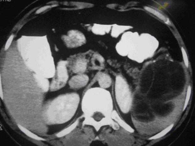
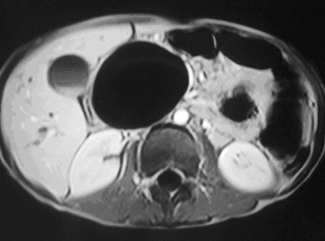
Splenectomy was required in three patients (Figure 1). Of those, the cyst attached to the spleen was totally removed in a case (#2) (Table 2). In a 14 year-old boy (#4), the cyst was located at the head of the pancreas causing abdominal pain and jaundice (Figure 2). Concerning the relatively high probability of morbidity and mortality after pancreaticoduodenectomy, a possible alternative intervention, partial pericystectomy was combined with a Roux-en-Y anastomosis in between the remaining cyst and jejunum, and a T-tube choledocostomy was performed in addition to cholecystectomy due to the connection of the distal common bile duct and Wirsung duct to the cyst. The intramuscularly located parasites were treated with total cyst removal (#6, 7 and 8) (Table 2). Scolicidal agents (20% hypertonic saline) were routinely used in order to decrease the risk of secondary disease in all patients.
Figure 1.

Computed tomography view of a large splenic cyst
Table 2.
The operative and long-term outcome details of the patients
| Patient no. | Operative approach | Hospital stay (day) | Follow-up period (month) | Long-term outcome |
|---|---|---|---|---|
| 1 | Splenectomy | 3 | 47 | Incisional hernia |
| 2 | Splenectomy, cystectomy | 5 | 69 | - |
| 3 | Splenectomy | 6 | 70 | - |
| 4 | Partial pericystectomy | 13 | 69 | - |
| Cystojejunostomy | ||||
| Jejunojejunostomy | ||||
| Cholecystectomy | ||||
| T-tube drainage | ||||
| 5 | Laparoscopic total cystectomy | 3 | 39 | - |
| 6 | Total cystectomy | 1 | 37 | - |
| 7 | Total cystectomy | 1 | 30 | - |
| 8 | Total cystectomy | 1 | 36 | - |
Figure 2.

Magnetic resonance imaging view of a pancreatic cyst
The postoperative periods were uneventful in all patients. The mean (±SD) hospital stay was 6.8±4.3 days in patients who had an intra-abdominally located disease. Albendazole (10–15 mg/ kg/day) was postoperatively used in six (85%) patients with monthly evaluation for hepatic toxicity. All cases with intramuscular cysts were discharged from the hospital on day 1. No recurrence was observed after a mean (±SD) follow-up period of 39.1 ±17.7 months. An incisional hernia was observed in a single patient (#1) who then underwent a hernia repair procedure (Table 2).
DISCUSSION
Hydatid disease is an endemic parasitic disease, and is still a problem showing worldwide distribution, especially in sheep-raising areas. The most frequent location of hydatid disease is the liver, because it is the first and the largest filter of parasitic embryos migrating from the intestine via the portal stream. If they escape the hepatic filter, the embryos enter into the systemic circulation and settle in the lungs or, rarely, in other organs (3). Extrahepatic localization of primary hydatid disease is rarely detected without hepatic involvement (5).
A serious diagnostic problem in this entity is an unusual location of primary hydatid disease (6, 7). The diagnosis is based on clinical symptoms and signs, laboratory and radiologic examinations. Non-complicated hydatid cysts are usually asymptomatic. The symptoms are generally non-specific and are related to local mass effect or cyst complication. The symptoms of hydatid cyst are organ-specific. In our patients, the presenting symptoms were abdominal pain (5 cases), jaundice (one case), and swelling (three cases) (Table 1).
Serology should be used to confirm a tentative diagnosis of hydatid disease (8). However, serologic examinations have the problems of low diagnostic sensitivity and specificity (9). IHA test was used for serologic confirmation in all patients (after surgery in two patients). Positive IHA test results were obtained in 50% (4/8) of cases.
The clinical features and immunologic test are not sufficient for a definite diagnosis in most cases. The preoperative diagnosis is based on radiologic examination (10). US, CT and MRI were used in the diagnosis of hydatid disease in our patients. The cystic lesion was detected in all patients. The images, clinical features and immunologic test were suspicious for hydatid cyst in 87.5% (7/8) of the patients. The patients underwent surgery for the cystic lesions and the histology evaluation confirmed the diagnosis of hydatid disease. The diagnosis was established after surgery by histological examination in a patient who had no sign of hydatid disease in preoperative clinical and radiologic examinations.
Surgery is still the treatment of choice with supplementary anti-helmintic chemotherapy. The surgical technique preferred was planned according to the location of the cysts. The optimal treatment is total cystectomy without entering into the cavity. But when total cystectomy is not possible because of anatomic location of the cyst or invasion to vital structures, anatomic resection, partial cystectomy, irrigation with scolicidal agent, and tube drainage of the cyst without contamination of the field are the treatments of choice. In our patients, splenectomy was performed for splenic hydatid disease and partial pericystectomy, Roux-en-Y cystojejunostomy, cholecystectomy and T-tube drainage for pancreatic hydatid disease. Scapular, lumbosacral and gluteal hydatid cysts were removed totally.
CONCLUSION
Primary extrahepatic hydatid cysts are rare and only a few sporadic cases have been reported. Although liver and lung are the commonly involved organs, hydatid disease can be seen in any organ or area throughout the body and suspicion of this disease should be justified in patients presenting with a cystic mass. In addition to clinical evaluation and laboratory examination, the diagnosis of hydatid disease is based on imaging. Especially CT should be performed before any surgical intervention in case of an uncommon location of hydatid disease. Surgery is still the treatment of choice. The surgical technique preferred should be planned according to the location of the cysts.
Footnotes
Informed Consent: There were no direct interactions with subjects and knowledge gained would not impact subject’s clinical care, thus, informed consent was not obtained.
Peer-review: Externally peer-reviewed.
Author Contributions: Concept – N.A., M.K., Y.E.A., N.O., M.Ö.; Design - N.A., M.K., Y.E.A., N.O., M.Ö.; Supervision - N.A., M.K., Y.E.A., N.O., M.Ö.; Data Collection and/or Processing – Y.E.A., N.O.; Analysis and/or Interpretation – N.A., M.K., N.O., M.Ö.; Literature Search – M.Ö., N.A.; Writing Manuscript – N.A., M.Ö.; Critical Review - N.A., M.K., Y.E.A., N.O., M.Ö.
Conflict of Interest: No conflict of interest was declared by the authors.
Financial Disclosure: The authors declared that this study has received no financial support.
REFERENCES
- 1.Col C, Col M, Lafci H. Unusual localizations of hydatid disease. Acta Med Aust. 2003;2:61–64. doi: 10.1046/j.1563-2571.2003.30081.x. http://dx.doi.org/10.1046/j.1563-2571.2003.30081.x. [DOI] [PubMed] [Google Scholar]
- 2.Makni A, Jouini M, Kacem M, Safta ZB. Extra-hepatic intra-abdominal hydatid cyst: which characteristic, compared to the hepatic location? Updates Surg. 2013;65:25–33. doi: 10.1007/s13304-012-0188-6. [DOI] [PubMed] [Google Scholar]
- 3.Balik AA, Çelebi F, Basoglu M, Ören D, Yildirgan I, Atamanalp SS. Intra-abdominal extrahepatic Echinococcosis. Surg Today. 2001;31:881–884. doi: 10.1007/s005950170027. http://dx.doi.org/10.1007/s005950170027. [DOI] [PubMed] [Google Scholar]
- 4.Guraya SY, Alzobydi AH, Guraya SS. Primary extrahepatic hydatid cyst of the soft tissue: a case report. J Med Case Rep. 2012;6:404. doi: 10.1186/1752-1947-6-404. http://dx.doi.org/10.1186/1752-1947-6-404. [DOI] [PMC free article] [PubMed] [Google Scholar]
- 5.Hamamci O, Besim H, Korkmaz A. Unusual locations of hydatid disease and surgical approach. ANZ J Surg. 2004;74:356–360. doi: 10.1111/j.1445-1433.2004.02981.x. http://dx.doi.org/10.1111/j.1445-1433.2004.02981.x. [DOI] [PubMed] [Google Scholar]
- 6.Tsaroucha AK, Polychronidis AC, Lyrantzopoulos N, Pitiakoudis MS, Karayiannakis A, Manolas KJ, et al. Hydatid disease of the abdomen and other locations. World J Surg. 2005;29:1161–1165 h. doi: 10.1007/s00268-005-7775-3. http://dx.doi.org/10.1007/s00268-005-7775-3. [DOI] [PubMed] [Google Scholar]
- 7.Polat P, Kantarci M, Alper F, Suma S, Koruyucu MB, Okur A. Hydatid disease from head to toe. Radiographics. 2003;23:475–494. doi: 10.1148/rg.232025704. http://dx.doi.org/10.1148/rg.232025704. [DOI] [PubMed] [Google Scholar]
- 8.Brunetti E, Kern P, Vuitton DA, Writing Panel for the WHO-IWGE Expert consensus for the diagnosis and treatment of cystic and alveolar echinococcosis in humans. Acta Trop. 2010;114:1–16. doi: 10.1016/j.actatropica.2009.11.001. http://dx.doi.org/10.1016/j.actatropica.2009.11.001. [DOI] [PubMed] [Google Scholar]
- 9.McManus DP, Zhang W, Li J, Bartley PB. Echinococcosis. Lancet. 2003;362:1295–1304. doi: 10.1016/S0140-6736(03)14573-4. http://dx.doi.org/10.1016/S0140-6736(03)14573-4. [DOI] [PubMed] [Google Scholar]
- 10.Mandal S, Mandal MD. Human cystic echinococcosis: epidemiologic, zoonotic, clinical, diagnostic and therapeutic aspects. Asian Pac J Trop Med. 2012;5:253–260. doi: 10.1016/S1995-7645(12)60035-2. http://dx.doi.org/10.1016/S1995-7645(12)60035-2. [DOI] [PubMed] [Google Scholar]


