Abstract
Appendix vermiformis duplex is an infrequent malformation. However if it is missed out, there might be some complications and medicolegal troubles. A surgeon must be aware of any other appendix during appendectomy. Therefore, the possible locations and shapes described in the Cave-Wallbridge classification should be considered by the surgeon. In this case report, we present a patient with a horseshoe-type dupplication of appendix in a perforated appendicitis diagnosed during an emergency laparotomy.
Keywords: Appendix vermiformis duplex, appendiceal duplication, perforated appendicitis, double appendicitis, double appendix
INTRODUCTION
Although appendicitis is frequently seen in surgical practice; duplication of appendix is quite rare. Fewer than 100 cases have been reported so far. The incidence of duplicated appendix was reported to range from 2 in 50,000 (0.004%) to 1 in 10,956 (0.009%) in seperate studies (1). Even though routine exploration of appendiceal duplication has not been suggested during appendectomy because of it’s rarity and increased complication rate, a synchronous second appendicitis or second appendicitis in a patient with appendectomy should not be overlooked due to the possible medical and legal complications in suspicious cases.
CASE PRESENTATION
A 52-year-old female presented with a chief complaint of belly ache and nausea for 5 days. She also complained about the loss of appetite. She had no previous health problems. On physical examination, the patient had fever recorded as 37.8°C (tympanic) accompanied by a rectal temperature of 38.3°C. Her abdominal examination revealed right lower quadrant pain with guarding and rebound tenderness and increased pain with coughing at Mc Burney’s point. Her bowel sounds were found to be normoactive. Complete blood count and urine analysis revealed normal findings. Her plain chest and abdominal radiographs had no abnormal findings. Abdominal ultrasonography showed that an inflamed bowel segment formed a mass of 31 × 35 mm with an increased echogenity due to edema in the surrounding mesentery in the right lower quadrant due to the marked periappendiceal fat echogenicity. The computed tomography scan with oral and intravenous contrast showed that an inflamed bowel segment formed a mass of 4 × 3 cm in size which mimicked a plastrone secondary to acute appendicitis and increased reticular density in periappendiceal mesenteric tissue in the right lower quadrant.
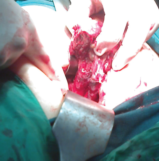
Surgery was planned with a prediagnosis of acute appendicitis. Laparotomy was performed via Mc Burney’s incision. The operative findings were two tubular structures surrounded by omentum at the top perforated with a purulent fluid around the lesion (Figure 1). Both of the tubular appendiceal structures were reaching out to cecum side to side. For both of the appendices, appendectomies were performed and proximal stumps were ligated twice. The postoperative course was uneventful. The patient was discharged without any complication after four days. The pathology report revealed an irregularly shaped browny fatty tissue with a size of 8 × 6 × 4 cm and appendices both measuring 8 cm in lenght and 1 cm in diameter within the previously described fatty tissue which was folded back like a horseshoe. There was a a perforation at one of the two tips and both inside and around the lumen of the perforated one.
Figure 1.

Intraoperative view of perforated horseshoe appendicitis
DISCUSSION
Preoperative diagnosis of a duplicated appendix is not easy at all for the surgeons and radiologists. Routine radiological imaging studies like ultrasonography and computerized tomography (CT) cannot distinguish this abnormal duplication of intestines from other pathologic lesions. If an experienced radiologist looks for this abnormality with a clinical suspicion in the CT of a patient with a right lower quadrant pain with a history of a previous appendectomy, the second lumen of the intestine could sometimes be visualized. In our case, preoperative radiological findings in ultrasonography and CT suggested a plastron secondary to acute appendicitis. When discussed with the radiologist about CT images after the operation, the radiologist could not distinguish two seperate lumens consistent with double appendix from the surrounding tissues.
Double appendix is generally recognized incidentally at surgery or on postmortem examination (1). It can rarely be picked up on barium enema for other reasons (2). Consequently, imaging studies is not useful for diagnosis of dupplicated appendix. Especially when duplication of appendix is encountered in childhood, other intestinal, genito-urinary or vertebral malformations must be explored (3).
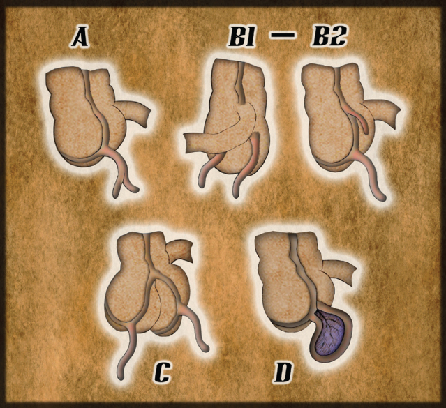
The possible locations and shapes of double appendix were described in the Cave-Wallbridge classification (Figure 2). The present case was Type D horseshoe type according to the Cave-Wallbridge classification. Localization of appendices may be either on taenia side to side as seen in our case or in distant places on both sides of ileocecal valve. In Kothari’s case, one of the appendices was located near ileocecal valve close to ileum and the other was located in the pelvis. That was type C dupplication with double cecum (4). In differential diagnosis, diverticulum of the cecum or appendix should be considered. As in our case, definitive diagnosis can be eventually made by histopathologic examination (5–7).
Figure 2.

Modified Cave-Wallbridge classification; Type A: Partial dupplication of appendix. Type B1: (Bird type) Two appendices are symmetrically placed on either side of the ileocecal valve. Type B2: (taenia coli-type) One appendix is at the usual site and the other one is far away along the lines of taenia. Type C: Dupplication of both cecum and appendix. Type D: (Horseshoe type) One appendix has two openings into the cecum
CONCLUSION
Since preoperative radiological identification of duplication of appendix is difficult, the surgeon’s intraoperative attention and awareness are vital in point of diagnosis. Macroscopic view and histopathological examination are substantially pathognomonic. This case has been alerting and awakening for surgeon about anatomic variations. Especially a “retrograde appendectomy” of an unrecognized case can easiliy result in an incomplete surgery when the surgeon misses the second opening to the cecum in difficult and laparoscopic cases which can create medicolegal issues.
Footnotes
Informed Consent: All necessary consents required by applicable law from the patient whose information is included in the article have been obtained in writing.
Peer-review: Externally peer-reviewed.
Author Contributions: Concept - N.C.; Design - S.P.B.; Supervision -M.A.; Data Collection and/or Processing - N.C., S.P.B.; Analysis and/or Interpretation - N.C., S.P.B.; Literature Review - S.P.B.; Writer - N.C., S.P.B.; Critical Review - M.A.
Conflict of Interest: No conflict of interest was declared by the authors.
Financial Disclosure: The authors declared that this study has received no financial support.
REFERENCES
- 1.Collins DC. A study of 50,000 specimens of human vermiform appendix. Surg Gynaecol Obstet. 1955;101:437–446. [PubMed] [Google Scholar]
- 2.Peddu P, Sidhu PS. Appearance of Type B duplex appendix on barium enema. Br J Radiol. 2004;77:248–249. doi: 10.1259/bjr/65763747. http://dx.doi.org/10.1259/bjr/65763747. [DOI] [PubMed] [Google Scholar]
- 3.Eroglu E, Erdogan E, Gundogdu G, Dervisoglu S, Yeker D. Dupplication of appendix vermiformis: a case in child. Tech Coloproctol. 2002;1:55–57. doi: 10.1007/s101510200010. http://dx.doi.org/10.1007/s101510200010. [DOI] [PubMed] [Google Scholar]
- 4.Kothari AA, Yagnik KR, Hathila VP. Dupplication of vermiform appendix. J Postgrad Med. 2004;50:285–286. [PubMed] [Google Scholar]
- 5.Mesko TW, Lugo R, Breitholtz T. Horseshoe anomaly of the appendix: a previously undescribed entity. Surgery. 1989;106:563–566. [PubMed] [Google Scholar]
- 6.Scarff JE, Harrold MW, Wylie JH. Duplication of the vermiform appendix. New variant of a rare anomaly. South Med J. 1982;75:860–862. doi: 10.1097/00007611-198207000-00024. http://dx.doi.org/10.1097/00007611-198207000-00024. [DOI] [PubMed] [Google Scholar]
- 7.Wallbridge PH. Double appendix. Br J Surg. 1963;50:346. doi: 10.1002/bjs.18005022124. http://dx.doi.org/10.1002/bjs.18005022124. [DOI] [PubMed] [Google Scholar]


