Abstract
Background
Trimethylamine N-oxide (TMAO), a gut microbe dependent metabolite of dietary choline and other trimethylamine containing nutrients, is both elevated in the circulation of patients suffering from heart failure (HF) and heralds worse overall prognosis. In animal studies, dietary choline or TMAO significantly accelerate atherosclerotic lesion development in ApoE deficient mice, and reduction in TMAO levels inhibits atherosclerosis development in the LDL receptor knockout mouse.
Methods and Results
C57BL6/J mice were fed either a control diet, a diet containing choline (1.2%) or a diet containing TMAO (0.12%) starting 3 weeks prior to surgical TAC. Mice were studied for 12 weeks following TAC. Cardiac function and left ventricular structure were monitored at 3-week intervals using echocardiography. Twelve weeks post-TAC myocardial tissues were collected to evaluate cardiac and vascular fibrosis, and blood samples were evaluated for cardiac BNP, choline, and TMAO levels. Pulmonary edema, cardiac enlargement, and left ventricular ejection fraction (LVEF) were significantly (p < 0.05, each) worse in mice fed either TMAO or choline supplemented diets compared to the control diet. In addition, myocardial fibrosis was also significantly greater (p < 0.01, each) in the TMAO and choline groups relative to controls.
Conclusions
Heart failure severity is significantly enhanced in mice fed diets supplemented in either choline or the gut microbe-dependent metabolite TMAO. The present results suggest that further studies are warranted examining whether gut microbiota and the dietary choline -> TMAO pathway contribute to increased heart failure susceptibility.
Keywords: metabolomics, left ventricular dysfunction, fibrosis, pulmonary edema, transverse aortic constriction, choline
Cardiovascular disease (CVD) represents the leading cause of death worldwide. A known environmental risk factor for the development of CVD is a diet rich in lipids and animal products1. In the past several years a role for gut microbiota in the pathogenesis of cardiovascular disease has been documented2–5. The interplay between diet and cardiovascular health has been well studied, establishing that diets high in fat and lipids lead to poor cardiac outcomes1. While historically a causal role for the dietary nutrients cholesterol and fat have been the primary focus, more recent studies have highlighted additional contributory roles of alternative trimethylamine containing dietary nutrients (e.g. choline, phosphatidylcholine, carnitine) and gut microbial metabolism6. Recent studies have shown that there is a mechanistic link between gut microbe dependent metabolism of dietary choline, phosphatidylcholine and carnitine and cardiovascular disease pathogenesis2, 4, 5, 7. Specifically, gut microbe metabolism of these dietary nutrients results in production of trimethylamine, which is rapidly converted by host hepatic flavin monooxygenase 3 (FMO3) into trimethylamine N-oxide (TMAO). Involvement of this meta-organismal (gut microbe and host) pathway in both TMAO formation and cardiovascular disease pathogenesis includes studies that demonstrate the choline-, gut microbiota- and TMAO-dependent acceleration in atherosclerosis in animal models2, 4, 7. Further, reduction in TMAO levels through antisense oligonucleotide mediated inhibition in host hepatic FMO3 significantly inhibited atherosclerosis development in the LDL receptor null mouse model, and the liver insulin receptor knockout mouse model8. Numerous mechanistic investigations demonstrate a central role for the choline->TMAO metaorganismal pathway in cholesterol and sterol metabolism, macrophage phenotype, and reverse cholesterol transport2, 7, 8,9. Importantly, recent microbial transplantation studies confirmed that both TMAO production and atherosclerosis susceptibility are transmissible traits5
Clinical studies have substantiated many of the above findings linking gut microbes and TMAO production to cardiovascular disease. First, the original discovery of the pathway began with an unbiased metabolomics study of plasma from subjects undergoing elective cardiac evaluations, revealing a striking association between plasma levels of choline, TMAO, and betaine with cardiovascular risks among subjects (n=1876)2. Examination of sequential subjects (n=1020) undergoing elective cardiac diagnostic catheterization show significantly increased circulating levels of TMAO in patients with more extensive angiographic evidence of coronary artery disease2. Furthermore, in a distinct cohort of subjects undergoing diagnostic cardiac catheterization (n=4007), elevated TMAO levels independently predicted increased risk of adverse cardiovascular outcomes including heart attack, stroke and death risk3. Elevated TMAO levels have also been shown to herald worse overall prognosis among subjects with (and without) impaired kidney function, and to predict enhanced atherosclerotic burden10, 11. Recently, TMAO levels have been shown to be associated with a three-fold enhanced risk for prevalent cardiovascular disease among a community based multi-ethnic population (hazard ratio (HR) 3.17; 95% confidence interval (CI) 1.05 to 9.51)12. Moreover, elevated TMAO plasma levels were reported in subjects (n=720) with stable heart failure (HF), with elevated levels predicting increased five year mortality risks (hazard ratio (HR) 2.2 (95% confidence interval (CI) 1.4 to 3.4), an association which remained significant independent of traditional risk factors and cardiorenal indices13. Elevated TMAO levels were also reported to predict increased risk of HF among both diabetic subjects (HR 4.6 (95% CI 2.0 to 10.7) and non-diabetic (HR 1.9 (95% CI 1.1 to 3.4) subjects alike14. In further studies, elevated TMAO levels were observed among subjects (n=112) with extensive serial echocardiography, and similarly were associated with more advanced left ventricular diastolic dysfunction and predicted poorer long-term adverse clinical outcomes independent of cardiac and renal biomarkers15. To date, no studies have tested whether dietary choline or TMAO directly promote development and progression of cardiac dysfunction and HF. In the present study, we investigated the effects of dietary choline or TMAO in the setting of cardiac hypertrophy and heart failure following transverse aortic constriction (TAC).
Methods
Experimental Animals
C57BL6/J male mice aged 10–12 weeks (Jackson Labs, Bar Harbor, ME) were utilized for these studies. All animals were housed and maintained in an on-site temperature-controlled animal facility adhering to a 12 hour light/dark cycle and provided with water and rodent chow ad libitum. All animals were humanely cared for in accordance with the Principles of Laboratory Animal Care dictated by the National Society of Medical Research and the Guide for the Care and Use of Laboratory Animals by the NIH (Publication No. 85-23, Revised 1996). All animal procedures were approved by IACUC of both The Louisiana State University Health Sciences Center and the Cleveland Clinic.
Experimental Diets
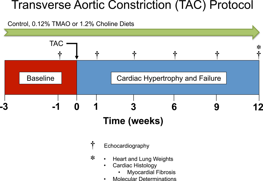
Mice were maintained on either a chemically defined control diet (TD.130104), a diet containing 0.12% TMAO added to the standard rodent chow (TD.07865), or a diet containing 1.2% choline added to the standard rodent chow (TD.09041) from Harlan Laboratories, Madison, WI as described previously. These diets were initiated at 3 weeks prior to transverse aortic constriction (TAC) surgery and maintained for an additional 12 weeks.
Transverse Aortic Constriction (TAC) Protocol
Cardiac pressure overload and HF was induced using TAC surgery as described previously16. The complete experimental protocol for these studies is depicted in Figure 1. Mice were anesthetized using xylazine (2 mg/kg) and ketamine (20 mg/kg) and maintained under anesthesia throughout the procedure by additional administration of xylazine (2 mg/kg) and ketamine (20 mg/kg) as needed. Mice were intubated and mechanically ventilated during the procedure using a Hugo Sachs type 845 minivent. Chest hair was removed from the mice by application of hair removal agent. The area was then cleaned using betadine and alcohol wipes alternating 3 times. An incision was made proximally left of the midline of the chest, below the clavicle. The aorta was exposed using blunt dissection. A ligature was placed between the brachiocephalic artery and the left common carotid artery and secured to induce pressure overload. The chest was closed in layers, closing the muscle layer first, then the skin. Mice were removed from ventilation after pedal reflex returned and it was determined respiration could be maintained autonomously. Mice were placed in a recovery cage and supplied with 100% O2 until normal behavior returned.
Figure 1.
Experimental protocol for transverse aortic constriction (TAC) studies in mice fed either control diet, trimethyamine N-oxide (TMAO), or choline. Mice were fed experimental diets starting at 3 weeks prior to TAC surgery and left ventricular (LV) function and structure were evaluated via two-dimensional echocardiography at 3-week intervals following baseline echocardiography studies at 1 week prior to TAC surgery. Upon completion of the study, mice were sacrificed at 12 weeks for heart and lung weights, cardiac histology and pathology as well as molecular determinations.
Echocardiography Assessment
One week prior to TAC procedure, baseline transthoracic echocardiogram was performed using 30-MHz probe on a Vevo 2100 (Visualsonics) while under light anesthesia with isoflurane (0.25 to 0.50%) supplemented with 100% O2, as described previously16. Following TAC procedure echocardiography was performed for 12 weeks at 3 week intervals.
Measurements of Serum Brain Natriuretic Peptide (BNP)
Brain-type natriuretic peptide (BNP EIA kit, Phoenix Pharmaceuticals, Inc.) serum levels were determined by ELISA at 12 weeks following TAC as described previously16.
Histology and Immunochemistry
Hearts were collected at the specified time points and fixed in 10% buffered formalin, embedded in paraffin, and stained with either Masson’s Trichome or Picrosirius Red to quantify fibrosis as described previously16.
Quantification of TMAO, Choline, Betaine and Carnitine
Stable isotope dilution LC/MS/MS was used for quantification of the total choline, TMA, TMAO, carnitine and betaine contents of diets, and the concentration of these analytes in plasma, as previously described4, 17. Briefly, TMAO, choline, betaine and carnitine were monitored in positive MRM MS mode using characteristic precursor–product ion transitions: m/z 76→58, m/z 104→60, m/z 118→59 and m/z 162→60, respectively. The internal standards d9(trimethyl)TMAO (d9-TMAO), d9(trimethyl)choline (d9-choline), d9(trimethyl)betaine and d3(methyl)carnitine (d3-carnitine), were added to serum samples before protein precipitation, and were similarly monitored in MRM mode at m/z 85→68, m/z 113→69, m/z 127→68, and m/z 165→63, respectively. Various concentrations of TMAO, choline, betaine and carnitine standards and a fixed amount of internal standards were spiked into control serum to prepare the calibration curves for quantification of serum analytes.
Statistical Analysis
All data are presented as median (interquartile range). Kruskal Wallis or Wilcoxon-Rank sum test was used for between group comparisons. A linear mixed effect model was used for repeated measure data to examine the change of all parameters during the 12-week study period. All analyses were performed using R 3.1.2 (Vienna, Austria) and p-values <0.05 were considered statistically significant.
Results
Impact of dietary TMAO or choline on plasma levels of metabolites
Analysis of plasma from mice in all groups at 12 weeks post TAC revealed that mice fed either TMAO or choline had significant (p < 0.0001) increase in the level of circulating TMAO compared to mice fed a control diet (Table). We also observed significant increases (p = 0.037) in plasma betaine levels in the mice fed TMAO.
Table.
Dietary trimethylamine n-oxide (TMAO) and choline 3 weeks before transverse aortic constriction increases circulating levels (µM) of metabolites indicated in the pathogenesis of cardiovascular disease at 12 weeks post TAC. Results presented as median and interquartile range, P valaues are comparisons between TMAO or Choline vs. Control using two-sample Wilcoxon tests.
| Choline | Betaine | TMAO | Carnitine | |
|---|---|---|---|---|
| Control (n = 10) | 22.9 (21.2 – 26.5) | 74.1 (59.5 – 96.8) | 1.7 (1.4 – 2.3) | 25.1 (23.3 – 27) |
| TMAO (n = 10) | 25.1 (19.6 – 25.8) (p = 0.853) | 56.2 (48.7 – 64.5) (p = 0.037) | 25.2 (16.2 – 41) (p < 0.0001) | 26.0 (21.9 – 26.3) (p = 0.65) |
| Choline (n = 12) | 33.4 (25.9 – 35.5) (p = 0.012) | 100.8 (83.6 – 126.3) (p = 0.138) | 26.6 (25.5 – 30.3) (p < 0.0001) | 27.4 (25.6 – 29.9) (p = 0.391) |
Dietary TMAO exacerbates cardiac dilatation and left ventricular dysfunction following TAC in mice
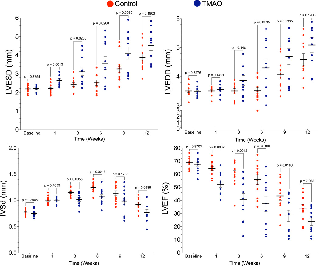
We evaluated the relationship between dietary TMAO and the pathogenesis of heart failure following TAC in mice. C57BL6/J mice were fed either standard chow diet, diet containing 0.12% TMAO or diet containing 1.2% choline starting three weeks prior to TAC procedure and maintained on diets for 12 weeks. (Figure 1). Chronic HF was produced by TAC resulting in pressure overload and cardiac hypertrophy. Both groups displayed significant (p < 0.0001 for LVSED, p < 0.0001 for LVEDD, p = 0.0382 for IVSd, and p < 0.0001 for LVEF) LV dilation, hypertrophy and reduced LV ejection fraction as compared to baseline. Mice that consumed 0.12% TMAO exhibited significantly worse cardiac function in all parameters measured following TAC (Figure 2), as compared to mice fed the control diet. An accelerated progression of cardiac dilatation, as evidenced by significantly increased left ventricular end-systolic diameter (LVESD) and end-diastolic diameter (LVEDD), was observed in the TMAO group (Figures 2A–B). LVESD was significantly greater at 1, 3, and 6 weeks (p = 0.0013, p = 0.0268, p = 0.0268 vs. control, respectively) following TAC. LVEDD was trending towards an increase in the TMAO group versus control group at 6 weeks post-TAC (p = 0.0595). In addition, we also observed worsened adverse ventricular remodeling as determined by interventricular septal dimension (p = 0.0056, p = 0.0045 vs. control) at 3 and 6 weeks (Figure 2C). Importantly, mice fed the TMAO diet exhibited significantly decreased left ventricular ejection fraction (EF) (p = 0.0007, p = 0.0013, p = 0.0188, p = 0.0188, p = 0.063 vs. control, respectively) at all time points investigated following TAC surgery (Figure 2D).
Figure 2.
Dietary trimethylamine N-oxide (TMAO) exacerbates cardiac dilation and dysfunction following transverse aortic constriction (TAC) in mice, as compared to a control diet. (A) Left ventricular (LV) end-systolic diameter (LVESD; in mm), (B) LV end-diastolic diameter (LVEDD; in mm), (C) Intraventricular septal wall diastolic diameter (IVSd; in mm) and (D) LV ejection fraction (%) from baseline to 12 weeks. Results are presented as mean ± SEM. A linear mixed effects model was used to evaluate the change of all parameters during the 12 week study period when compared to baseline. Two-sample Wilcoxon tests were used to compare between TMAO and control groups at different time points for 10 – 12 animals per group.
Dietary choline exacerbates cardiac dilatation and left ventricular dysfunction following (TAC) in mice
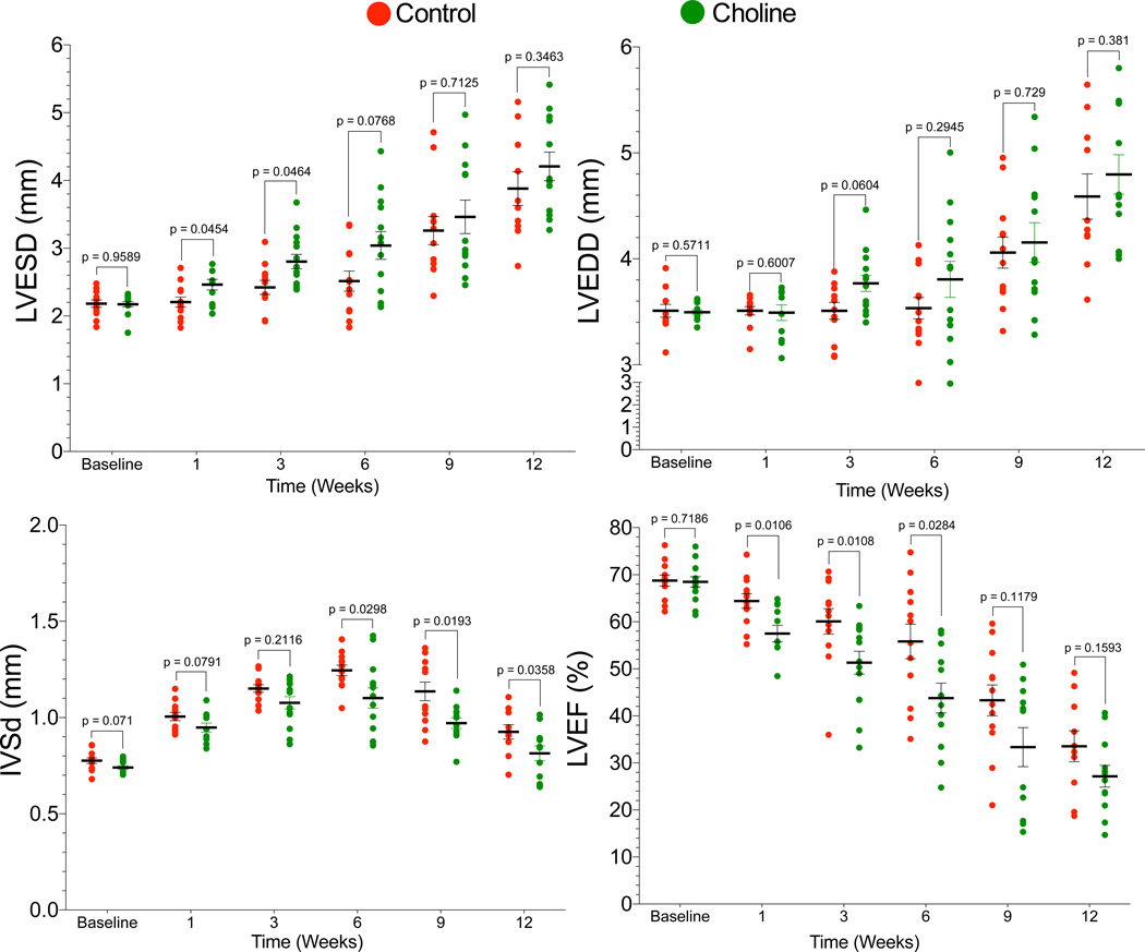
The effect of TMAO on adverse ventricular remodeling and accelerated HF progression following TAC suggested a potential link between dietary choline and HF severity. To investigate this possibility we fed mice either control chow or the same diet supplemented with 1.2% choline. Mice fed the control vs, choline supplemented diets were subjected to the same 12 week TAC experimental protocol. When compared to baseline both groups exhibited LV dilation, hypertrophy and decremented LVEF (p < 0.0001 for LVESD, p < .0001 for LVEDD, p = 0.0007 for IVSd, and p < 0.0001 for LVEF). Serial echocardiography analyses revealed that 1.2% dietary choline exerted similar effects on adverse cardiac remodeling as observed in mice that consumed TMAO supplemented diet (Figure 3). LVESD was significantly (p = 0.0454, p = 0.0464, respectively) greater at 1 and 3 weeks following TAC, and LVEDD was trending towards an increase (p = 0.0604) at 3 weeks compared to the control diet group (Figures 3A and 3B). LV Interventricular septal thickness was significantly (p = 0.0298, p = 0.0193, p = 0.0358, respectively) less at 6, 9, and 12 weeks following TAC surgery in the choline group suggesting greater adverse left ventricular remodeling (Figure 3C). Importantly, we also observed significantly worse left ventricular ejection fraction (Figure 3D) in mice fed a diet containing 1.2% choline, with significant (p = 0.0106, p = 0.0108, p = 0.0284, respectively) reduction noted at 1, 3, and 6 weeks following TAC as compared to the control diet group. Collectively, these data clearly demonstrate a detrimental effect of supplemental dietary choline on left ventricular structure and function in the setting of HF in the murine TAC model.
Figure 3.
Dietary choline (1.2%) exacerbates cardiac dilation and dysfunction following transverse aortic constriction (TAC) in mice, as compared to a control diet. (A) Left ventricular (LV) end-systolic diameter (LVESD; in mm), (B) LV end-diastolic diameter (LVEDD; in mm), (C) Intraventricular septal wall diameter (IVSd; in mm) and (D) LV ejection fraction (%) from baseline to 12 weeks. A linear mixed effects model was used to evaluate the change of all parameters during the 12 week study period when compared to baseline. Results are presented as mean ± SEM. Two-sample Wilcoxon tests were used to compare between choline and control groups at different time points for 10 – 12 animals per group.
Dietary TMAO and choline exacerbate cardiac enlargement and pulmonary edema following TAC
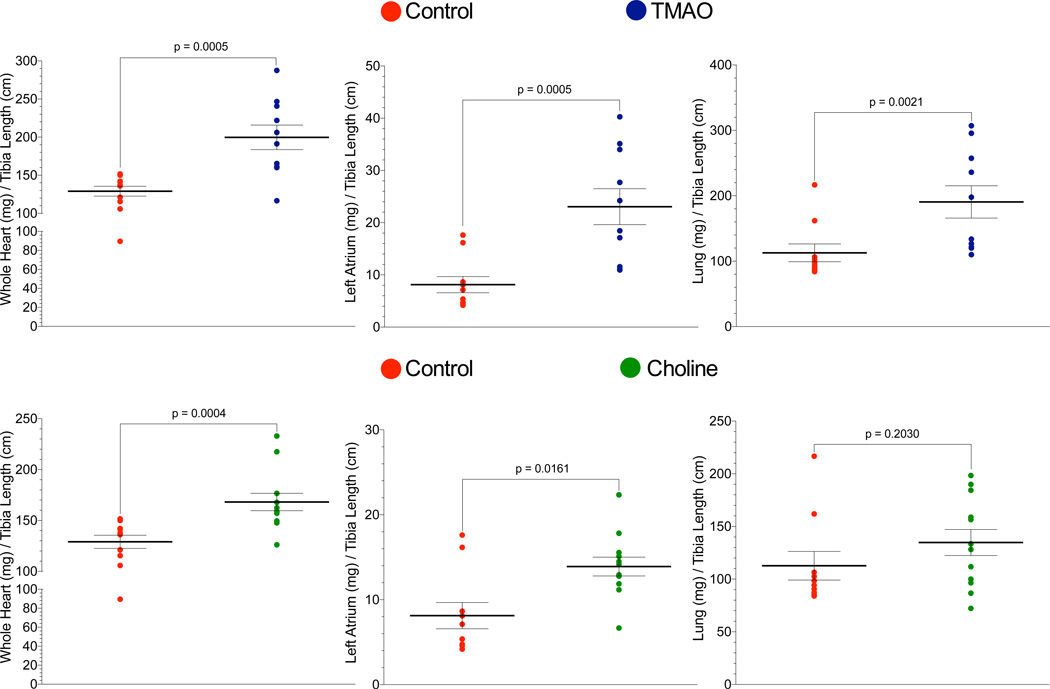
Body weights and percent survival following heart failure for mice among the three dietary groups revealed no statistically significant differences (Supplemental Figures 1 and 2). Since a major pathological consequence of HF is cardiac enlargement and pulmonary edema, we evaluated myocardial and pulmonary tissue weights as an index of cardiac enlargement and pulmonary edema in mice fed the different diets. First, comparison of control or TMAO diet groups, performed initially, is shown in Figure 4. Comparison of heart weight (normalized to tibia length) at time of sacrifice 12 weeks following TAC in TMAO fed and control mice showed a significant (p = 0.0005) increase in heart weight in the TMAO group (Figure 4A). Significant (p = 0.0005) increases in left atrial (LA) weight in the TMAO group as compared to the control group were also noted (Figure 4B). Pulmonary edema, as evidenced by lung weight/tibia length, was also significantly (p = 0.0021) increased in mice receiving dietary TMAO (Figure 4C). Parallel changes in cardiac and lung weights were observed in mice fed a diet containing 1.2% choline relative to control diet. Specifically, we observed significant increases in heart weight / tibia length (p = 0.0004) and LA / tibia length (p = 0.0161) in the choline group as compared to control diet (Figures 4D and E). However, while a trend toward increase lung weight was observed in the choline fed mice this difference failed to achieve statistical significance when compared to mice fed the control diet (Figure 4F).
Figure 4.
Dietary trimethylamine N-oxide (TMAO) and choline exacerbate cardiac enlargement and hypertrophy following transverse aortic constriction (TAC). (A) Ratio of heart weights to tibia length in TMAO vs. control. (B) Ratio of left atrial (LA) weights to tibia length in TMAO vs control. (C) Ratio of lung weights to tibia length in TMAO vs. control. (D) Ratio of heart weights to tibia length in choline vs. control. (E) Ratio of LA weights to tibia length in choline vs. control. (F), Ratio of lung weights to tibia length in choline vs. control. Results are presented as mean ± SEM. Two-sample Wilcoxon tests were used to compare between TMAO or Choline vs. control for 10 – 12 animals per group.
Dietary TMAO and choline increase levels of circulating brain natriuertic peptide (BNP) in mice following TAC
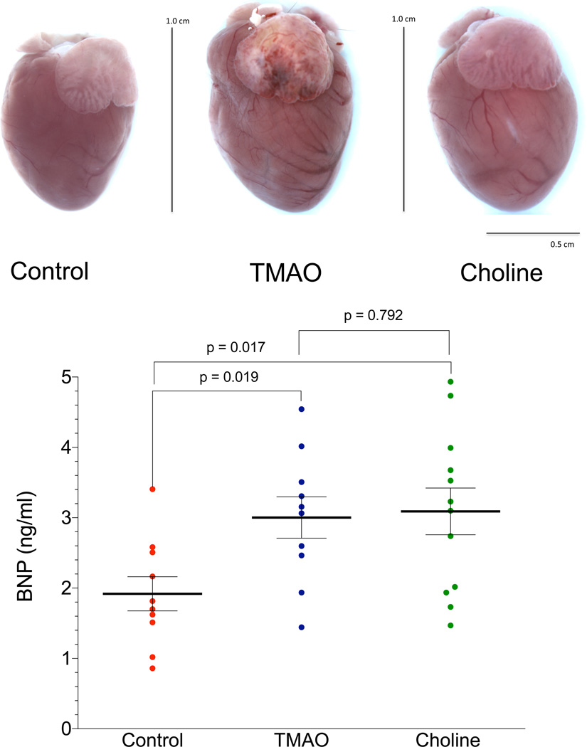
We evaluated circulating levels of B-type natriuretic peptide (BNP), a hormone released from myocardial tissues during enhanced stretch and associated with enhanced degree of heart failure severity, in mice at 12 weeks post TAC (Figure 5D). BNP levels were significantly increased in both the TMAO (p = 0.019) and choline (p = 0.017) groups compared to the mice fed a control diet, with similar BNP results between the TMAO and Choline groups (p = 0.792). These data correlate with our echocardiographic data suggesting that mice in the TMAO and choline groups experienced more severe left ventricular remodeling and reduction in left ventricular function compared to mice in the control group. Gross examination of the heart showed significant cardiac enlargement in mice within either the TMAO or choline supplemented dietary groups in comparison to controls (Figures 5A–5C).
Figure 5.
Increased circulating levels of brain natriuretic peptide (BNP) in mice following transverse aortic constriction (TAC) and the development of pressure overload induced heart failure. (A–C) Representative photomicrographs of control (TAC + control diet), trimethylamine n-oxide treated (TAC + TMAO diet), and choline treated (TAC + choline diet) hearts at 12 weeks following TAC. (D) Mean circulating BNP levels (ng/mL) in mice 12 weeks post TAC. Two-sample Wilcoxon tests were used for two group comparison for 10 – 12 animals per study group.
Dietary (TMAO) and choline (TAC) exacerbate interstitial and perivascular fibrosis following TAC
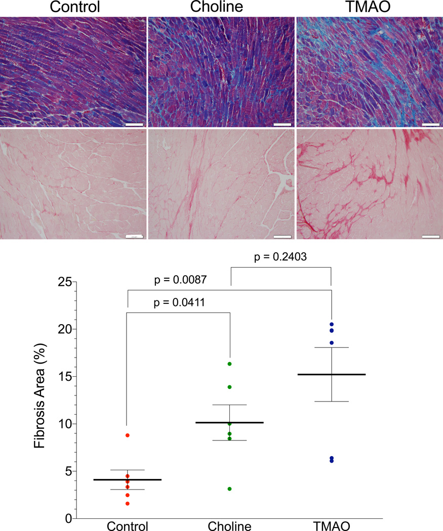
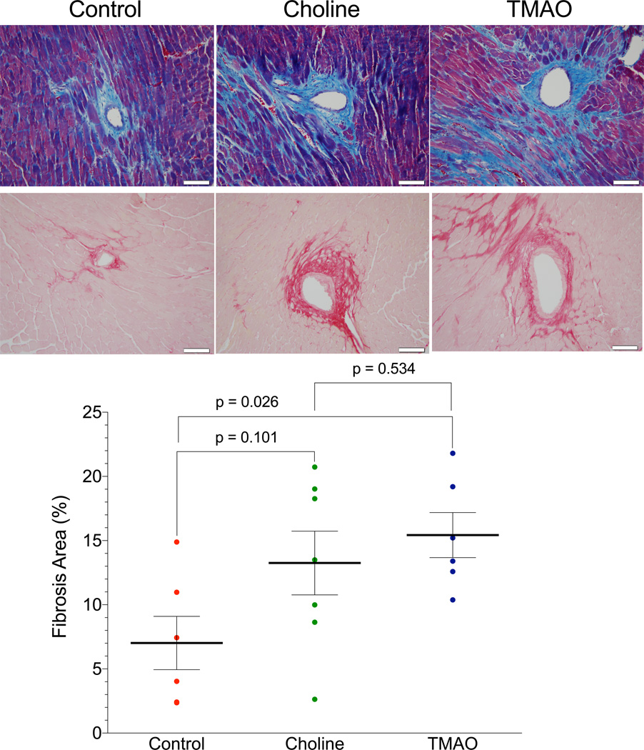
Myocardial connective tissue responds to cardiac hypertrophy and impaired heart function by the development of fibrosis, which results in left ventricular stiffening, loss of compliance, and further impairments in cardiac contraction and relaxation. To further evaluate cardiac remodeling we examined cardiac fibrosis formation utilizing two histological methods to detect collagen fibers in the heart following TAC (Figure 6). We performed both Masson’s trichrome and Pircrosirus red staining on cardiac tissues at 12 weeks following TAC. With each method of detecting enhanced collagen and fibrosis we observed significant increases in the hearts of mice fed either a diet supplemented in TMAO or choline relative to normal chow (control) diet. Representative photomicrographs are shown in Figures 6A and 6B for Control, choline, and TMAO and summary data are presented in Figures 6C. Computerized planimetry based quantification of the intensity of Picrosirius red staining (Figure 6C) affirmed significantly (p = 0.0087) greater level of intermuscular fibrosis in the TMAO group compared to the control group. Using the same method of tissue staining and analysis, we observed similar results in the perivascular sections of heart tissue collected 12 weeks post TAC in control, choline and TMAO fed mice (Figure 7). Depicted in Figures 7A and 7B, the photomicrographs show increased levels of perivascular fibrosis in the TMAO group compared to the control group, this increased significance (p = 0.026) is corroborated by quantification of the intensity of the Picrosirius red staining (Figure 7C).
Figure 6.
Dietary trimethylamine N-oxide (TMAO) and choline initiated at 3 weeks prior to transverse aortic constriction (TAC) worsens interstitial fibrosis. (A) Representative photomicrographs of Masson’s trichrome stained heart sections in Control, Choline and TMAO groups of mice illustrating intermuscular fibrosis at 12 weeks post TAC. (B) Representative photomicrographs of Picrosirius red stained heart sections in Control, Choline and TMAO mice illustrating intermuscular fibrosis at 12 weeks post TAC. (C) Cardiac fibrosis as a percent (%) of left ventricular area calculated from Picrosirius red stained heart. Scale bars on photomicrographs represent 500 micrometers. Results are presented as mean ± SEM. Two-sample Wilcoxon tests were used for two group comparison.for 6 – 7 animals per group.
Figure 7.
Dietary trimethylamine N-oxide (TMAO) and choline exacerbate perivascular fibrosis in the setting of transverse aortic constriction (TAC). (A) Representative photomicrographs of Masson’s trichrome stained heart sections in Control, Choline and TMAO groups of mice illustrating perivascular fibrosis at 12 weeks post TAC. (B) Representative photomicrographs of Picrosirius red stained heart sections in Control, Choline and TMAO mice illustrating perivascular fibrosis at 12 weeks post TAC. (C) Cardiac fibrosis as a percent (%) of left ventricular area calculated from Picrosirius red stained heart tissue. Scale bars on photomicrographs represent 500 micrometers. Results are presented as mean ± SEM. Two-sample Wilcoxon tests were used for two group comparison.for 6–7 animals per group.
Discussion
The critical importance of diet and overall metabolism in impacting cellular processes is growing in appreciation for its potential to impact the development and severity of cardiovascular diseases. Moreover, a growing body of data reveals strong associations between gut microbiota formation of the metabolite TMAO and adverse cardiovascular risks. For example, several recent clinical studies have shown that elevated circulating levels of the metabolite TMAO directly correlate with the severity of HF, adverse prognosis in HF subjects, and risk of HF among diabetic and non-diabetic subjects alike13, 14, 18. The present findings extend these human clinical associations, by demonstrating a remarkable effect of dietary supplementation with either choline, or its gut microbe-generated metabolite TMAO, and HF progression. Provision of either supplemental choline or TMAO each lead to greater propensity for adverse cardiac remodeling and left ventricular dysfunction in the setting of chronic pressure overload following TAC in naïve mice.
It is now appreciated that there are multiple microbes responsible for the conversion of choline to TMA, as identified in a recently published paper19. Romano, et al. show that there are numerous human commensals capable of making TMA from choline and then shown in vivo to produce TMA and TMAO when introduced into gnotobiotic mice. The human commensals examined are also known to inhabit the intestines of mice. Thus, it is not a case of a single species of microbe that forms TMA and TMAO but numerous microbes that exhibit the biochemical activity needed to convert different TMA containing nutrients into TMA.
Our study revealed that when TMAO levels are elevated in the circulation, susceptibility for development of HF is enhanced as evidenced by pathological left ventricular (LV) dilation, reduced LV ejection fraction, increased circulating BNP levels, increases in pulmonary edema, and increased myocardial fibrosis. It is interesting to note that recent animal model studies employing either choline or TMAO supplemented diets similarly revealed enhanced fibrosis in an alternative end organ, the kidney20. Specifically, exposure to both diets were noted to provoke tubulointerstitial fibrosis, and enhanced perivascular fibroisis in a manner dose dependently associated with circulating TMAO levels20. The enhanced myocardial fibrosis observed in the present study suggests a pro-fibrotic phenotype with high TMAO levels is a common observation, and likely plays a contributory role to the worse adverse ventricular remodeling observed with either choline or TMAO supplemented diet. Further, the enhanced myocardial fibrosis observed in the animal models herein is consistent with recently reported enhanced indices of diastolic dysfunction and adverse prognosis observed in subjects with HF and higher TMAO levels13, 14, 18.
Elevated levels of betaine were also observed in plasma of mice on the choline diet but not in the plasma of mice fed a 0.12% TMAO diet. In concordance, recent human studies have shown elevated betaine levels predict risk for HF14 and as such, we too observed elevated betaine and choline levels predicted poorer prognosis (mortality) in human subjects with HF. However, when TMAO was included in the model (with BNP, traditional CVD risk factors and renal function indices), only TMAO remained a significant predictor of adverse outcome.
Limitations of the present study are worth noting. The murine model is not fully comparable to human physiology due to significant metabolic differences. In our study, we used the TAC protocol to induce HF. While this is a reliable and consistent model for HF, it does not fully represent human HF and may not accurately predict progression of human disease. Of note, however, it is relevant that TMAO levels observed in the present study in mice (~25 µM) are well within the elevated levels observed among subjects with either HF or chronic kidney disease and adverse prognosis, where levels in excess of 100 µM can be observed10, 13. Another limitation of the present study is we do not know at the molecular level how TMAO exerts its adverse effects on cardiac remodeling, fibrosis, and function, and whether there is a specific “TMAO receptor” or is acting via its known effect on protein conformation and stability21, 22.
The “gut hypothesis” in heart failure has historically focused on the potential involvement of bowel edema with congestion, and gut microbiota generated toxins and lipopolysaccharide leak from reduced barrier function, leading to heightened inflammation and suppressed myocardial function19. The present studies, coupled with the recent recognition of elevated plasma TMAO levels as strong independent predictors of HF severity and adverse prognosis13, 14, suggests the potential importance of both diet and gut microbial pathways to HF susceptibility. Further studies aimed at manipulating this pathway and monitoring HF risks seem warranted.
Supplementary Material
Clinical Perspective.
It has been well documented in the literature that diet can affect cardiac outcomes. As the organ with the most direct contact with our environment, the importance of the gut and its microbiome in medicine is gaining appreciation. Recently the role of the gut microbiome in cardiovascular disease has emerged as a significant determinant of overall cardiovascular health and disease. It has been shown that heart failure patients have significantly increased levels of circulating trimethylamine n-oxide (TMAO), a metabolite formed by the metabolism of choline via gut microbes. TMAO has also been shown to accelerate atherosclerotic plaque formation. In the current study we examine the effects of dietary TMAO and choline on the development and progression of heart failure in a mouse model of transverse aortic constriction in order to better characterize this metabolite and its role in the pathogenesis of heart failure. Our data demonstrate that dietary TMAO and choline significantly exacerbate cardiac fibrosis and left ventricular adverse remodeling and dysfunction in the setting of cardiac hypertrophy and heart failure.
Acknowledgments
SOURCES OF FUNDING
This work was supported by grants from the National Heart, Lung, and Blood Institute (National Institutes of Health; 5R01 HL092141, 5R01 HL093579, 1R01 HL11657, and 1U24 HL 094373 to D.J.L.). This work was also supported by grants P20HL113452, R01HL103866 from the National Heart, Lung, and Blood Institute of the National Institutes of Health and the Office of Dietary Supplements (to SLH). S.L.H is also partially supported by the Leonard Krieger endowment. Mass spectrometry instrumentation used was housed within the Cleveland Clinic Mass Spectrometry Facility with partial support through a Center of Innovation by AB SCIEX.
SLH is named as the co-inventor on issued and pending patents held by the Cleveland Clinic relating to cardiovascular diagnostics and/or therapeutics; he is also paid as a consultant for the following companies: Cleveland Heart Laboratory, Esperion, and Procter & Gamble; and has received research funds from Astra Zeneca, Pfizer, Procter & Gamble, Roche Diagnostics Inc., and Takeda. SLH reports having the right to receive royalty payments for inventions or discoveries related to cardiovascular diagnostics and/or therapeutics from Cleveland Heart Laboratory, Siemens, Esperion, and Frantz Biomarkers, LLC.
Footnotes
DISCLOSURES
The remaining authors declare no conflict of interest.
References
- 1.Backhed F, et al. The gut microbiota as an environmental factor that regulates fat storage. PNAS. 2004;101:15718–15723. doi: 10.1073/pnas.0407076101. [DOI] [PMC free article] [PubMed] [Google Scholar]
- 2.Wang Z, Klipfell E, Bennett BJ, Koeth R, Levison BS, Dugar B, Feldstein AE, Britt EB, Fu X, Chung YM, Wu Y, Schauer P, Smith JD, Allayee H, Tang WH, DiDonato JA, Lusis AJ, Hazen SL. Gut flora metabolism of phosphatidylcholine promotes cardiovascular disease. Nature. 2011;472:57–63. doi: 10.1038/nature09922. [DOI] [PMC free article] [PubMed] [Google Scholar]
- 3.Tang WH, Wang Z, Levison BS, Koeth RA, Britt EB, Fu X, Wu Y, Hazen SL. Intestinal microbial metabolism of phosphatidylcholine and cardiovascular risk. The New England journal of medicine. 2013;368:1575–1584. doi: 10.1056/NEJMoa1109400. [DOI] [PMC free article] [PubMed] [Google Scholar]
- 4.Koeth RA, Levison BS, Culley MK, Buffa JA, Wang Z, Gregory JC, Org E, Wu Y, Li L, Smith JD, Tang WH, DiDonato JA, Lusis AJ, Hazen SL. Gamma-butyrobetaine is a proatherogenic intermediate in gut microbial metabolism of l-carnitine to tmao. Cell metabolism. 2014;20:799–812. doi: 10.1016/j.cmet.2014.10.006. [DOI] [PMC free article] [PubMed] [Google Scholar]
- 5.Gregory JC, Buffa JA, Org E, Wang Z, Levison BS, Zhu W, Wagner MA, Bennett BJ, Li L, DiDonato JA, Lusis AJ, Hazen SL. Transmission of atherosclerosis susceptibility with gut microbial transplantation. The Journal of biological chemistry. 2015;290:5647–5660. doi: 10.1074/jbc.M114.618249. [DOI] [PMC free article] [PubMed] [Google Scholar]
- 6.Brown JM, Hazen SL. The gut microbial endocrine organ: Bacterially derived signals driving cardiometabolic diseases. Annual review of medicine. 2015;66:343–359. doi: 10.1146/annurev-med-060513-093205. [DOI] [PMC free article] [PubMed] [Google Scholar]
- 7.Koeth RA, Wang Z, Levison BS, Buffa JA, Org E, Sheehy BT, Britt EB, Fu X, Wu Y, Li L, Smith JD, DiDonato JA, Chen J, Li H, Wu GD, Lewis JD, Warrier M, Brown JM, Krauss RM, Tang WH, Bushman FD, Lusis AJ, Hazen SL. Intestinal microbiota metabolism of l-carnitine, a nutrient in red meat, promotes atherosclerosis. Nature medicine. 2013;19:576–585. doi: 10.1038/nm.3145. [DOI] [PMC free article] [PubMed] [Google Scholar]
- 8.Shih DM, Wang Z, Lee R, Meng Y, Che N, Charugundla S, Qi H, Wu J, Pan C, Brown JM, Vallim T, Bennett BJ, Graham M, Hazen SL, Lusis AJ. Flavin containing monooxygenase 3 exerts broad effects on glucose and lipid metabolism and atherosclerosis. Journal of lipid research. 2015;56:22–37. doi: 10.1194/jlr.M051680. [DOI] [PMC free article] [PubMed] [Google Scholar]
- 9.Warrier M, Shih DM, Burrows AC, Ferguson D, Gromovsky AD, Brown AL, Marshall S, McDaniel A, Schugar RC, Wang Z, Sacks J, Rong X, Vallim TA, Chou J, Ivanova PT, Myers DS, Brown HA, Lee RG, Crooke RM, Graham MJ, Liu X, Parini P, Tontonoz P, Lusis AJ, Hazen SL, Temel RE, Brown JM. The tmao-generating enzyme flavin monooxygenase 3 is a central regulator of cholesterol balance. Cell reports. 2015;10:326–338. doi: 10.1016/j.celrep.2014.12.036. [DOI] [PMC free article] [PubMed] [Google Scholar]
- 10.Tang WH, Wang Z, Kennedy DJ, Wu Y, Buffa JA, Agatisa-Boyle B, Li XS, Levison BS, Hazen SL. Gut microbiota-dependent trimethylamine n-oxide (tmao) pathway contributes to both development of renal insufficiency and mortality risk in chronic kidney disease. Circulation research. 2015;116:448–455. doi: 10.1161/CIRCRESAHA.116.305360. [DOI] [PMC free article] [PubMed] [Google Scholar]
- 11.Stubbs JR, House JA, Ocque AJ, Zhang S, Johnson C, Kimber C, Schmidt K, Gupta A, Wetmore JB, Nolin TD, Spertus JA, Yu AS. Serum trimethylamine-n-oxide is elevated in ckd and correlates with coronary atherosclerosis burden. Journal of the American Society of Nephrology : JASN. 2015 Jul 30; doi: 10.1681/ASN.2014111063. pii: ASN.2014111063. [Epub ahead of print] doi: 10.1681/ASN.2014111063. [DOI] [PMC free article] [PubMed] [Google Scholar]
- 12.Mente A, Chalcraft K, Ak H, Davis AD, Lonn E, Miller R, Potter MA, Yusuf S, Anand SS, McQueen MJ. The relationship between trimethylamine-n-oxide and prevalent cardiovascular disease in a multiethnic population living in canada. The Canadian journal of cardiology. 2015;31:1189–1194. doi: 10.1016/j.cjca.2015.06.016. [DOI] [PubMed] [Google Scholar]
- 13.Tang WH, Wang Z, Fan Y, Levison B, Hazen JE, Donahue LM, Wu Y, Hazen SL. Prognostic value of elevated levels of intestinal microbe-generated metabolite trimethylamine-n-oxide in patients with heart failure: Refining the gut hypothesis. Journal of the American College of Cardiology. 2014;64:1908–1914. doi: 10.1016/j.jacc.2014.02.617. [DOI] [PMC free article] [PubMed] [Google Scholar]
- 14.Lever M, George PM, Slow S, Bellamy D, Young JM, Ho M, McEntyre CJ, Elmslie JL, Atkinson W, Molyneux SL, Troughton RW, Frampton CM, Richards AM, Chambers ST. Betaine and trimethylamine-n-oxide as predictors of cardiovascular outcomes show different patterns in diabetes mellitus: An observational study. PloS one. 2014;9:e114969. doi: 10.1371/journal.pone.0114969. [DOI] [PMC free article] [PubMed] [Google Scholar]
- 15.Tang WH, Wang Z, Shrestha K, Borowski AG, Wu Y, Troughton RW, Klein AL, Hazen SL. Intestinal microbiota-dependent phosphatidylcholine metabolites, diastolic dysfunction and adverse clinical outcomes in chronic systolic heart failure. Journal of cardiac failure. 2014;21:91–96. doi: 10.1016/j.cardfail.2014.11.006. [DOI] [PMC free article] [PubMed] [Google Scholar]
- 16.Bhushan S, Kondo K, Polhemus DJ, Otsuka H, Nicholson CK, Tao YX, Huang H, Georgiopoulou VV, Murohara T, Calvert JW, Butler J, Lefer DJ. Nitrite therapy improves left ventricular function during heart failure via restoration of nitric oxide-mediated cytoprotective signaling. Circulation research. 2014;114:1281–1291. doi: 10.1161/CIRCRESAHA.114.301475. [DOI] [PMC free article] [PubMed] [Google Scholar]
- 17.Wang Z, Levison BS, Hazen JE, Donahue L, Li XM, Hazen SL. Measurement of trimethylamine-n-oxide by stable isotope dilution liquid chromatography tandem mass spectrometry. Analytical biochemistry. 2014;455:35–40. doi: 10.1016/j.ab.2014.03.016. [DOI] [PMC free article] [PubMed] [Google Scholar]
- 18.Tang WH, Dupont M, Hernandez AF, Voors AA, Hsu AP, Felker GM, Butler J, Metra M, Anker SD, Troughton RW, Gottlieb SS, McMurray JJ, Armstrong PW, Massie BM, Califf RM, O'Connor CM, Starling RC. Comparative assessment of short-term adverse events in acute heart failure with cystatin c and other estimates of renal function: Results from the ascend-hf trial. JACC. Heart failure. 2015;3:40–49. doi: 10.1016/j.jchf.2014.06.014. [DOI] [PubMed] [Google Scholar]
- 19.Romano KA, Vivas EI, Amador-Noguez D, Rey FE. Intestinal microbiota composition modulates choline bioavailability from diet and accumulation of the proatherogenic metabolite trimethylamine-n-oxide. mBio. 2015;6:e02481. doi: 10.1128/mBio.02481-14. [DOI] [PMC free article] [PubMed] [Google Scholar]
- 20.Miao J, Ling AV, Manthena PV, Gearing ME, Graham MJ, Crooke RM, Croce KJ, Esquejo RM, Clish CB, Vicent D, Biddinger SB Morbid Obesity Study G. Flavin-containing monooxygenase 3 as a potential player in diabetes-associated atherosclerosis. Nature communications. 2015;6:6498. doi: 10.1038/ncomms7498. [DOI] [PMC free article] [PubMed] [Google Scholar]
- 21.Mondal J, Halverson D, Li IT, Stirnemann G, Walker GC, Berne BJ. How osmolytes influence hydrophobic polymer conformations: A unified view from experiment and theory. Proceedings of the National Academy of Sciences of the United States of America. 2015;112:9270–9275. doi: 10.1073/pnas.1511780112. [DOI] [PMC free article] [PubMed] [Google Scholar]
- 22.Ma J, Pazos IM, Gai F. Microscopic insights into the protein-stabilizing effect of trimethylamine n-oxide (tmao) Proceedings of the National Academy of Sciences of the United States of America. 2014;111:8476–8481. doi: 10.1073/pnas.1403224111. [DOI] [PMC free article] [PubMed] [Google Scholar]
Associated Data
This section collects any data citations, data availability statements, or supplementary materials included in this article.


