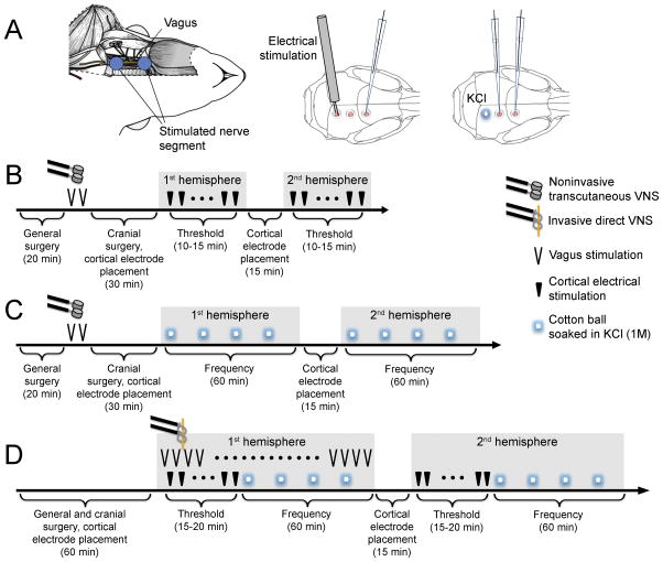
Fig. 1. Experimental setup and VNS paradigms.
A) Left: Surgical exposure of the vagus nerve for iVNS and the vagus nerve segment stimulated using the helical electrode are shown. For nVNS, transcutaneous disc electrodes were placed where indicated by blue circles. Middle and right: Craniotomies were prepared on occipital cortex for electrical or KCl stimulation, and on parietal and frontal cortices for electrophysiological recordings. B) Experimental timeline to test nVNS on electrical CSD threshold. C) Experimental timeline to test nVNS on KCl-induced CSD frequency. D) Experimental timeline to test iVNS on electrical CSD threshold and KCl-induced CSD frequency. All symbols are defined on the right. Details of stimulation parameters are provided in the Methods.

