Abstract
Sepsis-induced cholestasis is a complication of infection. Infections cause systemic and intrahepatic increase in proinflammatory cytokines which result in impaired bile flow ie. cholestasis. Several other mediators of impairment in bile flow have been identified under conditions of sepsis such as increased nitric oxide production and decreased aquaporin channels. The development of cholestasis may also further worsen inflammation. The molecular basis of normal bile flow and mechanisms of impairment in sepsis are discussed.
Keywords: Cirrhosis, Sepsis, Cholestasis, Bile acids, Bilirubin, Tumor necrosis factor-α, Acute phase reaction, Bacterial translocation, Acute of chronic liver failure, Bilirubinostasis, Review
2. INTRODUCTION
The production of bile is one of the most important functions of the liver. Bile removes excess cholesterol, other endogenous toxic compounds e.g. bilirubin and the metabolic byproducts of a variety of xenobiotics from the body. It also delivers bile salts to the small intestine where they play a key role in the digestion and absorption of ingested lipids. In recent years, it has also been recognized that bile salts act as metabolic sensors and help regulate the homeostatic responses to caloric intake and the availability of specific nutrients (1). In addition, bile salts affect the incretin response to various stimuli and are likely to play a role in maintaining the integrity of the gut and pancreatic beta cell mass (2). There is also a complex interplay between the intestinal microbiome and the specific bile salts that are present in the intestinal lumen that maintains a symbiotic balance between the host and their intestinal flora (3). It is therefore not surprising that cholestasis i.e. a failure of bile flow has important consequences for the afflicted individual. In this review, we will focus on the molecular events underlying the cholestatic response to sepsis.
3. PLACING SEPSIS AND CHOLESTASIS IN CLINICAL PERSPECTIVE
Bacterial infections and sepsis may lead to cholestasis. In infants, this is a common cause of cholestasis, with urinary tract infections as a common precipitant (4). In hospitalized adult patients with jaundice, approximately 20% of cases were attributed to infection in one case series (5). Bilirubin can increase prior to documented infection (6). Both gram-negative and gram-positive bacterial infections have been reported to cause jaundice (6-8). In most instances of sepsis-induced jaundice, the infection is intra-abdominal and may include biliary infection, urinary tract infections or intra-abdominal abscesses (8). However, jaundice has also been reported to be associated with pneumonia, meningitis and bacterial endocarditis (9, 10).
Jaundice may result either directly from bacterial products or as a consequence of the host response to infection. Frequently, both factors contribute to the development of jaundice. In addition, specific infections that target the liver may cause jaundice due to the liver injury associated with hepatic infection. While jaundice may be an isolated abnormality, it is often associated with features of cholestasis. In critically ill patients, the development of jaundice and/or cholestasis complicates the clinical picture and poses a clinical challenge both in terms of diagnostic evaluation as well as management. The differential diagnosis often includes the infection itself, presence of pylephlebitis, biliary obstruction by gallstones or tumor, development of hepatic abscesses and drug-induced liver injury. Table 1 outlines the differential diagnosis of jaundice in critically ill patients (11). A clear understanding of the mechanisms contributing to this cholestatic response to infections and their consequences is thus important for the optimal management of a given patient.
Table 1.
Differential diagnosis of jaundice in critical illness
| Biliary Tract Disease |
| • Cholecystitis |
| • Cholangitis |
| • Biliary tract obstruction |
| Liver Disease |
| • Hepatitis |
| • Hepatic ischemia |
| • Liver abscess |
| Systemic Infection |
| Hemolysis |
| • Infections |
| • Drugs |
| • Blood transfusions |
| Hepatotoxic drugs/toxins |
| • Antibiotics |
| • Tylenol |
| • Total Parenteral Nutrition |
4. THE MOLECULAR PHYSIOLOGY OF BILE FLOW
The development of cholestasis in sepsis should be considered in the context of normal physiology of bile production and flow. Bile is produced by the secretion of osmotically active substances in to the biliary canaliculus and the paracellular transport of water in to the canaliculi. Bile salts are the principal solutes secreted in to the canaliculi and are the primary drivers of bile formation and flow. Bile salts can be synthesized de novo in the hepatocytes or taken up from sinusoidal blood. They are transported and then secreted in to biliary canaliculi from where bile flow carries them to the gallbladder and, in the post-prandial state, to the intestine. They are actively reabsorbed from the terminal ileum and are taken up from the portal blood in the hepatic sinusoids at the basolateral membranes of hepatocytes only to be secreted in to the canaliculus again. Thus bile salts undergo entero-hepatic circulation approximately 4-5 times daily. Defects at virtually any of these steps can interrupt this enterohepatic recycling of bile salts and slow down the flow of bile.
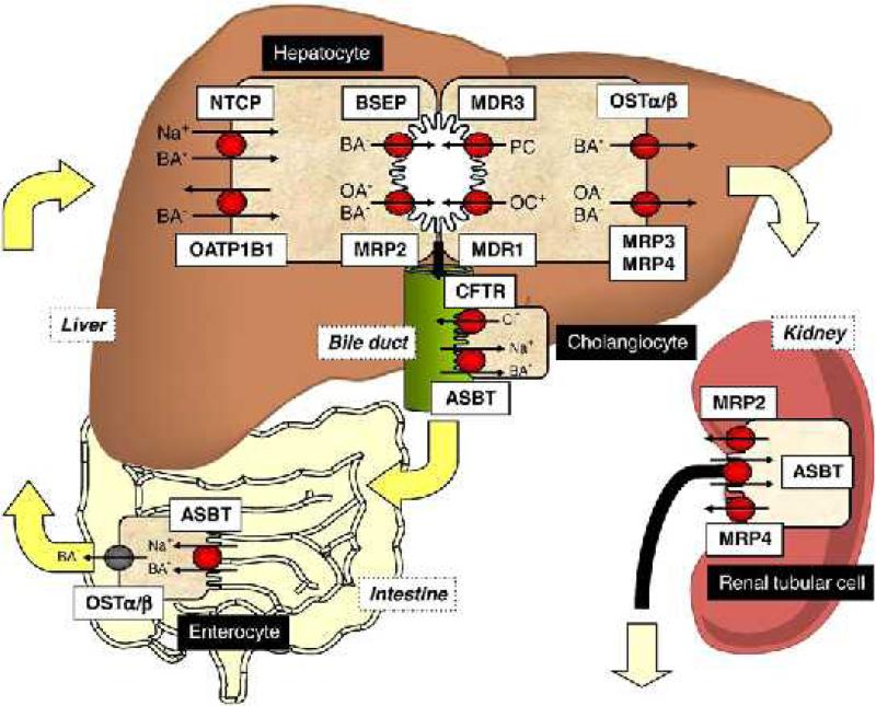
The transport of bile acids across the hepatocytes has been studied. (Figure 1) outlines the key transporters involved in bile acid transport in the hepatobilliary system. At the basolateral (sinusoidal) membrane, conjugated bile acids are transported from the portal blood supply into hepatocytes by the Sodium Taurocholate Co-transporting Polypeptide, NTCP (SLC10A1, human) (12). This process is aided by the basolateral Na/K/ATPase pump which creates an electrochemical gradient to draw in sodium and bile salt (13). Unconjugated bile acids are transported across the basolateral membrane by members of the Organic Anion Transport Proteins, OATP's (SLC21A6, human) (14). The basolateral membrane also has efflux systems for bile acids, including the Multidrug Resistance Proteins (MRP) and OSTalpha/OSTbeta which transport bile acids back into the circulation (13, 15). The rate limiting step in bile formation is the transport of bile acids into the biliary canaliculus (13). At the canalicular membrane, monovalent bile acids are exported by the Bile Salt Export Pump, BSEP (ABCB11, human) and divalent bile acids are exported by the Multi-Drug-Resistance-associated Protein 2, MRP2 (ABCC2, human) (16, 17) At the canalicular membrane of hepatocytes, transport of other substances is carried out by specific transporters. The Multi Drug Resistance Protein 3, MDR3, (ABCB4, human) transports phospholipids, ABCG5 and ABCG8 transports cholesterol and bilirubin is transported across the apical membrane by MRP1 and MRP2 (mouse) (18-20). The chloride-bicarbonate Anion Exchange Isoform 2, AE2, (SLC4A2, human) secretes bicarbonate (21). Water enters bile though diffusion across the tight junctions (22).
Figure 1.
Bile acid transporters in the hepatobilliary system, intestine and kidney (74). Bile acids are transported from portal blood into hepatocytes by NTCP and OATP. They are effluxed at the canalicular membrane by BSEP and MRP2. MRP3, MRP4 and OSTalpha/beta efflux bile acids at the basolateral membrane back to the systemic circulation. MDR3 transports phospholipids and MDR1 transports cations including drugs across the apical membrane. Bile acid reabsorption can occur at the level of the cholangiocyte, via ASBT, the terminal ileum via ASBT and within the enterocyte, bile acids are effluxed by OSTalpha/beta. Bile acids can also be reabsorbed in the proximal renal tubule via ASBT. Bile acids may be excreted into urine via MRP2 and MRP4. Reproduced with permission from (74).
Once bile enters the bile duct, it travels to the gallbladder and then is released and enters the small intestine. In the bile duct, the cystic fibrosis transmembrane regulator channel transports chloride and theAE2 channel is responsible for secretion of bicarbonate into bile to maintain pH and fluidity of bile (21). Bile acid reabsorption primarily occurs in the terminal ileum by the transporter ASBT (SLC10a2, human) into the enterocyte, where the ileal bile acid binding protein, I-Babp, binds the bile acids and plays a role in transcellular bile acid transport, and at the basolateral membrane, the OSTalpha/OSTbeta transporter exports bile acids into the portal circulation and back to the liver (23-26). Bile acids are also reabsorbed at the level of the bile duct, where apical sodium-dependent bile acid transporter ASBT transports bile acids into cholangiocytes and they are effluxed by OSTalpha/OSTbeta (27, 28)
Bile acids are returned to the liver for first pass clearance via uptake by the NTCP transporter at the basolateral membrane of hepatocytes. Some bile acids escape this and they are reabsorbed at the level of the kidney. Bile acids are reabsorbed in the proximal renal tubular cells by the apical transporter ASBT (human) and then enter the systemic circulation via the basolateral membrane transporter OSTalpha/OSTbeta (27, 29, 30).
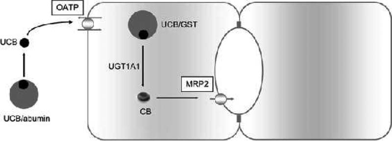
Bilirubin is an end product of heme metabolism and is the predominant bile pigment. Unconjugated bilirubin is primarily bound to albumin, and a small amount is bound to HDL (31). Unconjugated bilirubin at hepatocyte basolateral membrane dissociates from albumin, and is transported in the hepatocyte by OATP-2 (human) (32). Within the hepatocytes, bilirubin is bound to cytosolic glutathione-S-transferases, to prevent efflux. Unconjugated bilirubin is conjugated by UGT1A1. The now hydrophilic molecules of conjugated bilirubin are transported across the canalicular membrane into bile by the apical export pump, MRP2 (human) (33). (Figure 2) illustrates bilirubin metabolism and transport in the hepatocyte (70).
Figure 2.
Bilirubin metabolism and transport in the hepatocyte. At the hepatocyte basolateral membrane, unconjugated bilirubin (UCB) dissociates from albumin and is transported into the hepatocyte by OATP. Within the hepatocyte, UCB binds to glutathione-S-transferase and is conjugated by UGT1A1. The unconjugated bilirubin is transported across the canalicular membrane by the apical export pump, MRP2. Reproduced with permission from (75).
5. THE HEPATIC RESPONSE TO SEPSIS
The liver can be involved in several ways during sepsis. It may be a site of bacterial seeding and a primary focus of infection e.g. a hepatic abscess and biliary sepsis. The release of cytokines due to focal infection in the liver or due to the systemic release of cytokines also induces complex changes that activate inflammatory responses within the liver. Hepatic inflammation involves activation of sinusoidal endothelial cells, margination and migration of leukocytes in to the liver. Activation of resident macrophages is yet another source of cytokines that activate proinflammatory signaling within the liver. These have profound effects on the function of the liver and bile flow. Clinically this often manifests as cholestasis. The molecular basis for this is discussed below:
5.1. Hepatocellular and ductal mechanism of cholestasis
Cholestasis in sepsis can result from a defect in bile formation at the hepatocyte due to reduced transporter protein expression or impaired bile flow at the bile duct level. The molecular mechanisms of cholestasis in sepsis have been studied in animal models.
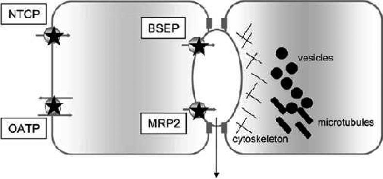
Lipopolysaccharide (LPS) induces Kupffer cells to release pro-inflammatory cytokines which lead to downregulation of transporters involved in bile flow, coordinated by nuclear receptors and transcription factors (34). The key transporters that are downregulated in sepsis include NTCP (mouse), and the cannalicular transporters, MRP2 (human) and bile salt export pump BSEP (human), as illustrated in (Figure 3) (35, 36).
Figure 3.
Molecular mechanisms of cholestasis in sepsis. In the setting of sepsis, bile flow is mainly impaired due to changes in bile acid transport. There is a downregulation of the basolateral bile acid transporters, NTCP and OATP and canalicular bile acid transporters, BSEP and MRP2. In addition, localization of transporters to the cell membrane may be disrupted due to changes in microtubules. Reproduced with permission from (75).
Endotoxin leads the production of inflammatory cytokines including TNFalpha and IL-1beta, which lead to a reduction in the activation of the NTCP (rat) promoter (36). NTCP (rat) expression is downregulated by decreased binding of the transcription factor Hnf1alpha and the RXRalpha:RARalpha heterodimer to the NTCP promoter (37). Endotoxin treatment also decreased Na-K-ATPase activity at the hepatocyte, which is important in the transport of bile acids across the sinusoidal membrane via the NTCP (rat/mouse) (36). There is also a reduction in basolateral OATP (rat) transporter expression (38). Canalicular transport proteins are also reduced in sepsis models. BSEP (rat) and MRP2 (rat) are reduced with LPS exposure (38). IL-1B has been demonstrated to downregulate the RXRalpha-RARalpha transactivators of the promoters for NTCP and MRP2 (in cell culture) (39). In addition to transcriptional modifications, transporter changes can occur due to change in transporter localization, as demonstrated by LPS induced MRP-2 retrieval from the canalicular membrane. Post transcriptional change has also been demonstrated as a mechanism to decrease MRP2 and BSEP in LPS exposure to human and rat liver specimens (35, 40).
In addition to the bile acid transporters, aquaporins, membrane water channels, also play a role in cholestasis. 95% of bile is water and thus aquaporins play an important role in bile production (41). In a rodent model, LPS reduced expression of hepatocyte canalicular AQP8. This was mediated by TNFalpha, and was a result of posttranscriptional downregulation of AQP8 expression (42). In addition to this, it has been demonstrated in a mouse model that leukocyte infiltration, mediated by p-selectin, plays a role in reduction of bile flow and hepatocellular apoptosis and necrosis in sepsis-associated cholestasis (43).
5.2. The role of nitric oxide in cholestasis-associated sepsis
Nitric oxide has also been implicated in playing a role in cholestasis in sepsis. High levels of nitric oxide may be harmful by promoting hypotension and decreased liver circulation. LPS exposure increases inducible nitric oxide synthase, iNOS, in Kupffer and endothelial cells (44). It has been demonstrated in cell culture that nitric oxide inhibits cAMP production by adenylyl cyclase which plays a key role in chloride and bicarbonate secretion by the CFTR and AE-2 transporters in the bile duct to maintain bile fluidity (45). Nitric oxide secretion also increases tight junction permeability in hepatocytes by decreasing zona occludens, key proteins to maintain tight junctions (46). Tight junctions are important in maintaining the osmotic gradient for bile production. Nitric oxide has also been demonstrated to impair bile canalicular contraction, which can lead to cholestasis (46, 47). Nitric oxide is a key player in sepsis associated cholestasis.
5.3. Secondary changes that promote sepsis and cholestasis
The accumulation of bile acids within hepatocytes may themselves contribute to cholestasis. Hydrophobic bile acids such as taurolithocholic acid induce cholestasis at least in part by inducing retraction of the NTCP (rodent) and MRP2 (rat) from the plasma membrane in to the cytosol (48, 49). These functions are mediated by activation of protein kinase c epsilon (48). This system is counterbalanced by the effects of cAMP and phosphoinositol 3 kinase mediated activation of protein kinase c delta which promotes translocation of these transporters to the plasma membrane (50). As cholestasis develops, the accumulation of bile acids may further worsen cholestasis by tilting this balance towards cytosolic retraction of these transporters.
In the intestine, the binding of bile acids to FXR has been found to have an anti-inflammatory effect (51). In experimental models, decreased FXR activation has been found to be associated with increased inflammation (51, 52). Under conditions of cholestasis, the intestinal bile acid concentrations decrease. Consequently, one could assume that there would be decreased FXR activation which would promote intestinal inflammation; if so, the intestinally-derived cytokines would be expected to activate the innate and adaptive immune systems and also promote a systemic pro-inflammatory reaction. This however remains to be experimentally verified.
5.4. Bacterial translocation in cholestasis
Bacterial translocation has been demonstrated in animal models of cholestasis (53, 54). In an animal model, cholestasis leads to structural changes in the enterocytes and increased space between enterocytes, which was associated with bacterial translocation (55). Similarly, increased intestinal permeability has been demonstrated in patients with cholestasis (56). Furthermore, bacterial translocation has been implicated as a precipitant of sepsis (57). Cholestasis may lead to bacterial translocation, which may cause sepsis and then inflammatory cytokines which lead to changes in bile acid formation and perpetuate the cycle of cholestasis. Cholestasis in sepsis is reversible once the infection has been treated.
5.5. Bilirubinostasis associated with cholestasis of sepsis
A common clinical manifestation of cholestasis is hyperbilirubinemia. This is typically due to increased levels of conjugated bilirubin. The rate-limiting step in bilirubin transport i.e. the canalicular secretion of conjugated bilirubin is mediated by the MRP2 transporter. The expression of MRP2 (human) is decreased by several pro-inflammatory cytokines such as IL-6 (58). Inflammation induced oxidative stress also alters the protein kinase A to protein kinase C activity. In turn, these cause retraction of MRP2 (rat) from the canalicular membrane to the cytosol thereby further decreasing the ability of the hepatocyte to transport bilirubin (59). Importantly, correction of this intrahepatic oxidative stress allows reversal of this process via both a microtubule-independent and a microtubule-dependent pathway regulated by protein kinases A and C (59).
6. ADAPTIVE MECHANISMS THAT LIMIT CHOLESTASIS AND INFLAMMATION IN THE LIVER
Cholestasis leads to adaptive protective responses in the hepatobiliary system. The adaptive responses have primarily been investigated in obstructive cholestasis, and include decreased bile acid production in the liver, increasing efflux of bile acids from hepatocytes with subsequent renal excretion and decreased bile acid reabsorption.
6.1. Bile acid synthesis
Bile acids are synthesized by two pathways from cholesterol. The classic pathway produces cholic acid (CA) and chenodeoxycholic acid (CDCA) and the rate limiting step in this cascade is cholesterol 7alpha-hydroxylase (CYP7A1). In the alternative pathway, the enzyme sterol 27-hydrozylase (CYP27A1) is the first enzyme responsible for the synthesis of CDCA. These key enzymes in bile acid synthesis are downregulated in cholestasis (60, 61). FXR, a nuclear bile acid receptor, plays a key role in the regulation of bile acid synthesis. It has been demonstrated that FXR induces FGF19, a protein that represses CYP7A1 (62). FGF19 is up-regulated in cholestasis, and this may be a player in adaptive responses to cholestasis to limit bile acid synthesis (63)
6.2. Hepatocellular bile acid transport
Basolateral bile acid uptake is reduced in sepsis, as evidenced by a reduction in the expression of NTCP and OATPs due to inflammatory cytokines. In addition, in cholestasis, basolateral bile acid efflux proteins are upregulated in cholestasis to prevent bile acid accumulation in hepatocytes. MRP3 (rat) and MRP4 (rat) are upregulated and efflux conjugated bile acids into the portal blood which can then be filtered and excreted by the kidneys (64, 65).
6.3. Bile acid hydroxylation and conjugation
In hepatocytes, unconjugated bile acids are hydroxylated by CYP3A4 and then conjugated with sulfate or glucuronidate by UDP-glucuronosyltransferases and dehydroepiandrosterone-sulfotransferase. In a rodent model of cholestasis, CYP3a11 (the rodent homologue of CYP3A4) is upregulated (66). Similarly, in cholestasis, an increase in the UDP-glucuronosyltransferases has been demonstrated (67). Through increased activity of hydroxylation and conjugation of bile acids, bile acids become less toxic and are able to be excreted into urine. The upregulation of the enzymes involved in detoxification of bile acids may be protective in cholestasis.
6.4. Bile acid reabsorption and excretion
In cholestasis, the small intestine and kidneys also play roles in eliminating bile acids. Both the terminal ileum and renal proximal tubule have the bile acid transporter ASBT (human, rat) and in cholestasis this transporter is downregulated in the small intestine and upregulated in the kidney, promoting bile acid excretion (29, 68). In addition, active renal tubular secretion via MRP2 (rat) and MRP4 (human) may also eliminate bile acids into the urine (69-71).
6.5. Adaptive mechanisms that reduce bilirubinostasis
Several nuclear hormone receptors have been shown to have important effects on the transport functions of the hepatocyte and integrate the metabolic functions of the liver with its transport functions. Two key receptors are the pregnane-X-receptor (PXR) and the constitutive androstane receptor (CAR). The PXR-CAR system can be activated by FXR which is activated by retained bile acids. The key targets of PXR-CAR include transporters for bilirubin such as MRP2, and PXR and CAR can stimulate bile acid hydroxylation enzymes Cyp3a11 and Cyp2b10 which make bile acids less toxic (72). Activation of PXR-CAR also allow a greater capacity for the liver to handle xenobiotics under conditions of stress via induction of cytochrome p450 3A (Cyp3A) (72). PXR can also be directly activated by lithocholic acid (73).
7. SUMMARY
In summary, sepsis induces a profound alteration in the hepatic ability to transport bile acids and bilirubin in to the hepatic canaliculi thereby causing cholestasis. The cholestatic response can itself trigger inflammatory responses within the liver to further exaggerate the cholestatic response. On the other hand, there are also adaptive mechanisms that come in to play that can ameliorate the cholestatic response. Better understanding of the balance between the cholestatic and adaptive mechanisms are likely to provide insights that will allow development of targeted therapy for sepsis induced cholestasis.
ACKNOWLEDGEMENTS
This manuscript is an original review paper and this work is not under consideration for publication elsewhere. The authors express their gratitude to Dr. Michael Fuchs for his constructive critique and assistance with manuscript development. Support: T32 DK 007150-35.
Footnotes
Conflicts of Interest: None to report for this manuscript
REFERENCES
- 1.Pols TW, Noriega LG, Nomura M, Auwerx J, Schoonjans K. The bile acid membrane receptor TGR5 as an emerging target in metabolism and inflammation. J Hepatol. 2011;54:1263–72. doi: 10.1016/j.jhep.2010.12.004. [DOI] [PMC free article] [PubMed] [Google Scholar]
- 2.Trauner M, Claudel T, Fickert P, Moustafa T, Wagner M. Bile acids as regulators of hepatic lipid and glucose metabolism. Dig Dis. 2010;28:220–4. doi: 10.1159/000282091. [DOI] [PubMed] [Google Scholar]
- 3.Swann JR, Want EJ, Geier FM, Spagou K, Wilson ID, Sidaway JE, Nicholson JK, Holmes E. Systemic gut microbial modulation of bile acid metabolism in host tissue compartments. Proc Natl Acad Sci U S A. 2011;108:4523–30. doi: 10.1073/pnas.1006734107. [DOI] [PMC free article] [PubMed] [Google Scholar]
- 4.Garcia FJ, Nager AL. Jaundice as an early diagnostic sign of urinary tract infection in infancy. Pediatrics. 2002;109(5):846–51. doi: 10.1542/peds.109.5.846. [DOI] [PubMed] [Google Scholar]
- 5.Whitehead MW, Hainsworth I, Kingham JG. The causes of obvious jaundice in South West Wales: perceptions versus reality. Gut. 2001;48:409–13. doi: 10.1136/gut.48.3.409. [DOI] [PMC free article] [PubMed] [Google Scholar]
- 6.Franson TR, LaBrecque DR, Buggy BP, Harris GJ, Hoffmann RG. Serial bilirubin determinations as a prognostic marker in clinical infections. Am J Med Sci. 1989;297:149–52. doi: 10.1097/00000441-198903000-00003. [DOI] [PubMed] [Google Scholar]
- 7.Bernstein J, Brown AK. Sepsis and jaundice in early infancy. Pediatrics. 1962;29:873–82. [PubMed] [Google Scholar]
- 8.Vermillion SE, Gregg JA, Baggenstoss AH, Bartholomew LG. Jaundice associated with bacteremia. Archives of Internal Medicine. 1969;124:611–618. [PubMed] [Google Scholar]
- 9.Miller DJ, Keeton DG, Webber BL, Pathol FF, Saunders SJ. Jaundice in severe bacterial infection. Gastroenterology. 1976;71:94–7. [PubMed] [Google Scholar]
- 10.Rooney JC, Hill DJ, Danks DM. Jaundice associated with bacterial infection in the newborn. Am J Dis Child. 1971;122:39–41. doi: 10.1001/archpedi.1971.02110010075012. [DOI] [PubMed] [Google Scholar]
- 11.Chand N, Sanyal AJ. Sepsis-induced cholestasis. Hepatology. 2007;45:230–41. doi: 10.1002/hep.21480. [DOI] [PubMed] [Google Scholar]
- 12.Hagenbuch B, Dawson P. The sodium bile salt cotransport family SLC10. Pflugers Arch. 2004;447:566–70. doi: 10.1007/s00424-003-1130-z. [DOI] [PubMed] [Google Scholar]
- 13.Trauner M, Boyer JL. Bile salt transporters: molecular characterization, function, and regulation. Physiol Rev. 2003;83:633–71. doi: 10.1152/physrev.00027.2002. [DOI] [PubMed] [Google Scholar]
- 14.Kullak-Ublick GA, B Hagenbuch, Stieger B, Schteingart CD, Hofmann AF, Wolkoff AW, Meier PJ. Molecular and functional characterization of an organic anion transporting polypeptide cloned from human liver. Gastroenterology. 1995;109:1274–82. doi: 10.1016/0016-5085(95)90588-x. [DOI] [PubMed] [Google Scholar]
- 15.Boyer JL, Trauner M, Mennone A, Soroka CJ, Cai SY, Moustafa T, Zollner G, Lee JY, Ballatori N. Upregulation of a basolateral FXR-dependent bile acid efflux transporter OSTalpha-OSTbeta in cholestasis in humans and rodents. Am J Physiol Gastrointest Liver Physiol. 2006;290:G1124–30. doi: 10.1152/ajpgi.00539.2005. [DOI] [PubMed] [Google Scholar]
- 16.Strautnieks SS, Bull LN, Knisely AS, Kocoshis SA, Dahl N, Arnell H, Sokal E, Dahan K, Childs S, Ling V, Tanner MS, Kagalwalla AF, Nemeth A, Pawlowska J, Baker A, Mieli-Vergani G, Freimer NB, Gardiner RM, Thompson RJ. A gene encoding a liver-specific ABC transporter is mutated in progressive familial intrahepatic cholestasis. Nat Genet. 1998;20:233–8. doi: 10.1038/3034. [DOI] [PubMed] [Google Scholar]
- 17.Paulusma CC, Oude Elferink RP. The canalicular multispecific organic anion transporter and conjugated hyperbilirubinemia in rat and man. J Mol Med (Berl) 1997;75:420–8. doi: 10.1007/s001090050127. [DOI] [PubMed] [Google Scholar]
- 18.Jedlitschky G, Leier I, Buchholz U, Hummel- Eisenbeiss J, Burchell B, Keppler D. ATP-dependent transport of bilirubin glucuronides by the multidrug resistance protein MRP1 and its hepatocyte canalicular isoform MRP2. Biochem J. 1997;327:305–10. doi: 10.1042/bj3270305. [DOI] [PMC free article] [PubMed] [Google Scholar]
- 19.Repa JJ, Berge KE, Pomajzl C, Richardson JA, Hobbs H, Mangelsdorf DJ. Regulation of ATP-binding cassette sterol transporters ABCG5 and ABCG8 by the liver X receptors alpha and beta. J Biol Chem. 2002;277:18793–800. doi: 10.1074/jbc.M109927200. [DOI] [PubMed] [Google Scholar]
- 20.Oude Elferink RP, Ottenhoff R, van Wijland M, Smit JJ, Schinkel AH, Groen AK. Regulation of biliary lipid secretion by mdr2 P-glycoprotein in the mouse. J Clin Invest. 1995;95:31–8. doi: 10.1172/JCI117658. [DOI] [PMC free article] [PubMed] [Google Scholar]
- 21.Martinez-Anso E, Castillo JE, Diez J, Medina JF, Prieto J. Immunohistochemical detection of chloride/bicarbonate anion exchangers in human liver. Hepatology. 1994;19:1400–6. [PubMed] [Google Scholar]
- 22.Arrese M, Trauner M. Molecular aspects of bile formation and cholestasis. Trends Mol Med. 2003;9(12):558–64. doi: 10.1016/j.molmed.2003.10.002. [DOI] [PubMed] [Google Scholar]
- 23.Dawson PA, Hubbert M, Haywood J, Craddock AL, Zerangue N, Christian WV, Ballatori N. The heteromeric organic solute transporter alpha-beta, Ostalpha-Ostbeta, is an ileal basolateral bile acid transporter. J Biol Chem. 2005;280:6960–8. doi: 10.1074/jbc.M412752200. [DOI] [PMC free article] [PubMed] [Google Scholar]
- 24.Gong YZ, Everett ET, Schwartz DA, Norris JS, Wilson FA. Molecular cloning, tissue distribution, and expression of a 14-kDa bile acid-binding protein from rat ileal cytosol. Proc Natl Acad Sci U S A. 1994;91:4741–5. doi: 10.1073/pnas.91.11.4741. [DOI] [PMC free article] [PubMed] [Google Scholar]
- 25.Wong MH, Oelkers P, Dawson PA. Identification of a mutation in the ileal sodium-dependent bile acid transporter gene that abolishes transport activity. J Biol Chem. 1995;270:27228–34. doi: 10.1074/jbc.270.45.27228. [DOI] [PubMed] [Google Scholar]
- 26.Wong MH, Oelkers P, Craddock AL, Dawson PA. Expression cloning and characterization of the hamster ileal sodium-dependent bile acid transporter. J Biol Chem. 1994;269:1340–7. [PubMed] [Google Scholar]
- 27.Ballatori N, Christian WV, Lee JY, Dawson PA, Soroka CJ, Boyer JL, Madejczyk MS, Li N. OSTalpha- OSTbeta: a major basolateral bile acid and steroid transporter in human intestinal, renal, and biliary epithelia. Hepatology. 2005;42:1270–9. doi: 10.1002/hep.20961. [DOI] [PubMed] [Google Scholar]
- 28.Lazaridis KN, Pham L, Tietz P, Marinelli RA, deGroen PC, Levine S, Dawson PA, LaRusso NF. Rat cholangiocytes absorb bile acids at their apical domain via the ileal sodium-dependent bile acid transporter. J Clin Invest. 1997;100:2714–21. doi: 10.1172/JCI119816. [DOI] [PMC free article] [PubMed] [Google Scholar]
- 29.Schlattjan JH, Winter C, Greven J. Regulation of renal tubular bile acid transport in the early phase of an obstructive cholestasis in the rat. Nephron Physiol. 2003;95:49–56. doi: 10.1159/000074330. [DOI] [PubMed] [Google Scholar]
- 30.Burckhardt G, Kramer W, Kurz G, Wilson FA. Photoaffinity labeling studies of the rat renal sodium bile salt cotransport system. Biochem Biophys Res Commun. 1987;143:1018–23. doi: 10.1016/0006-291x(87)90353-6. [DOI] [PubMed] [Google Scholar]
- 31.Goessling W, Zucker SD. Role of apolipoprotein D in the transport of bilirubin in plasma. Am J Physiol Gastrointest Liver Physiol. 2000;279:G356–65. doi: 10.1152/ajpgi.2000.279.2.G356. [DOI] [PubMed] [Google Scholar]
- 32.Cui Y, Konig J, Leier I, Buchholz U, Keppler D. Hepatic uptake of bilirubin and its conjugates by the human organic anion transporter SLC21A6. J Biol Chem. 2001;276:9626–30. doi: 10.1074/jbc.M004968200. [DOI] [PubMed] [Google Scholar]
- 33.Kamisako T, Leier I, Cui Y, Konig J, Buchholz U, Hummel-Eisenbeiss J, Keppler D. Transport of monoglucuronosyl and bisglucuronosyl bilirubin by recombinant human and rat multidrug resistance protein. 2 Hepatology. 1999;30:485–90. doi: 10.1002/hep.510300220. [DOI] [PubMed] [Google Scholar]
- 34.Geier A, Fickert P, Trauner M. Mechanisms of disease: mechanisms and clinical implications of cholestasis in sepsis. Nature clinical practice.Gastroenterology & hepatology. 2006;3:574–585. doi: 10.1038/ncpgasthep0602. [DOI] [PubMed] [Google Scholar]
- 35.Elferink MG, Olinga P, Draaisma AL, Merema MT, Faber KN, Slooff MJ, Meijer DK, Groothuis GM. LPSinduced downregulation of MRP2 and BSEP in human liver is due to a posttranscriptional process. Am J Physiol Gastrointest Liver Physiol. 2004;287(5):G1008–16. doi: 10.1152/ajpgi.00071.2004. [DOI] [PubMed] [Google Scholar]
- 36.Green RM, Beier D, Gollan JL. Regulation of hepatocyte bile salt transporters by endotoxin and inflammatory cytokines in rodents. Gastroenterology. 1996;111:193–198. doi: 10.1053/gast.1996.v111.pm8698199. [DOI] [PubMed] [Google Scholar]
- 37.Geier A, Dietrich CG, Voigt S, Kim SK, Gerloff T, Kullak-Ublick GA, Lorenzen J, Matern S, Gartung C. Effects of proinflammatory cytokines on rat organic anion transporters during toxic liver injury and cholestasis. Hepatology. 2003;38:345–54. doi: 10.1053/jhep.2003.50317. [DOI] [PubMed] [Google Scholar]
- 38.Cherrington NJ, Slitt AL, Li N, Klaassen CD. Lipopolysaccharide-mediated regulation of hepatic transporter mRNA levels in rats. Drug Metab Dispos. 2004;32:734–41. doi: 10.1124/dmd.32.7.734. [DOI] [PubMed] [Google Scholar]
- 39.Denson LA, Auld KL, Schiek DS, McClure MH, Mangelsdorf DJ, Karpen SJ. Interleukin-1beta suppresses retinoid transactivation of two hepatic transporter genes involved in bile formation. J Biol Chem. 2000;275:8835–43. doi: 10.1074/jbc.275.12.8835. [DOI] [PubMed] [Google Scholar]
- 40.Kubitz R, Wettstein M, Warskulat U, Haussinger D. Regulation of the multidrug resistance protein 2 in the rat liver by lipopolysaccharide and dexamethasone. Gastroenterology. 1999;116:401–10. doi: 10.1016/s0016-5085(99)70138-1. [DOI] [PubMed] [Google Scholar]
- 41.Lehmann GL, Larocca MC, Soria LR, Marinelli RA. Aquaporins: their role in cholestatic liver disease. World J Gastroenterol. 2008;14:7059–67. doi: 10.3748/wjg.14.7059. [DOI] [PMC free article] [PubMed] [Google Scholar]
- 42.Lehmann GL, Carreras FI, Soria LR, Gradilone SA, Marinelli RA. LPS induces the TNF-alpha-mediated downregulation of rat liver aquaporin-8: role in sepsisassociated cholestasis. Am J Physiol Gastrointest Liver Physiol. 2008;294:567–75. doi: 10.1152/ajpgi.00232.2007. [DOI] [PubMed] [Google Scholar]
- 43.Laschke MW, Menger MD, Wang Y, Lindell G, Jeppsson B, Thorlacius H. Sepsis-associated cholestasis is critically dependent on P-selectin-dependent leukocyte recruitment in mice. American journal of physiology.Gastrointestinal and liver physiology. 2007;292:G1396–402. doi: 10.1152/ajpgi.00539.2006. [DOI] [PubMed] [Google Scholar]
- 44.Aono K, Isobe K, Kiuchi K, Fan ZH, Ito M, Takeuchi A, Miyachi M, Nakashima I, Nimura Y. In vitro and in vivo expression of inducible nitric oxide synthase during experimental endotoxemia: involvement of other cytokines. J Cell Biochem. 1997;65:349–58. [PubMed] [Google Scholar]
- 45.Spirli C, Fabris L, Duner E, Fiorotto R, Ballardini G, Roskams T, Larusso NF, Sonzogni A, Okolicsanyi L, Strazzabosco M. Cytokine-stimulated nitric oxide production inhibits adenylyl cyclase and cAMP-dependent secretion in cholangiocytes. Gastroenterology. 2003;124:737–53. doi: 10.1053/gast.2003.50100. [DOI] [PubMed] [Google Scholar]
- 46.Han X, Fink MP, Uchiyama T, Yang R, Delude RL. Increased iNOS activity is essential for hepatic epithelial tight junction dysfunction in endotoxemic mice. Am J Physiol Gastrointest Liver Physio. 2004;286:G126–36. doi: 10.1152/ajpgi.00231.2003. [DOI] [PubMed] [Google Scholar]
- 47.Dufour JF, Turner TJ, Arias IM. Nitric oxide blocks bile canalicular contraction by inhibiting inositol trisphosphate-dependent calcium mobilization. Gastroenterology. 1995;108:841–9. doi: 10.1016/0016-5085(95)90459-x. [DOI] [PubMed] [Google Scholar]
- 48.Beuers U, Denk GU, Soroka CJ, Wimmer R, Rust C, Paumgartner G, Boyer JL. Taurolithocholic acid exerts cholestatic effects via phosphatidylinositol 3-kinasedependent mechanisms in perfused rat livers and rat hepatocyte couplets. J Biol Chem. 2003;278:17810–8. doi: 10.1074/jbc.M209898200. [DOI] [PubMed] [Google Scholar]
- 49.Geier A, Dietrich CG, Voigt S, Ananthanarayanan M, Lammert F, Schmitz A, Trauner M, Wasmuth HE, Boraschi D, Balasubramaniyan N, Suchy FJ, Matern S, Gartung C. Cytokine-dependent regulation of hepatic organic anion transporter gene transactivators in mouse liver. Am J Physiol Gastrointest Liver Physiol. 2005;289:G831–41. doi: 10.1152/ajpgi.00307.2004. [DOI] [PubMed] [Google Scholar]
- 50.Schonhoff CM, Gillin H, Webster CR, Anwer MS. Protein kinase Cdelta mediates cyclic adenosine monophosphate-stimulated translocation of sodium taurocholate cotransporting polypeptide and multidrug resistant associated protein 2 in rat hepatocytes. Hepatology. 2008;47:1309–16. doi: 10.1002/hep.22162. [DOI] [PubMed] [Google Scholar]
- 51.Vavassori P, Mencarelli A, Renga B, Distrutti E, Fiorucci S. The bile acid receptor FXR is a modulator of intestinal innate immunity. J Immunol. 2009;183:6251–61. doi: 10.4049/jimmunol.0803978. [DOI] [PubMed] [Google Scholar]
- 52.Gadaleta RM, van Erpecum KJ, Oldenburg B, Willemsen EC, Renooij W, Murzilli S, Klomp LW, Siersema PD, Schipper ME, Danese S, Penna G, Laverny G, Adorini L, Moschetta A, Mil SW. Farnesoid X receptor activation inhibits inflammation and preserves the intestinal barrier in inflammatory bowel disease. Gut. 2010;60:463–72. doi: 10.1136/gut.2010.212159. [DOI] [PubMed] [Google Scholar]
- 53.Parks RW, Clements WD, Pope C, Halliday MI, Rowlands BJ, Diamond T. Bacterial translocation and gut microflora in obstructive jaundice. J Anat. 1996;189:561–5. [PMC free article] [PubMed] [Google Scholar]
- 54.White JS, Hoper M, Parks RW, Clements WD, Diamond T. Patterns of bacterial translocation in experimental biliary obstruction. J Surg Res. 2006;132:80–4. doi: 10.1016/j.jss.2005.07.026. [DOI] [PubMed] [Google Scholar]
- 55.Parks RW, Stuart Cameron CH, Gannon CD, Pope C, Diamond T, Rowlands BJ. Changes in gastrointestinal morphology associated with obstructive jaundice. J Pathol. 2000;192:526–32. doi: 10.1002/1096-9896(2000)9999:9999<::AID-PATH787>3.0.CO;2-D. [DOI] [PubMed] [Google Scholar]
- 56.Welsh FK, Ramsden CW, MacLennan K, Sheridan MB, Barclay GR, Guillou PJ, Reynolds JV. Increased intestinal permeability and altered mucosal immunity in cholestatic jaundice. Ann Surg. 1998;227:205–12. doi: 10.1097/00000658-199802000-00009. [DOI] [PMC free article] [PubMed] [Google Scholar]
- 57.Van Leeuwen PA, Boermeester MA, Houdijk AP, Ferwerda CC, Cuesta MA, Meyer S, Wesdorp RI. Clinical significance of translocation. Gut. 1994;35:28–34. doi: 10.1136/gut.35.1_suppl.s28. [DOI] [PMC free article] [PubMed] [Google Scholar]
- 58.Vee ML, Lecureur V, Stieger B, Fardel O. Regulation of drug transporter expression in human hepatocytes exposed to the proinflammatory cytokines tumor necrosis factor-alpha or interleukin-6. Drug Metab Dispos. 2009;37:685–93. doi: 10.1124/dmd.108.023630. [DOI] [PubMed] [Google Scholar]
- 59.Sekine S, Ito K, Horie T. Canalicular Mrp2 localization is reversibly regulated by the intracellular redox status. Am J Physiol Gastrointest Liver Physiol. 2008;295:1035–41. doi: 10.1152/ajpgi.90404.2008. [DOI] [PubMed] [Google Scholar]
- 60.Matsuzaki Y, Bouscarel B, Ikegami T, Honda A, Doy M, Ceryak S, Fukushima S, Yoshida S, Shoda J, Tanaka N. Selective inhibition of CYP27A1 and of chenodeoxycholic acid synthesis in cholestatic hamster liver. Biochim Biophys Acta. 2002;1588:139–48. doi: 10.1016/s0925-4439(02)00157-6. [DOI] [PubMed] [Google Scholar]
- 61.T Li, Jahan A, Chiang JY. Bile acids and cytokines inhibit the human cholesterol 7 alpha-hydroxylase gene via the JNK/c-jun pathway in human liver cells. Hepatology. 2006;43:1202–10. doi: 10.1002/hep.21183. [DOI] [PMC free article] [PubMed] [Google Scholar]
- 62.Holt JA, Luo G, Billin AN, Bisi J, McNeill YY, Kozarsky KF, Donahee M, Wang DY, Mansfield TA, Kliewer SA, Goodwin B, Jones SA. Definition of a novel growth factor-dependent signal cascade for the suppression of bile acid biosynthesis. Genes Dev. 2003;17:1581–91. doi: 10.1101/gad.1083503. [DOI] [PMC free article] [PubMed] [Google Scholar]
- 63.Schaap FG, van der Gaag NA, Gouma DJ, Jansen PL. High expression of the bile salt-homeostatic hormone fibroblast growth factor 19 in the liver of patients with extrahepatic cholestasis. Hepatology. 2009;49:1228–35. doi: 10.1002/hep.22771. [DOI] [PubMed] [Google Scholar]
- 64.Donner MG, Keppler D. Up-regulation of basolateral multidrug resistance protein 3 (Mrp3) in cholestatic rat liver. Hepatology. 2001;34:351–9. doi: 10.1053/jhep.2001.26213. [DOI] [PubMed] [Google Scholar]
- 65.Denk GU, Soroka CJ, Takeyama Y, Chen WS, Schuetz JD, Boyer JL. Multidrug resistance-associated protein 4 is up-regulated in liver but down-regulated in kidney in obstructive cholestasis in the rat. J Hepatol. 2004;40:585–91. doi: 10.1016/j.jhep.2003.12.001. [DOI] [PubMed] [Google Scholar]
- 66.Cho JY, Matsubara T, Kang DW, Ahn SH, Krausz KW, Idle JR, Luecke H, Gonzalez FJ. Urinary metabolomics in Fxr-null mice reveals activated adaptive metabolic pathways upon bile acid challenge. J Lipid Res. 2010;51:1063–74. doi: 10.1194/jlr.M002923. [DOI] [PMC free article] [PubMed] [Google Scholar]
- 67.Hasegawa Y, Kishimoto S, Takahashi H, Inotsume N, Takeuchi Y, Fukushima S. Altered expression of nuclear receptors affects the expression of metabolic enzymes and transporters in a rat model of cholestasis. Biol Pharm Bull. 2009;32:2046–52. doi: 10.1248/bpb.32.2046. [DOI] [PubMed] [Google Scholar]
- 68.Hruz P, Zimmermann C, Gutmann H, Degen L, Beuers U, Terracciano L, Drewe J, Beglinger C. Adaptive regulation of the ileal apical sodium dependent bile acid transporter (ASBT) in patients with obstructive cholestasis. Gut. 2006;55:395–402. doi: 10.1136/gut.2005.067389. [DOI] [PMC free article] [PubMed] [Google Scholar]
- 69.Wagner M, Trauner M. Transcriptional regulation of hepatobiliary transport systems in health and disease: implications for a rationale approach to the treatment of intrahepatic cholestasis. Ann Hepatol. 2005;4:77–99. [PubMed] [Google Scholar]
- 70.van Aubel RA, Smeets PH, Peters JG, Bindels RJ, Russel FG. The MRP4/ABCC4 gene encodes a novel apical organic anion transporter in human kidney proximal tubules: putative efflux pump for urinary cAMP and cGMP. J Am Soc Nephrol. 2002;13:595–603. doi: 10.1681/ASN.V133595. [DOI] [PubMed] [Google Scholar]
- 71.Schaub P, Kartenbeck J, Konig J, Vogel O, Witzgall R, Kriz W, Keppler D. Expression of the conjugate export pump encoded by the mrp2 gene in the apical membrane of kidney proximal tubules. J Am Soc Nephrol. 1997;8:1213–21. doi: 10.1681/ASN.V881213. [DOI] [PubMed] [Google Scholar]
- 72.Wagner M, Halilbasic E, Marschall HU, Zollner G, Fickert P, Langner C, Zatloukal K, Denk H, Trauner M. CAR and PXR agonists stimulate hepatic bile acid and bilirubin detoxification and elimination pathways in mice. Hepatology. 2005;42:420–30. doi: 10.1002/hep.20784. [DOI] [PubMed] [Google Scholar]
- 73.Staudinger JL, Goodwin B, Jones SA, Hawkins-Brown D, MacKenzie KI, LaTour A, Liu Y, Klaassen CD, Brown KK, Reinhard J, Willson TM, Koller BH, Kliewer SA. The nuclear receptor PXR is a lithocholic acid sensor that protects against liver toxicity. Proc Natl Acad Sci U S A. 2001;98:3369–74. doi: 10.1073/pnas.051551698. [DOI] [PMC free article] [PubMed] [Google Scholar]
- 74.Zollner G, Wagner M, Trauner M. Nuclear receptors as drug targets in cholestasis and druginduced hepatotoxicity. Pharmacol Ther. 2010;126:228–43. doi: 10.1016/j.pharmthera.2010.03.005. [DOI] [PubMed] [Google Scholar]
- 75.Fuchs M, Sanyal AJ. Sepsis and cholestasis. Clinics in liver disease. 2008;12:151–72. doi: 10.1016/j.cld.2007.11.002. [DOI] [PubMed] [Google Scholar]


