Abstract
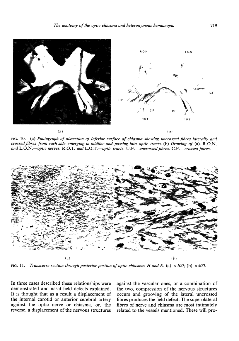
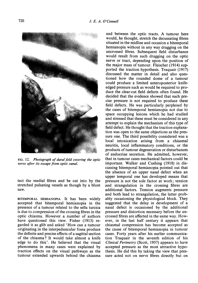
The gross anatomy of the optic nerves and chiasma has been studied, and differences in the tension in the crossed and uncrossed fibres after chiasmal displacement have been investigated. The anterior and posterior attachments of the medial and lateral fibres of the nerves have been studied. The chiasma has been dissected under low power microscopy and a three dimensional picture of it developed. Bitemporal hemianopia, as well as associated or independent hemianopic scotomata, results from stretching of the crossing fibres in the chiasma. Binasal hemianopia results from compression of the uncrossed fibres in the optic nerve or chiasma by the anterior cerebral or internal carotid arteries. The compression is effective because it is sharply localized and, probably as a result of pulsation, deeply grooves the nerve with a resulting acute distortion of fibres; it is likely that the lax lateral fibres would be less affected by a more widely spread compression. When this defect develops on top of an existing bitemporal hemianopia, it is believed that its usual cause remains the same. The crossed and uncrossed fibres of the optic chiasma differ not only anatomically in the areas of retina in which they arise but also physically. Tension is the force which occasions bitemporal hemianopia and pressure that which produces nasal field defects.
Full text
PDF


Images in this article
Selected References
These references are in PubMed. This may not be the complete list of references from this article.
- BULL J. The normal variations in the position of the optic recess of the third ventricle. Acta radiol. 1956 Jul-Aug;46(1-2):72–80. doi: 10.3109/00016925609170814. [DOI] [PubMed] [Google Scholar]
- DAWSON B. H. The blood vessels of the human optic chiasma and their relation to those of the hypophysis and hypothalamus. Brain. 1958 Jun;81(2):207–217. doi: 10.1093/brain/81.2.207. [DOI] [PubMed] [Google Scholar]
- GAMBLE H. J. COMPARATIVE ELECTRON-MICROSCOPIC OBSERVATIONS ON THE CONNECTIVE TISSUES OF A PERIPHERAL NERVE AND A SPINAL NERVE ROOT IN THE RAT. J Anat. 1964 Jan;98:17–26. [PMC free article] [PubMed] [Google Scholar]
- Kupfer C., Chumbley L., Downer J. C. Quantitative histology of optic nerve, optic tract and lateral geniculate nucleus of man. J Anat. 1967 Jun;101(Pt 3):393–401. [PMC free article] [PubMed] [Google Scholar]
- O'Connell J. E., Du Boulay E. P. Binasal hemianopia. J Neurol Neurosurg Psychiatry. 1973 Oct;36(5):697–709. doi: 10.1136/jnnp.36.5.697. [DOI] [PMC free article] [PubMed] [Google Scholar]
- O'connell J. E. The Intraneural Plexus and its Significance. J Anat. 1936 Jul;70(Pt 4):468–497. [PMC free article] [PubMed] [Google Scholar]
- Traquair H. M. BITEMPORAL HEMIOPIA: THE LATER STAGES AND THE SPECIAL FEATURES OF THE SCOTOMA: With an examination of current theories of the mechanism of production of the field defects. Br J Ophthalmol. 1917 May;1(5):281–294. doi: 10.1136/bjo.1.5.281. [DOI] [PMC free article] [PubMed] [Google Scholar]


