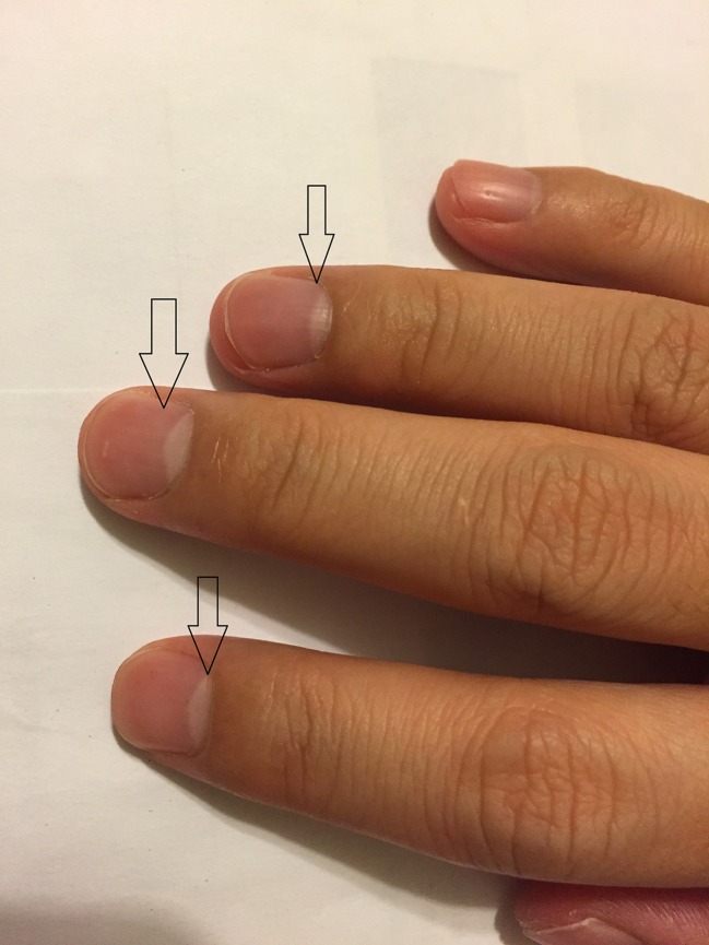
A 66-year-old man with diabetes and cirrhosis due to chronic hepatitis C infection (HCV RNA >1,000,000 IU/mL) developed a creatinine rise from 0.5 mg /deciliter to 2.6 mg/deciliter. The patient’s fingernails demonstrated significant changes compared to a normal fingernail, with a white band (lunula) occupying more than 50 % of the nail bed proximally, suggestive of Lindsay’s nail. (Figs. 1 and 2) Renal biopsy demonstrated membranoproliferative glomerulopathy and he was started on hemodialysis.
Figure 1.
Photograph demonstrating Lindsay’s nail or half-and-half nail characterized by proximal nail bed whiteness and distal nail bed red, pink or brown band occupying 20 % to 60 % of nail bed.
Figure 2.
Photograph demonstrating normal fingernail in healthy individual.
A clinical differentiation between Lindsay’s nail (half-and-half nail) and Terry’s nail is difficult. In Lindsay’s nail, the proximal part of the nail is white, while the distal portion occupying 20 % to 60 % of nail bed is reddish-brown and does not fade with pressure.1,2 The cause of Lindsay’s nail is unclear, but the distal reddish-brown band might be the result of an increased concentration of β-melanocyte–stimulating hormone.2 This condition can be found in up to 40 % of patients of chronic kidney disease.1 On the other hand, Terry’s nail is defined as a 0.5–3.0 mm brown to pink distal band with proximal nail bed whiteness occupying approximately 80 % of nail bed.3 This condition is frequently associated with cirrhosis, chronic congestive heart failure, and adult-onset diabetes mellitus.3
Compliance with Ethical Standards
Conflict of Interest
The authors declare that they do not have a conflict of interest.
REFERENCES
- 1.Lindsay PG. The half-and-half nail. Arch Intern Med. 1967;119(6):583–587. doi: 10.1001/archinte.1967.00290240105007. [DOI] [PubMed] [Google Scholar]
- 2.Dermatological Manifestations of Kidney Disease. Place of Publication Not Identified: Springer, 2015. Print.
- 3.Holzberg M, Walker HK. Terry’s nails: revised definition and new correlations. Lancet. 1984;1(8382):896–899. doi: 10.1016/S0140-6736(84)91351-5. [DOI] [PubMed] [Google Scholar]


