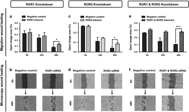
Figure 3.
ROR1 and ROR2 silencing inhibits wound healing migration. (a) Wound healing cell migration is significantly decreased following ROR1 knockdown in A2780-cis cells. Results represent an average of three experiments. Error bars represent the s.d. of the mean. *P<0.05. (b) Representative images of wound healing analysis showing dark shading as ‘open image area' where no cells are present. Increased dark shading is seen in the ROR1 knockdown cells after 48 h incubation. (c) Cell migration is significantly decreased following ROR2 knockdown in A2780-cis cells. Results represent an average of three experiments. Error bars represent the s.d. of the mean. *P<0.05. (d) Representative images of wound healing analysis showing dark shading as ‘open image area' where no cells are present. Increased dark shading is seen in the ROR2 knockdown cells after 48 h incubation. (e) Cell migration is most significantly decreased following double ROR1 and ROR2 knockdown in A2780-cis cells. Results represent an average of three experiments. Error bars represent the s.d. of the mean. ***P<0.001. (f) Representative images of wound healing analysis showing dark shading as ‘open image area' where no cells are present. The darkest shading is seen in the double ROR1 and ROR2 knockdown cells after 48 h incubation.

