

Abstract
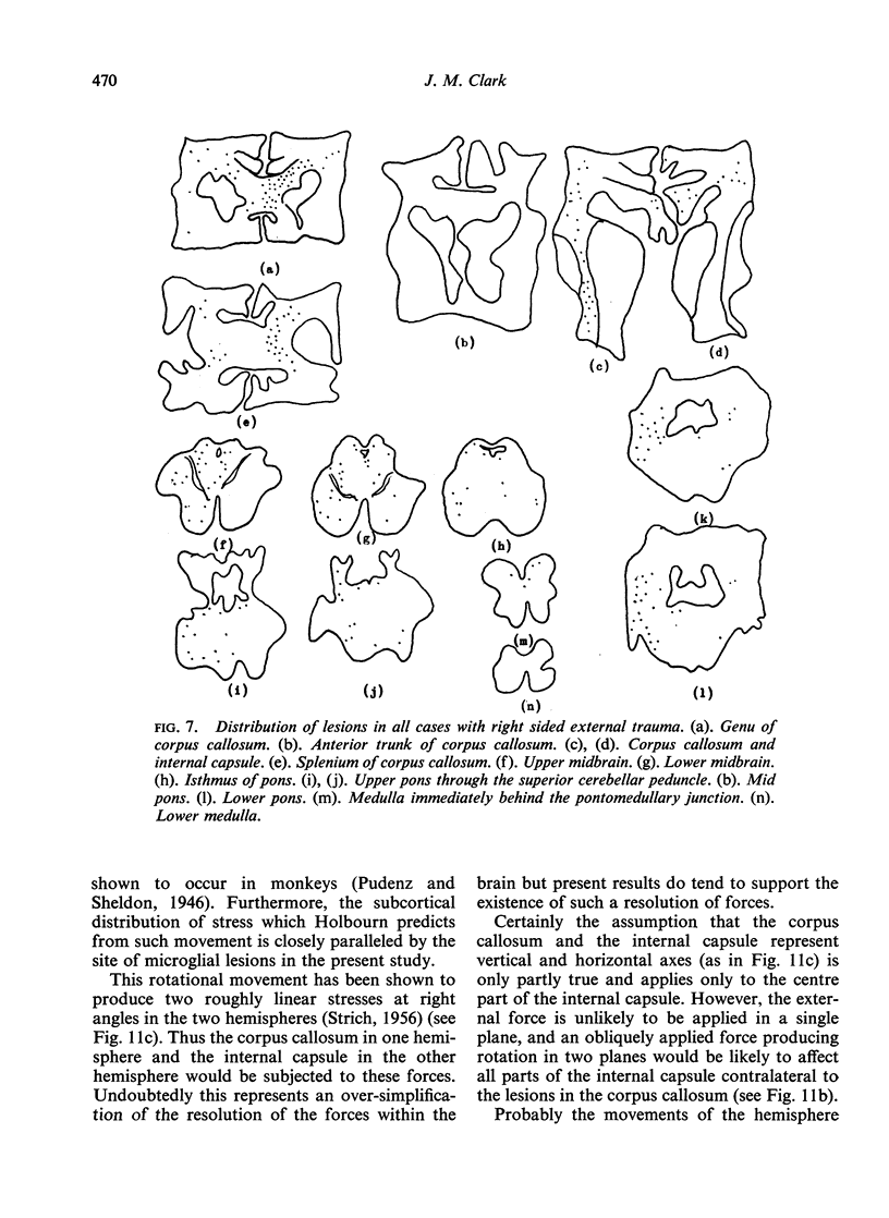
The brains of 12 cases of head injury have been submitted to gross pathological study and microscopic examination by the Weil-Davenport method, with special reference to the corpus callosum, internal capsules, and brain-stem in each case. Microglial clusters were observed in 11 out of 12 cases, the most common sites for these being the corpus callosum ipsilateral to the external applied force and the internal capsule and brain-stem contralateral to this applied force. This pattern of distribution of lesions remained constant in all cases. The nature, aetiology, and distribution of these lesions is discussed and it is concluded that such lesions arise from the formation of definite patterns of shearing forces which snap axons. These forces arise from the rotational movements set up within the skull resulting from the relative delay of movement of the brain with respect to the skull and dura mater.
Full text
PDF


Images in this article


Selected References
These references are in PubMed. This may not be the complete list of references from this article.
- Lindenberg R., Freytag E. Brainstem lesions characteristic of traumatic hyperextension of the head. Arch Pathol. 1970 Dec;90(6):509–515. [PubMed] [Google Scholar]
- Oppenheimer D. R. Microscopic lesions in the brain following head injury. J Neurol Neurosurg Psychiatry. 1968 Aug;31(4):299–306. doi: 10.1136/jnnp.31.4.299. [DOI] [PMC free article] [PubMed] [Google Scholar]
- STRICH S. J. Diffuse degeneration of the cerebral white matter in severe dementia following head injury. J Neurol Neurosurg Psychiatry. 1956 Aug;19(3):163–185. doi: 10.1136/jnnp.19.3.163. [DOI] [PMC free article] [PubMed] [Google Scholar]