

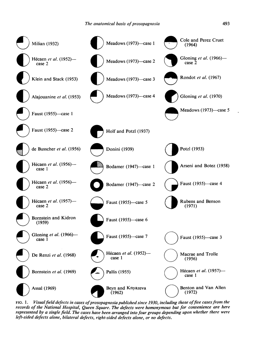
Abstract
Evidence is presented that patients with prosopagnosia have right anterior inferior occipital lesions in the region of the occipital temporal junction. Many if not all cases have an additional lesion in the left hemisphere; this is often but apparently not always symmetrical with the right hemisphere lesion. This evidence is discussed in relation to the anatomical connections of these regions and the results of experiments in animals.
Full text
PDF
Images in this article
Selected References
These references are in PubMed. This may not be the complete list of references from this article.
- ALAJOUANINE T., BOUCHET M., PIALOUX P., LHERMITTE F. La paralysie dilatateurs de la glotte dans la sclérose latérale amyotrophique. Rev Neurol (Paris) 1953;89(2):157–158. [PubMed] [Google Scholar]
- Assal G. Régression des troubles de la reconnaissance des physionomies et de la mémoire topographique chez un malade opéré d'un hématome intracérébral pariéto-temporal droit. Rev Neurol (Paris) 1969 Aug;121(2):184–185. [PubMed] [Google Scholar]
- BODAMER J. Die Prosop-Agnosie; die Agnosie des Physiognomieerkennens. Arch Psychiatr Nervenkr Z Gesamte Neurol Psychiatr. 1947;118(1-2):6–53. doi: 10.1007/BF00352849. [DOI] [PubMed] [Google Scholar]
- BORNSTEIN B., KIDRON D. P. Prosopagnosia. J Neurol Neurosurg Psychiatry. 1959 May;22(2):124–131. doi: 10.1136/jnnp.22.2.124. [DOI] [PMC free article] [PubMed] [Google Scholar]
- Benson D. F., Greenberg J. P. Visual form agnosia. A specific defect in visual discrimination. Arch Neurol. 1969 Jan;20(1):82–89. doi: 10.1001/archneur.1969.00480070092010. [DOI] [PubMed] [Google Scholar]
- Benton A. L., Van Allen M. W. Prosopagnosia and facial discrimination. J Neurol Sci. 1972 Feb;15(2):167–172. doi: 10.1016/0022-510x(72)90004-4. [DOI] [PubMed] [Google Scholar]
- Bornstein B., Sroka H., Munitz H. Prosopagnosia with animal face agnosia. Cortex. 1969 Jun;5(2):164–169. doi: 10.1016/s0010-9452(69)80027-4. [DOI] [PubMed] [Google Scholar]
- Critchley M. Acquired anomalies of colour perception of central origin. Brain. 1965 Nov;88(4):711–724. doi: 10.1093/brain/88.4.711. [DOI] [PubMed] [Google Scholar]
- DE BUSSCHER J., HOFFMANN G., KLUYSKENS J. Agnosie visuelle temporaire pour les personnes et optico-spatiale pour les objets à la suite d'un ictus unique. Acta Neurol Psychiatr Belg. 1956 Mar;56(3):162–176. [PubMed] [Google Scholar]
- De Renzi E., Spinnler H. Facial recognition in brain-damaged patients. An experimental approach. Neurology. 1966 Feb;16(2):145–152. doi: 10.1212/wnl.16.2_part_1.145. [DOI] [PubMed] [Google Scholar]
- Garland H., Pearce J. Neurological complications of carbon monoxide poisoning. Q J Med. 1967 Oct;36(144):445–455. [PubMed] [Google Scholar]
- Geschwind N. Disconnexion syndromes in animals and man. II. Brain. 1965 Sep;88(3):585–644. doi: 10.1093/brain/88.3.585. [DOI] [PubMed] [Google Scholar]
- Gloning I., Gloning K., Jellinger K., Quatember R. A case of "prosopagnosia" with necropsy findings. Neuropsychologia. 1970 Apr;8(2):199–204. doi: 10.1016/0028-3932(70)90007-2. [DOI] [PubMed] [Google Scholar]
- Gross C. G., Rocha-Miranda C. E., Bender D. B. Visual properties of neurons in inferotemporal cortex of the Macaque. J Neurophysiol. 1972 Jan;35(1):96–111. doi: 10.1152/jn.1972.35.1.96. [DOI] [PubMed] [Google Scholar]
- HECAEN H., ANGELERGUES R. Agnosia for faces (prosopagnosia). Arch Neurol. 1962 Aug;7:92–100. doi: 10.1001/archneur.1962.04210020014002. [DOI] [PubMed] [Google Scholar]
- HECAEN H., ANGELERGUES R., BERNHARDT C., CHIARELLI J. Essai de distinction des modalités cliniques de l'agnosie des physionomies. Rev Neurol (Paris) 1957 Feb;96(2):125–144. [PubMed] [Google Scholar]
- HECAEN H., DE AJURIAGUERRA J., MAGIS C., ANGELERGUES R. Le problème de l'agnosie des physionomies. Encephale. 1952;41(4):322–355. [PubMed] [Google Scholar]
- HECAEN H., PENFIELD W., BERTRAND C., MALMO R. The syndrome of apractognosia due to lesions of the minor cerebral hemisphere. AMA Arch Neurol Psychiatry. 1956 Apr;75(4):400–434. [PubMed] [Google Scholar]
- HUBEL D. H., WIESEL T. N. RECEPTIVE FIELDS AND FUNCTIONAL ARCHITECTURE IN TWO NONSTRIATE VISUAL AREAS (18 AND 19) OF THE CAT. J Neurophysiol. 1965 Mar;28:229–289. doi: 10.1152/jn.1965.28.2.229. [DOI] [PubMed] [Google Scholar]
- HUBEL D. H., WIESEL T. N. Receptive fields, binocular interaction and functional architecture in the cat's visual cortex. J Physiol. 1962 Jan;160:106–154. doi: 10.1113/jphysiol.1962.sp006837. [DOI] [PMC free article] [PubMed] [Google Scholar]
- Hubel D. H., Wiesel T. N. Receptive fields and functional architecture of monkey striate cortex. J Physiol. 1968 Mar;195(1):215–243. doi: 10.1113/jphysiol.1968.sp008455. [DOI] [PMC free article] [PubMed] [Google Scholar]
- KLEIN R., STACK J. J. Visual agnosia and alternating dominance; analysis of a case. J Ment Sci. 1953 Oct;99(417):749–762. doi: 10.1192/bjp.99.417.749. [DOI] [PubMed] [Google Scholar]
- Levy J., Trevarthen C., Sperry R. W. Reception of bilateral chimeric figures following hemispheric deconnexion. Brain. 1972;95(1):61–78. doi: 10.1093/brain/95.1.61. [DOI] [PubMed] [Google Scholar]
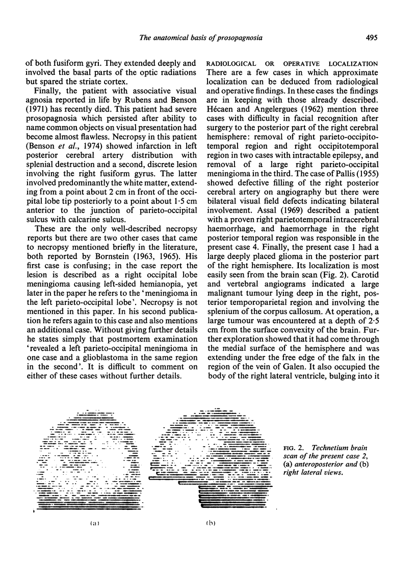
- Lhermitte F., Chain F., Escourolle R., Ducarne B., Pillon B. Etude anatomo-clinique d'un cas de prosopagnosie. Rev Neurol (Paris) 1972 May;126(5):329–346. [PubMed] [Google Scholar]
- MACRAE D., TROLLE E. The defect of function in visual agnosia. Brain. 1956 Mar;79(1):94–110. doi: 10.1093/brain/79.1.94. [DOI] [PubMed] [Google Scholar]
- POTZL O. Zur Agnosie des Physiogomiegedächtnisses. Wien Z Nervenheilkd Grenzgeb. 1953;6(4):335–354. [PubMed] [Google Scholar]
- Rubens A. B., Benson D. F. Associative visual agnosia. Arch Neurol. 1971 Apr;24(4):305–316. doi: 10.1001/archneur.1971.00480340037003. [DOI] [PubMed] [Google Scholar]
- Taylor A., Warrington E. K. Visual agnosia: a single case report. Cortex. 1971 Jun;7(2):152–161. doi: 10.1016/s0010-9452(71)80011-4. [DOI] [PubMed] [Google Scholar]
- Tzavaras A., Hécaen H., Le Bras H. Le probleme de la specificite du deficit de la reconnaissance du visage humain lors des lesions hemispheriques unilateales. Neuropsychologia. 1970 Nov;8(4):403–416. doi: 10.1016/0028-3932(70)90037-0. [DOI] [PubMed] [Google Scholar]
- Yin R. K. Face recognition by brain-injured patients: a dissociable ability? Neuropsychologia. 1970 Nov;8(4):395–402. doi: 10.1016/0028-3932(70)90036-9. [DOI] [PubMed] [Google Scholar]