Abstract
Teriparatide (TPTD) is the only currently available therapeutic agent that increases the formation of new bone tissue and can provide some remediation of the architectural defects in the osteoporotic skeleton. The use of teriparatide clinically is limited to 24 months. We review clinical findings during daily teriparatide treatment over time. Teriparatide appears to increase bone formation more than bone resorption as determined biochemically and histologically. Teriparatide exerts its positive effects on bone formation in two distinct fashions. The first is direct stimulation of bone formation that occurs within active remodeling sites (remodeling-based bone formation) and on surfaces of bone previously inactive (modeling-based bone formation). The second is an increase in the initiation of new remodeling sites. Both processes contribute to the final increase in bone density observed by non-invasive tools such as DXA. Remodeling is the repair process by which skeletal tissue is maintained in a young healthy state, and when stimulated by TPTD is associated with a positive bone balance within each remodeling cavity. It seems likely therefore that this component will contribute to the anti-fracture efficacy of TPTD. Teriparatide reduces the risk of fracture, and this effect appears to increase with longer duration of therapy. The use of novel treatment regimens, including shorter courses, should be held in abeyance until controlled clinical trials are completed to define the relative fracture benefits of such approaches in comparison to the 24-month daily use of the agent.
Summary In patients with osteoporosis at high risk for fracture, the full continuous 24-month course with teriparatide results in improved skeletal health and outcomes than shorter time periods.
Keywords: Bone histomorphometry, Fracture, Osteoporosis, Teriparatide
Introduction
In post-menopausal women, osteoporosis is the result of excessive bone resorption not accompanied by equally increased bone formation. Namely, targeted remodeling removes a volume of bone which is not fully compensated. The resulting volume deficit increases strain in neighboring bone [1]. Accordingly, pharmacologic treatment of this disease has focused on either reducing bone resorption or increasing bone formation. Various anti-resorptive treatments for osteoporosis are available, while teriparatide (TPTD) (recombinant human parathyroid hormone, rhPTH) [1–34] is the first approved anabolic agent that stimulates osteoblastic bone formation to improve bone quality and bone mass [2]. Depending on geography, teriparatide has been approved to treat post-menopausal women with osteoporosis, men with hypogonadal or idiopathic osteoporosis, and men and women with glucocorticoid-induced osteoporosis who are at high risk for fracture [3].
In the teriparatide phase 3 Fracture Prevention Trial [4], the planned duration of treatment with teriparatide versus placebo was 36 months, but the study was stopped early because of rat toxicology findings of osteosarcoma [5, 6]. Thus, the Fracture Prevention Trial analyzed the effects of a median 19 months of teriparatide versus placebo and the maximum duration of teriparatide treatment was 24 months [4]. As a consequence, the approved lifetime duration of treatment with teriparatide is 24 months [3]. Because teriparatide is approved in patients at high risk for fracture who are generally of increased clinical concern, the correct use of teriparatide may be important to achieve optimal outcomes. There are considerations that might suggest a shorter course, including the cost of the medication, the requirement for daily subcutaneous injections, and a perceived lack of certainty about whether a longer course might confer greater benefits than a shorter course.
Because there is some uncertainty about how long patients should be treated with teriparatide to achieve the best outcomes, we were motivated to review the available information regarding continuous therapy with teriparatide for 24 months. We reviewed the effect of teriparatide on biochemical markers of bone turnover. Also, we reviewed the long-term effects of teriparatide on human bone tissue, including the ability to stimulate the overfilling of resorption sites, an important effect in the setting of high bone resorption. Furthermore, we reviewed bone imaging techniques indicating continuous bone formation effects and increasing bone mass during continuous teriparatide treatment. Additionally, we reviewed the clinical data on fracture outcomes during the 24-month treatment course. Finally, we reviewed the long-term safety information for teriparatide.
Methods
The authors identified references through literature searches using the search word teriparatide along with knowledge of the field and continuous monitoring of the teriparatide literature during the past 20 years. On October 15, 2015, a search of Pubmed with the terms teriparatide and “24 months” revealed 62 references, and a search with teriparatide and “2 years” revealed 38 references. All of the relevant references from these searches are included in this review. Additionally, some previously unpublished information and images are provided as indicated.
Biochemical markers of bone turnover
Biochemical markers of bone turnover have been studied in many teriparatide clinical trials. The reference standard of biochemical marker of bone formation is procollagen type I N-terminal propeptide (PINP) [7, 8], and the effects of teriparatide on this marker have recently been reviewed [9]. Serum C-terminal cross-linking telopeptide of type I collagen (βCTXI [CTX]) is considered the reference standard for bone resorption [7].
During the first months of teriparatide treatment, there is a rapid rise in biochemical markers of bone formation, reflecting stimulation of osteoid formation by bone multicellular units (BMUs) in their formation phase, direct modeling-based bone formation, or both, without an accompanying increase in bone resorption, so bone formation exceeds bone resorption by a large margin early during the treatment course [10, 11]. Subsequently, serum levels of PINP and CTX peak after 6–12 months of TPTD therapy, followed by a gradual decrease in both processes [12–16].
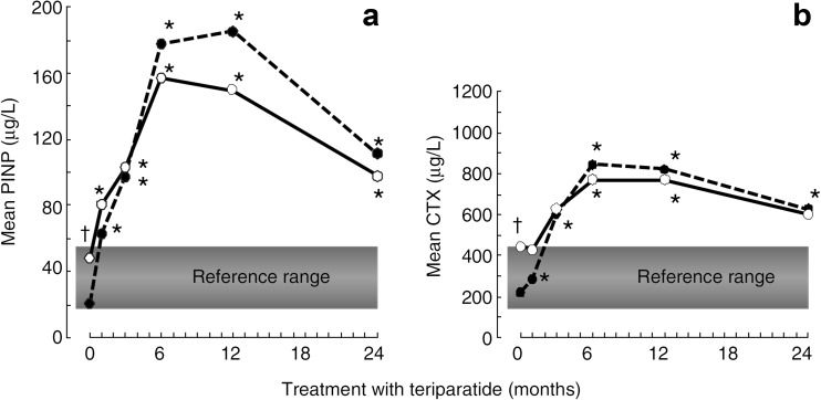
Although most studies of teriparatide have measured biochemical markers of bone turnover for less than 24 months, some studies have included measurements of biochemical markers of bone turnover during a full 24-month treatment course. For example, in a study of post-menopausal women with osteoporosis, increases in PINP exceeded increases in CTX during the entire 24-month treatment course, both in women previously treated with alendronate and in women who were osteoporosis drug naïve (Fig. 1) [15]. In another study of post-menopausal women treated with teriparatide for 24 months, the increase in bone formation markers PINP and osteocalcin exceeded the increase in CTX throughout the 24-month treatment course [16, 17]. Most compellingly, in a study of patients with glucocorticoid-induced osteoporosis, increases in biochemical markers of bone formation exceeded those of resorption during a full 36 months of treatment [14]. Thus, available clinical trial data shows that bone formation exceeds bone resorption during the full 24 months of teriparatide treatment. Another interpretation of these data is that the anabolic effect of teriparatide is maximal early during treatment when formation is increased and resorption is not. However, if teriparatide results in overfilling of resorption sites, then ongoing teriparatide even in the setting of high levels of bone resorption will result not only in replacement of old bone with a similar amount of new healthy bone but will result in ongoing anabolism [18].
Fig. 1.
Changes in biochemical markers of bone turnover in patients treated with teriparatide. Solid lines indicate treatment-naïve patients (n = 16), and dotted lines indicate alendronate-pre-treated patients (n = 29). X axes have been scaled to allow comparisons of relative changes to reference ranges. *p < 0.05 versus from baseline, † p < 0.05 between groups. PINP procollagen I N-terminal propeptide, CTX type 1 collagen cross-linked C-telopeptide. Reproduced with permission from Stepan JJ et al. [15]
We are not aware of any published evidence that bone turnover markers are useful for determining the duration that individual patients should be treated with teriparatide, and for this purpose, markers have several limitations. Among other factors, the concentrations of markers result from the whole skeleton amount of bone formed and resorbed, the amount of the marker accessing the circulation, and the clearance of the marker from the circulation [19]. Given that multiple factors are involved in determining the concentration of each marker, it is difficult to compare changes in different markers and reach definitive information regarding the balance of formation to resorption. Additionally, biochemical markers of bone turnover do not provide detailed information regarding particular skeletal site [20] or bone compartments or at individual bone formation units. Thus, if the effects of teriparatide over time differ in different compartments of the skeleton, the more general and global information from markers may miss this information. Dynamic bone histomorphometry parameters such as percent of mineralization surface/bone surface (MS/BS%) correlates with serum PINP during teriparatide treatment [15, 21, 22], and both PINP and the histologically obtained data reviewed below suggest a continuous effect of teriparatide both on trabecular and cortical bone new bone formation, even later during treatment.
Bone histomorphometry
Anabolic therapy would be expected to involve bone formation exceeding bone resorption in all bone envelopes, leading to accrual of bone and an increase in bone mass. Here, we review the effects of 24 months of teriparatide on histomorphometric variables in trabecular and cortical bone.
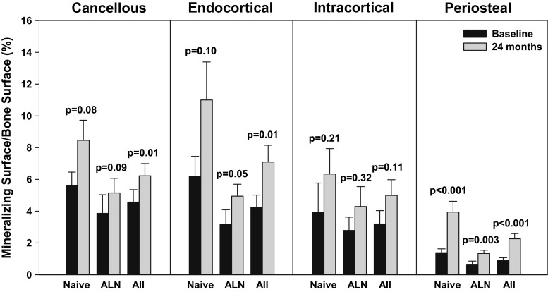
The most important study included 29 alendronate-pre-treated and 16 treatment-naïve post-menopausal women who were treated with teriparatide and underwent an iliac crest biopsy at baseline and after 24 months of teriparatide [15, 23]. This study is highlighted because it included baseline and 24-month biopsies with no gap between teriparatide treatment and the biopsy procedure. An array of histomorphometry parameters were collected, but we focus on mineralizing surface (MS/BS%), a measure of the proportion of bone surface on which new mineralized bone is being deposited (at the time of tetracycline labeling) as the primary bone formation parameter [24]. MS/BS directly measured is used in the calculation of a number of derived indices such as bone formation rate (BFR) [25] and is sensitive to drug effects [22]. In this study, with pooling of the treatment groups, the increase in bone formation from baseline to 24 months was statistically significant in all compartments except for intracortical (Fig. 2). Also, the point estimates of MS/BS increased from baseline in the cancellous, endocortical, intracortical, and periosteal surface in the drug-naïve and alendronate pre-treated patients, with statistically significant results at the periosteal surfaces (Fig. 2) [15, 23]. These findings suggest an ongoing effect of teriparatide at 24 months to increase bone formation. The highest MS/BS% values after 24 months were observed on the endocortical surfaces as compared with baseline. However, larger MS/BS% values were observed after 1 month [26] and after 6 month of teriparatide treatment [22], which is in agreement with the dynamics of serum PINP values (Fig. 1).
Fig. 2.
Bone formation in post-menopausal women with osteoporosis treated with teriparatide as reflected by mineralizing surface divided by bone surface (MS/BS%) at cancellous, endocortical, intracortical, and periosteal compartments. The study included 29 patients previously treated with alendronate and 16 patients naive to previous osteoporosis treatment. Results are means ± SEM shown for each group and for the two groups pooled together (the latter previously unpublished). P values are for comparisons of baseline versus the 24-month treatment period. The data is from the study published by Stepan et al. [15] and Ma et al. [23]
Increased remodeling of the cortex during teriparatide treatment is reflected by an increased cortical porosity and increased intracortical (MS/BS%; Fig. 2). The presence of ongoing increases in intracortical remodeling after 2 years of TPTD treatment indicates a persistent teriparatide effect during the full treatment course. Importantly, the cortical voids (pores) formed during teriparatide treatment show tetracycline double labels. Although cortical voids represent a weak area in the bone, these voids due to remodeling are transient, represent a small percentage of the cortical section area, and are mainly located at the endocortical level; overfilling of these voids may result in an increase in cortical bone mass, and cortical remodeling results in the replacement of older and potentially damaged bone with new bone [27].
Direct evidence supporting long-term modeling-based bone formation by TPTD was provided by studies of periosteal bone apposition. The periosteum contains progenitor cells capable of generating new bone in response to mechanical stimuli [28–30]. Thus, the increase in MS/BS at 24 months suggests an increase in periosteal modeling or the formation of new bone uncoupled from previous resorption. Modeling adds new bone to the bone surface and is an efficient means to increase bone mass, but old bone is not removed and remains beneath the newly formed bone. In studies of 1-month and of 12 to 24-month durations, teriparatide has also been shown to induce modeling in both the trabecular and endocortical envelopes [26, 31, 32].
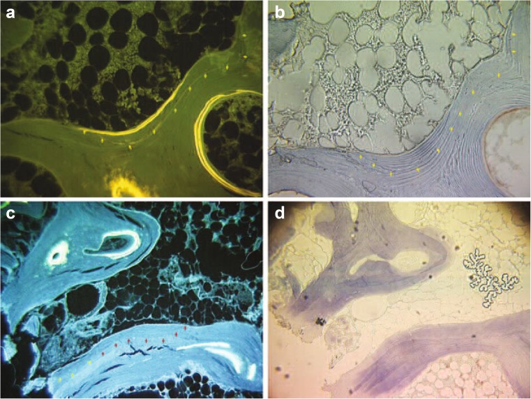
In histomorphometry studies, double labels overlying an eroded (scalloped) cement line are considered to represent remodeling-based formation (bone formation on a previously resorbed bone surface) [33]. During teriparatide treatment, the stimulation of remodeling with overfilling of resorption sites may be the predominant effect of teriparatide both after 1 month and after 12 to 24 months of treatment [31, 32]. This effect involves the removal of old bone and overreplacement with new bone. The positive balance of formation to resorption increases bone mass, and the replacement of old and damaged bone with new organic matrix may result in improved bone microarchitecture, reduced microcrack accumulation [27, 34], and improved quality and elasticity of bone organic matrix. Indeed, bone collagen maturation measured as the ratio between α-CTX and β-CTX showed that teriparatide treatment induced a collagen profile consistent with young matrix [13, 35]. Patients administered teriparatide exhibited significantly lower matrix mineralization, mineral crystallinity, and collagen cross-link ratio when compared with placebo, indicating that the bone-forming effect of TPTD results in reduced bone age [36, 37]. Thus, while the removal of old bone could transiently result in focal weakness, the overfilling of resorption sites results in an increase in bone mass and replacement of old bone with new bone. That teriparatide results in overfilling of resorption sites suggests that teriparatide should be continued rather than discontinued in the setting of increased bone resorption to increase bone mass and quality. Figure 3 illustrates both modeling- and remodeling-based bone formation in a patient treated with teriparatide.
Fig. 3.
This figure shows previously unpublished iliac crest bone biopsy specimens from a patient treated with teriparatide 20 mcg/day to illustrate two fundamental anabolic actions: a unstained fluorescence light image and b transmitted light for toluidine blue staining image specific for cement lines. Newly formed lamellar bone is indicated by the yellow arrows. Smooth cement lines and parallel collagen fiber orientation versus the adjacent bone tissue, without interruption, indicate that this bone formation is related to bone modeling or the deposition of new bone on a previously quiescent bone surface. c Unstained fluorescence light image and d transmitted light for toluidine blue staining image specific for cement lines. Scalloped cement line shown by red arrows represents previous resorption surface, and the smooth cement line in the adjacent bone illustrated by yellow arrows illustrates the spilling of new bone formation onto an adjacent surface. The mixture of scalloped cement lines transitioning to smooth cement lines is indicative of overfilling of a remodeling site onto the adjacent bone surface. These previously unpublished images from the study described by Ma et al. [32] are courtesy of Dr. Linda Ma
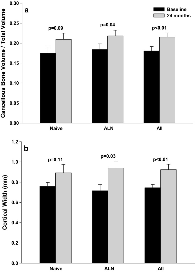
Increased bone formation would be anticipated to increase cortical and trabecular bone mass. Indeed, both cortical thickness and trabecular bone volume/total bone volume increased in the 24-month paired biopsy study (Fig. 4). Increases in these parameters have been reported in other studies as well [38].
Fig. 4.
Increase in cancellous bone volume divided by total volume and cortical thickness from baseline through 24 months of teriparatide treatment and included patients previously treated with alendronate and patients naïve to previous osteoporosis treatment. Results are means ± SEM shown for each group and for the two groups pooled together (the latter previously unpublished). P values are for comparisons of baseline versus the 24-month treatment period. Results are from Stepan et al. [15] and Ma et al. [23]
Effects of full course of teriparatide on areal bone mineral density using dual-energy X-ray absorptiometry
The majority of the larger clinical trials after the introduction of recombinant teriparatide (hPTH (1-34); DNA origin), including the pivotal, phase 3 trial [4], were of approximately 18-month duration. However, more recent studies have analyzed the effects of the full 24-month treatment course. Accordingly, this section will focus on trials including teriparatide at the approved dose of 20 μg/day for 24 months, and the results of those studies are provided in Table 1. Other studies including an open-label clinical trial with synthetic teriparatide ∼25 μg/day in combination with estrogen replacement therapy for 3 years [43, 44] and studies in men and post-menopausal women with low bone mineral density (BMD) treated with synthetic teriparatide at a daily dose of approximately 37 μg/day for 24 months [45, 46] have also been published but will not be reviewed here.
Table 1.
Areal bone mineral density (BMD) results in subjects treated for 24 months or longer with daily teriparatide at the approved dose (20 μg/day) in clinical trials
| Reference | Study population (na) | Prior osteoporosis drug treatment (mean duration) | Teriparatide treatment duration | Δ BMD lumbar spine | Δ BMD total hip | Δ BMD femoral neck | Δ BMD distal radius |
|---|---|---|---|---|---|---|---|
| [39] | Menopausal women (n = 503) | Anti-resorptives 86.3 % (11 to 54 months)b | 24 months | +13.1 % (naïve), +9.8 to +10.2 % (pre-AR) | +3.8 % (naïve), +2.3 % (pre-AR) | +4.8 % (naïve), +3.4 to +3.9 % (pre-AR) | |
| [15] | Menopausal women (n = 66) | Alendronate 57.5 % (64.5 months) | 24 months | +10.0 % (naïve), +6.5 % (pre-ALN) | +1.0 % (naïve), +1.0 % (pre-ALN) | ||
| [40] | Menopausal women and men (n = 96) | Anti-resorptives 36.8 % (NR) | 24 months | +13.96 % | +3.69 % | +3.25 % | |
| [17] | Menopausal women (n = 28) | Anti-resorptives 42 % (40 months) | 24 months | +9.5 % | +2.0 % | +2.8 % | −1.7 % |
| [41] | Menopausal women (n = 73) | Alendronate 41 % (5.9 years) | 24 months | +8.8 % (naïve), +7.5 % (pre-ALN) | +4.0 % (naïve), +3.0 % (pre-ALN) | +2.9 % (naïve), +3.0 % (pre-ALN) | −4.2 % (naïve), −0.7 % (pre-ALN) |
| [14] | Women and men with GIO (n = 123) | Bisphosphonates 9 % (NR) | 36 months | +11.0 % | +5.2 % | +6.3 % | |
| [42] | Pre-menopausal women (n = 21) | None | 24 months | +10.8 % | +6.2 % | +7.6 % |
Comparative arm data, including combination therapies, are not shown
ALN alendronate, AR anti-resorptives, GIO glucocorticoid-induced osteoporosis, NR not reported
aTeriparatide arms only
bMedian
cLateral spine view
In the European Forsteo® Study (EuroFORS) in patients who were either naïve to osteoporosis drugs or previously treated with different anti-resorptive (AR) drugs, 503 post-menopausal women with osteoporosis received teriparatide for 24 months, including 84 subjects (16.7 %) who were osteoporosis treatment naïve [39]. The change in lumbar spine BMD from baseline to 24 months was +10.5 %. At this site, the increases were +13.1 % in the osteoporosis treatment-naïve patients compared to +9.8 to +10.2 % in previously AR-treated patients, with the difference between naïve and pre-treated being statistically significant. The change in femoral neck BMD from baseline to 24 months was, on average, +3.9 % for all teriparatide-treated patients, and +4.8 %, and between +3.4 and +3.9 % for the treatment-naïve and the AR pre-treated subgroups, respectively. Similar results were observed at the total hip (Table 1). Most relevantly, the BMD values showed a statistically significant increase between 18 and 24 months of daily teriparatide treatment at the spine and the proximal femur. Indeed, in the AR-pre-treated subjects, the BMD increment between 18 and 24 months at the hip and femoral neck was approximately the same as the increment from the first 18 months of therapy. These results highlight the importance of the full course of teriparatide treatment to increase cortical bone BMD, especially in subjects who previously were treated long term with AR [39, 47–49].
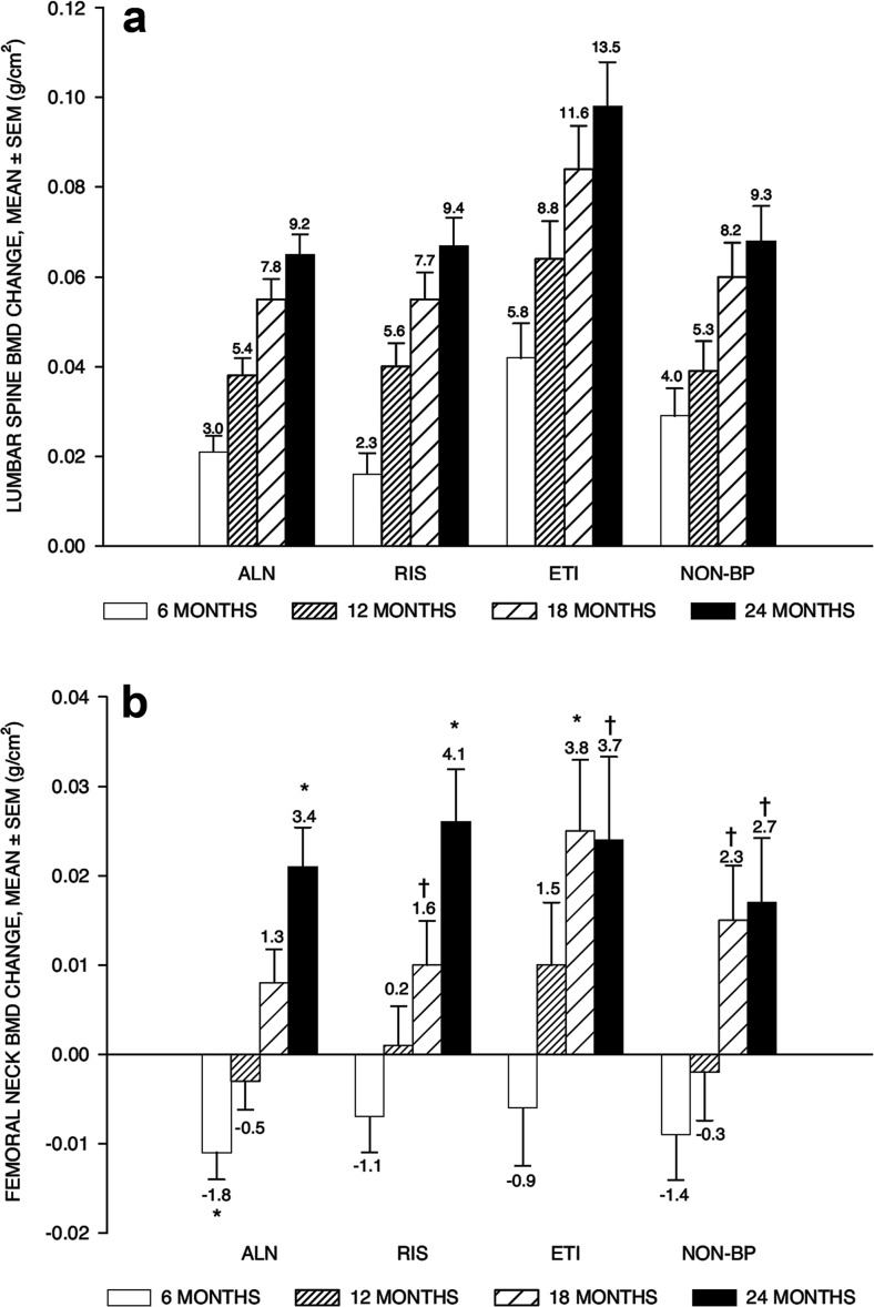
The effect of different types of AR on the BMD response after 24 month of continuous teriparatide treatment was also analyzed in the EuroFORS cohort [50]. The skeletal responses at the lumbar spine were similar among previous AR therapy groups at each time point during the study, although previous users of etidronate showed a higher increase, probably reflecting its weaker anti-remodeling activity (Fig. 5).
Fig. 5.
Adjusted mean BMD changes from baseline after 24 months of continuous teriparatide treatment stratified by previous predominant treatment. Numbers at top vertical bars indicate percent change from baseline. a Lumbar spine, p < 0.001 for all within-treatment group changes from baseline at all time points. Between-group comparisons at 6, 12, 18, and 24 months, p < 0.05 etidronate (ETI) versus alendronate (ALN) and ETI versus risedronate (RIS) and p < 0.05 ETI versus non-bisphosphonate (NONBP) at 18 and 24 months. All other between-group comparisons were not statistically significant. b Femoral neck, p < 0.05 for ALN versus ETI between-group comparison at 18 months. All other between-group comparisons were not statistically significant. Error bars indicate SEM. *p < 0.001 for within-treatment group change from baseline, † p < 0.05 within-treatment group change from baseline. Reproduced with permission from Boonen et al. [50]
The BMD effects after 24 months of treatment with teriparatide 20 μg daily were also analyzed in 66 patients who were either treatment-naïve (n = 28) or had lower bone turnover initially due to previous alendronate therapy (n = 38) in the Prague-Graz study [15]. After 24 months of treatment with teriparatide, lumbar spine and femoral neck BMD values increased significantly in both patient groups. Similar to the results in the EuroFORS trial, the increase in spine BMD in treatment-naïve subjects was statistically higher (+10.0 %) than in alendronate prior-treated patients (+6.5 %). The increase in femoral neck BMD was not different between the two groups (+5.0 and +3.3 %, respectively; p < 0.05 vs baseline).
A 24-month duration study of teriparatide treatment in Asian subjects was reported by Miyauchi et al. [40]. Daily injections of 20 μg of teriparatide in 96 Japanese men and women with low bone mass revealed significant increases from baseline in areal BMD at the spine (+13.42 %), total hip (+3.67 %), and femoral neck (+3.26 %) [40].
The comparative effects of 24-month treatment with teriparatide, denosumab, and the combination of the two drugs have been recently reported [17]. In this open-label trial in 83 post-menopausal women with osteoporosis, the areal BMD increases observed in the teriparatide-only arm were +9.5, +2.8, and +2.0 % at the spine, femoral neck, and total hip, respectively. Of note, 13 of the 31 patients (42 %) in the teriparatide arm had received prior bisphosphonate treatment for a mean of 40 months [17]. The decrease in the distal radius BMD in the teriparatide group (−1.7 %) was not statistically significant compared to baseline but differed from the changes observed with denosumab (+2.1 %) or the combination group (+2.2 %).
Cosman et al. [41] have compared the effects of daily teriparatide for 24 months or four 3-month teriparatide cycles, each followed by 3 months off (12-month total teriparatide) in two groups of subjects. In osteoporosis treatment-naïve patients (n = 86), increases in BMD were twofold greater in daily versus cyclic treatment (spine +8.8 vs +4.8 %, total hip +4.0 vs +2.1 %, and femoral neck +2.9 vs 1.2 %; p < 0.05), but radius BMD declined more in the daily therapy group (−4.2 vs −2.1 %; p = 0.08). In alendronate-pre-treated women (n = 64, average therapy duration 5.9 years), there were no group differences ([daily vs cyclic teriparatide] spine +7.5 vs +6.0 %, total hip +3.0 vs 2.5 %, femoral neck +3.0 vs 1.5 %, and radius −0.7 vs −1.4 %).
In subjects with glucocorticoid-induced osteoporosis, Saag et al. [14] have reported the BMD results of continuous treatment for 36 months with teriparatide in a double-blind, comparative trial with alendronate (Table 1). Within the teriparatide group, the mean percent changes in BMD were significantly increased between 24 and 36 months at the lumbar spine (+9.76 vs +10.95 %; p < 0.01) and at the femoral neck (+4.76 vs +6.34 %; p < 0.001) but not at the total hip (+4.75 vs 5.22 %; p = 0.115).
Finally, Cohen et al. [42] have reported 24-month areal BMD results in a series of 21 osteoporosis treatment-naïve pre-menopausal women with unexplained fragility fractures or low BMD, treated with teriparatide. At 24 months, the largest increase in BMD was at the lumbar spine (+10.8 %). Significant increases also occurred at the femoral neck (+7.6 %) and the total hip (+6.2 %; all p < 0.001). Similar to the results of the EuroFORS trial, the 24-month increases in BMD at the proximal femur were approximately twice that seen after the initial 18 months of therapy.
The BMD effectiveness of daily teriparatide in the context of regular clinical practice has been reported in two single-center, prospective, observational studies. In 60 women with osteoporosis who completed a 24-month treatment course, the BMD changes compared to baseline at the lumbar spine, total hip, and distal radius were +10.9, +3.9, and −2.4 % (all p < 0.05) [51]. In a Scottish cohort of 217 severe osteoporotic patients treated for approximately 21 months, the increases in lumbar spine and femoral BMD were +13.7 and +2.3 %, respectively [52]. Prior use of osteoporosis drugs, mainly oral bisphosphonates, was reported in 85 and 56 % of the subjects included in these two studies, respectively [51]. Finally, a retrospective analysis of 65 women who received teriparatide during a mean of 23 months as part of their routine osteoporosis management showed a +7.6 % BMD increase at the spine and a +4.3 % increase in the trabecular bone score (TBS), a novel technique based on areal DXA technology that can be used for the non-invasive assessment of intravertebral trabecular bone microarchitecture [53]. Of note, 95 % of these patients had prior therapy with an oral or IV bisphosphonate. In general, BMD results of these real-world studies were similar to those observed in randomized controlled trials.
Long-term effects of teriparatide on volumetric BMD using quantitative computed tomography, including finite element analysis and strength
The changes in bone distribution, geometry, and bone strength based on 3D quantitative computed tomography (QCT) of the femoral neck in a group of 52 subjects with osteoporosis receiving 24-month continuous teriparatide (44 of them having received previous AR therapies) have been reported [54]. After 24 months of treatment, areal and volumetric femoral neck BMD increased significantly by +4.0 and +3.0 %, respectively, compared with baseline. Cortical cross-sectional area (CSA) increased by +4.3 %, whereas total CSA remained unchanged over the study duration, indicating that endosteal but not periosteal apposition was detectable with this technique. Hip QCT revealed different temporal response patterns of cortical and trabecular bone envelopes that cannot be separated by DXA. At 6 months of treatment, cortical volumetric BMD (vBMD) decreased significantly, whereas trabecular vBMD showed a significant increase. This led to a significant decrease in total vBMD of −1.4 % (p < 0.05). However, in the remaining 18 months, cortical vBMD remained stable, whereas cortical areas increased, which led to a net gain of +6.7 % (p < 0.0001) in cortical bone mineral content (BMC). These results support the importance of continuous full-course treatment with teriparatide, especially in AR pre-treated subjects.
Strength parameters for buckling did not change at 6 or 12 months but improved significantly at 24 months (−4.3 %), indicating an improvement in cortical stability [54]. Although minimal and maximal section moduli of the femoral neck showed a non-statistically significant increase compared to baseline at 24 months (+2.3 and +1.9 %, respectively; p < 0.1), it was noted that the changes in the second year of continuous teriparatide treatment, buckling strength, and bending strength indices increased significantly compared to 12 months (buckling ratio −4.6 %, p < 0.0001; Zmin +2.7 %, p < 0.05; Zmax +3.9 %, p < 0.01).
Using a novel CT image processing technique on 65 women with baseline and 24-month paired hip scans from the EuroFORS cohort, Poole et al. [55] showed that teriparatide treatment increased cortical thickening. This increase was higher at sites of hip mechanical loading including the infero-medial cortex and the calcar femorale regions, reaching significant increases of +6 to +8 % compared to baseline (p < 0.05). These results suggest a possible synergistic effect between habitual loading and teriparatide in the human proximal femur, since peak effects were seen at sites that are stressed by walking. No regions of cortical thinning were apparent.
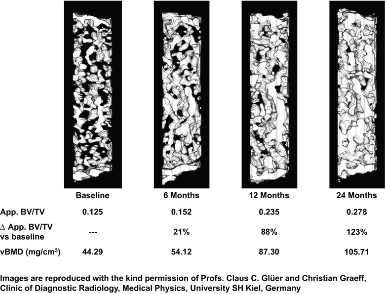
Another study has applied high-resolution QCT and finite element (FE) analysis at the 12th thoracic vertebra to evaluate the effects of 24 months of continuous treatment with teriparatide in a subset of 44 patients of the EuroFORS trial, most of them pre-treated with AR drugs [56]. After 24 months, bone strength was increased in compression by +28.1 %, in bending by +28.3 %, whereas the apparent bone volume fraction (BV/TV) was increased by +54.7 %, volumetric BMD by +19.1 %, and areal BMD of L1–L4 by +10.2 %. This analysis included standardized changes to compare the performance of the different densitometry and strength variables. Results after 24 months of treatment showed that the standardized changes were similar for volumetric and areal BMD (+0.56 and +0.70 standard deviation increases compared to baseline, respectively). Interestingly, the standardized increases in variables of the compression test simulation were statistically larger than those of both the densitometry measures (ranging between +1.07 and 1.18 SD compared to baseline). However, the largest standardized increase was for the trabecular BV/TV, a surrogate marker of bone tissue quantity, which showed a +1.27 standard deviation increase after 24 months of treatment. The increase in bone mass in a patient with a robust response to teriparatide is shown over time in Fig. 6.
Fig. 6.
Rectangular “virtual biopsies” taken using high-resolution CT scan images of the 12th thoracic vertebra at baseline and after 6, 12, and 24 months of treatment with teriparatide show a progressive increase in bone mass. These previously unpublished images kindly provided by Professors Claus C. Glüer and Christian Graeff, University SH Kiel, Germany, are from the study described by Graeff et al. [56]
The FE analysis technique also allowed evaluating the damage distribution within the vertebral body after applying different forces. The analysis of the areas with higher damage risk indicated that 24 months of teriparatide treatment significantly reduced the volume of damage from a similar load from 2.7 to 0.1 % in compression and from 2.2 to 0.02 % after bending [56]. Interestingly, CSA of the body of the 12th thoracic vertebra increased by +0.7 % (5.7 mm2; p < 0.001), whereas the CSA of the spinal canal increased marginally by +0.9 % (2.6 mm2) over 24 months (p < 0.001). None of the patients showed a trend for narrowing of the spinal canal which could result in spinal stenosis [57].
Finally, the longest study where the effects of teriparatide on the distal radius and tibia were analyzed using high-resolution peripheral QCT have been reported in abstract form from the DATA cohort [58]. Volumetric cortical BMD was decreased by almost −4.0 % after 24 months of treatment with teriparatide at both sites. Cortical thickness was unchanged in this study. Similar results have been published using this technique in 18-month trial duration. In a longitudinal study using HR-pQCT in 11 post-menopausal women, treatment with teriparatide decreased cortical vBMD at distal radius (−4.5 %; p < 0.1) and tibia (−1.3 %; p < 0.05), whereas a non-significant increase was seen in cortical porosity at both sites [59]. Hansen et al. [60] performed an open-label observational study comparing effects of 18 months of treatment with either teriparatide 20 μg/day (n = 18), rhPTH(1-84), or zoledronic acid. In response to treatment with teriparatide, cortical vBMD at the distal radius and tibia decreased (−2.4 and −1.6 %, respectively; p < 0.05) due to a significantly increased cortical porosity (+32.4 and +13 %), while cortical thickness increased at both sites (+2.0 and +3.8 %; p < 0.05). Despite these findings, these studies showed no reduction in bone strength as assessed by FE analyses in response to teriparatide [58–60].
The apparent decrement in radial vBMD may be a combination of several effects induced by teriparatide that occur simultaneously, including increased endocortical remodeling, increased remodeling space within the cortical haversian systems, and an increase in measured area due to periosteal bone apposition [2]. Moreover, the deposits of new osteoid upon bone surfaces actively participating in remodeling will not attenuate photons until it is mineralized, so true cortical apposition remains undetected since the mineral density is below the threshold level that qualifies as bone [61, 62]. Since cortical thickness is obtained by dividing the cortical volume by the outer bone surface (i.e., the cortical area by the perimeter of each slice), and the boundary between the cortical and cancellous region is defined by a drop in mineral density, changes in the degree of bone mineralization, particularly at the endocortical region—as observed with teriparatide—are likely to affect the evaluation of cortical thickness and BMD. Moreover, the high correlation that is normally reported between cortical thickness and cortical density in HR-pQCT studies suggest that partial volume effects limit the reliability of cortical density measurements [63]. Another limitation of this technology to assess the effects of teriparatide on cortical bone is the issue of repositioning of the scanning region in longitudinal studies. The HR-pQCT software incorporates a CSA registration method to correct axial misplacement between successive scans, but possible tilt of the limb with even small angular deviations or changes induced by therapy on the CSA could lead to considerable mismatch in the selected region [64].
Teriparatide fracture data
The Fracture Prevention Trial (FPT) was the pivotal phase 3 fracture trial for teriparatide. This was a randomized, double-blind, placebo-controlled study of 1637 post-menopausal women with prior fractures treated with daily subcutaneous teriparatide 20 or 40 μg or placebo [4]. The median duration of treatment was 19 months, and the median duration of observation was 21 months. Patients received daily supplements of calcium 1000 mg and vitamin D 400 to 1200 IU.
Vertebral fracture
Lateral spine radiographs were obtained at baseline and at study end point [4]. The first report of teriparatide efficacy from the Fracture Prevention Trial defined vertebral fractures by a single visual semiquantitative (SQ) reading. Readers were blinded to group assignment but not to order of the radiographs. By this method, a total of 64 out of 448 patients (14 %) in the placebo group had new vertebral fractures (22 mild and 42 moderate/severe) and 22 out of 444 patients (5 %) in the 20-μg/day group had new vertebral fractures (18 mild and 4 moderate/severe). The overall relative risk reduction in the teriparatide 20-μg/day group was 65 % and the absolute risk reduction was 9 %.
After stopping study drug in the FPT, 1262 patients were enrolled in a follow-up study and lateral spine radiographs were repeated 18 months later. During this follow-up study, other osteoporosis drugs were used by more patients in the former placebo group than the former teriparatide group (p = 0.04). Even so, the reduction in vertebral fracture risk associated with previous treatment with teriparatide 20 μg/day was 41 % (p = 0.004) during the follow-up study. The absolute risk reduction from the FPT baseline to the 18-month follow-up was 13 % [65]. Thus, the difference in vertebral fracture incidence between the teriparatide and placebo groups increased further after study drug was discontinued.
The method used to define vertebral fractures in many osteoporosis trials has been a quantitative morphometry method requiring quantitative decreases in vertebral height plus confirmation of fracture by the semiquantitative visual methodology. Lateral spine radiographs from the FPT teriparatide 20 μg/day and placebo groups were re-assessed in blinded fashion using this methodology, revealing that a total of 51 out of 448 (11.4 %) patients in the placebo group had new vertebral fractures compared to 8 out of 444 (1.8 %) in the teriparatide 20-μg/day group. By this stricter fracture assessment methodology, the relative risk reduction in the teriparatide 20-μg/day group was 84 % and the absolute risk reduction was 9.6 %.
In the placebo group of the Fracture Prevention Trial, by either the SQ methodology or the QM with SQ confirmation methodology, the number and severity of prevalent vertebral fractures at baseline were significantly predictive of the risk for incident vertebral fractures (p < 0.001) [66, 67], a phenomenon termed a “fracture cascade.” The absolute vertebral fracture benefit of teriparatide may be greatest in patients with higher burdens of vertebral fracture. For example, among patients with two prevalent vertebral fractures, 15/84 (17.9 %) in the placebo group and 1/85 (1.2 %) in the teriparatide group had incident vertebral fractures, representing a relative risk reduction of 93 % (p < 0.001) and an absolute risk reduction of 17 %. Among those with three or more prevalent vertebral fractures, 24/69 (34.8 %) in the placebo group and three of 60 (5.0 %) in the teriparatide group had incident vertebral fractures, representing a relative risk reduction of 86 % (p < 0.001) and an absolute risk reduction of 30 % [66]. In patients with baseline severe vertebral fractures, new vertebral fractures were reported for 27.7 % of the placebo group and 1.2 % of the teriparatide group, representing a relative risk reduction of 96 % (p < 0.001) and an absolute risk reduction of 27 %. These results indicate that anabolic therapy with teriparatide alters the natural history of the progression of osteoporosis.
In a post hoc analysis of the Fracture Prevention Trial, there were approximately half as many clinical vertebral fractures in the teriparatide 20 μg/day versus placebo group during the first 7 months of observation; during subsequent intervals, the incidence of clinical vertebral fractures increased further in the placebo group but was very low in the teriparatide group (Table 2) [68]. Thus, during additional intervals of observation, increasingly large differences in fracture incidence were observed between the teriparatide and placebo group. These results suggest that greater duration of treatment appeared to be associated with greater reduction in the incidence clinical vertebral fractures.
Table 2.
Teriparatide 20 μg/day fracture data from clinical trials, observational studies, and claim database studies
| Reference | Interval duration | Fracture type | Treatment | Fracture rates (%) | |||
|---|---|---|---|---|---|---|---|
| Intervals | |||||||
| 1 | 2 | 3 | 4 | ||||
| Clinical trial | |||||||
| [27, 68] | 7 months | Non-vertebral | TPTD | 1.3 % | 0.83 % | 0.66 %** | |
| Placebo | 1.5 % | 1.8 % | 2.8 % | ||||
| Clinical vertebral | TPTD | 0.9 % | 0.0 % | 0.3 % | |||
| Placebo | 1.8 % | 2.6 % | 3.1 % | ||||
| Observational studies | |||||||
| [69] | 6 months | Non-vertebral (all) | TPTD | 1.42 % | 0.91 %* | 0.70 %* | 0.81 %* |
| Non-vertebral (female) | TPTD | 1.49 % | 0.94 %* | 0.74 %* | 0.90 %* | ||
| Non-vertebral (male) | TPTD | 0.81 % | 0.66 % | 0.38 % | 0.0 % | ||
| [27] | 6 months | Hip | TPTD | 0.27 % | 0.07 %* | 0.15 % | 0.0 %* |
| [70] | 6 months | Clinical fractures | TPTD | 4.6 % | 3.5 %* | 2.8 %* | |
| Clinical vertebral | TPTD | 1.8 % | 1.3 % | 0.7 %* | |||
| Non-vertebral | TPTD | 2.9 % | 2.2 % | 2.1 % | |||
| Main non-vertebral | TPTD | 2.4 % | 1.8 % | 1.7 %* | |||
| [71] | Clinical fractures | TPTD | 3.0 % | 2.0 % | 1.7 %* | 1.6 %* | |
| Clinical vertebral | TPTD | 1.1 % | 0.1 %* | 0.2 %* | 0.3 %* | ||
| Non-vertebral | TPTD | 2.0 % | 1.9 % | 1.5 % | 1.3 % | ||
| Claim database study | Teriparatide treatment duration | ||||||
| <6 | 6–12 | 13–18 | 19–24 | ||||
| [72]*** | 6 months | Any clinical | TPTD | 101.1 | 75.9 | 72.5 | 51.5 |
| Vertebral | TPTD | 24.7 | 13.5 | 1.3 | 6.1 | ||
| Hip | TPTD | 10.0 | 8.4 | 6.6 | 5.2 | ||
| Non-hip/non-vertebral | TPTD | 66.4 | 54.0 | 54.6 | 40.2 | ||
To illustrate the effect of teriparatide over time, fracture rates are shown by different intervals of observation. For example, in the Lindsay et al. analysis at the top of the table, the fracture rates during months 0–7, 7–14, and >14 were reported and are reflected in the table as interval duration 7 months and fracture rates are provided for intervals 1, 2, and 3. For clinical trials and observational studies, the fracture rates are percentages determined as the number of patients with fractures among the total observed patients during the interval. For the Yu et al. [72] claim database study, patients had differing durations of teriparatide treatment but the same overall follow-up of 24 months post-teriparatide initiation
TPTD teriparatide
*p < 0.05 versus the first interval
**The relative hazard of non-vertebral fragility fractures decreased by 7.3 % for each additional month of teriparatide 20 μg versus placebo (p = 0.009)
***Fracture incidence is shown as fractures per 1000 patient-years of observation
Non-vertebral fracture
In the Fracture Prevention Trial, non-vertebral fractures were confirmed by a review of a radiograph or radiology report. For the pre-specified non-vertebral fracture end point, pathological and traumatic fractures and fractures of the face, skull, metacarpals, fingers, and toes were excluded [4]. In the placebo group, non-vertebral fractures were observed for 30 of 544 patients (5.5 %) and for 14 of 541 (2.6 %) teriparatide 20 μg/day patients. Treatment with teriparatide 20 μg/day reduced the risk of non-vertebral fractures by 53 % compared with placebo after a median treatment of 19 months (p = 0.02).
In the Fracture Prevention Trial, placebo-treated patients with zero, one, or two or more prior non-vertebral fractures, the incidence of non-vertebral fractures were 4, 8, and 18 %, respectively (Cochran-Armitage trend test, p < 0.001), illustrating a non-vertebral fracture cascade. In the teriparatide 20-μg/day group, new non-vertebral fractures occurred in 2.7, 0, and 3.9 % of patients with zero, one, and two or more prior non-vertebral fragility fractures, respectively (Cochran-Armitage trend test, p = 0.96). Thus, in the teriparatide group, there was no significant increase in non-vertebral fracture risk with increasing number of prior non-vertebral fractures, indicating that teriparatide favorably impacted the natural history of osteoporosis progression [67].
Non-vertebral fractures at specific sites in the teriparatide 20-μg/day group (n = 541) versus placebo group (n = 544) included 1 versus 4 hip, 2 versus 7 wrist, 1 versus 3 ankle, 2 versus 2 humerus, 3 versus 5 rib, 0 versus 1 foot, 0 versus 3 pelvis, and 6 versus 8 other [4]. Although the trial was not powered for detecting significant differences at individual fracture sites, this pattern suggests that teriparatide might have a consistent effect across different skeletal sites.
Additional analyses from the Fracture Prevention Trial have shown the effects of teriparatide on various non-vertebral fracture end points with inclusion or exclusion of traumatic fractures; results showed greater relative risk reductions in the teriparatide group for non-vertebral fracture end points most likely to be of osteoporotic origin [73]. For example, for any non-vertebral fracture including those classified by investigators as traumatic, the relative risk reduction in the teriparatide 20 μg/day versus placebo group was 35 %. On the other hand, for major non-vertebral fractures (a set of fracture sites believed to be related to osteoporosis) and with exclusion of traumatic fractures, the relative risk reduction was 62 %. Thus, teriparatide appears to have greater efficacy to reduce the incidence of osteoporotic versus traumatic fractures.
Inspection of the Kaplan-Meier curve for non-vertebral fracture shows that the placebo and teriparatide 20 μg/group diverged after approximately 9 months with the gap between the groups subsequently increasing with longer duration of treatment [4]. During the 30-month follow-up study after study drugs were stopped, the gap between the placebo and teriparatide group further increased, indicating that the skeletal effect of teriparatide was maintained after study drug cessation [74]. Thus, non-vertebral fragility fractures occurred in 55 cases (13.3 %) treated with placebo during the double-blind phase and in 37 cases (8.5 %) in the former teriparatide 20 μg-treated group. Most of the patients received any type of AR after stopping the study medication. The relative risk reduction was 38 % (p = 0.022).
The relationship between duration of teriparatide treatment and reduction in non-vertebral fracture risk in the Fracture Prevention Trial has been further explored [68]. Fracture incidence during the first, second, and third 7 months of the study, corresponding to approximately one thirds of the median observation, was assessed (Table 2). Patients entered each subsequent observation period if they remained on study treatment and had not previously fractured. During the initial 7 months of observation, the incidence of non-vertebral fractures was similar in the placebo and teriparatide 20-μg/day groups. Compared to the placebo group, during the next 7 months, there were approximately half as many fractures in the teriparatide group, and during the final 7 months, there were about a fourth as many fractures in the teriparatide group [68]. The risk for non-vertebral fracture in the teriparatide 20 μg/day versus placebo group over time was modeled, controlling for baseline age, vertebral T-score, and multiple vertebral fractures. The results showed that the relative hazard of non-vertebral fragility fractures was reduced by 7.3 % for each additional month of teriparatide (p = 0.009). Thus, the hazard ratio after 1 month of therapy was 92.7 % of that at baseline, after two months was 85.9 %, and after three months was 79.7 %, etc. Graphical representation of these results showed that after 24 months of teriparatide, the predicted hazard for non-vertebral fracture in the teriparatide versus placebo group was approximately 20 %. Thus, early during teriparatide treatment, the incidence of non-vertebral fracture may not be different from placebo; thereafter, increased duration of teriparatide treatment appeared to result in progressive decreases in non-vertebral fracture risk.
A post hoc analysis was conducted regarding an osteoporotic fracture end point including pooled fractures of the spine and low-trauma non-vertebral fractures [75]. This end point was assessed for Fracture Prevention Trial plus the follow-up study through 18 months, at which time patients underwent lateral spine imaging. During the 39 total months of observation, the relative risk reduction for any fracture in the teriparatide 20-μg/day group was 46 %, the absolute risk reduction (ARR) was 12.7 % (p < 0.0001), and the number needed to treat (NNT) to prevent a new fracture was 8. Patients with more severe osteoporosis had greater absolute benefit. For example, in patients with low BMD and baseline vertebral fractures, the relative risk reduction was 56 %, ARR was 21.7 % (p < 0.0001), and NNT was 5. Thus, for the osteoporotic fracture end point, teriparatide showed efficacy over a duration including the Fracture Prevention Trial plus 18 months of follow-up and the greatest absolute benefit was among the patients with most severe osteoporosis [75].
Observational studies
Several large observational studies of osteoporosis patients treated with teriparatide 20 μg/day have been conducted. In these studies, patients are treated with teriparatide as part of their normal clinical care, and so, the patient populations are more representative of “real-world” patients than those in randomized controlled trials which include many inclusion and exclusion criteria. However, these observational studies of teriparatide have not included control groups so that fractures in patients treated with teriparatide cannot be compared to fractures in patients treated with other treatments. Instead, these studies have been designed to compare fracture incidence over time. The basis for this design is the finding in the Fracture Prevention Trial described above that the incidence of non-vertebral fractures in patients treated with placebo and teriparatide is similar during the initial approximately 9 months, so that the fracture incidence during the initial months of teriparatide treatment provides a reference fracture incidence which might be approximately similar to a placebo fracture incidence. Fracture incidence later during treatment can then be compared to fracture incidence during the reference interval.
For example, the Direct Assessment of Non-Vertebral Fractures in Community Experience (DANCE) study evaluated 4085 men and women treated with teriparatide for 24 months and then observed for another 24 months [69]. Using the same methodology as that used to examine the effect of teriparatide over time in the Fracture Prevention Trial, the incidence of non-vertebral fragility fracture were 1.42, 0.91, 0.70, and 0.81 % for the four 6-month teriparatide treatment periods (Table 2). The odds of fracture during each 6 month interval were lower than the first 6-month reference period. Compared to the reference interval, the odds of fracture was 43 % lower in the last 6-month period of teriparatide treatment. After stopping teriparatide, the incidence of fracture during the subsequent 6-month intervals after treatment cessation were 0.80, 0.68, 0.33, and 0.33 %. The results from this study indicated that after an initial 6-month reference period, non-vertebral fracture incidence was reduced, and this reduction persisted after teriparatide was stopped. The incidence of hip fractures showed a similar pattern over time [27].
In an observational study of 1581 European post-menopausal women treated with teriparatide for 18 months and then observed for another 18 months (European Forsteo Observational Study (EFOS)), a 74 % decrease in the adjusted odds of any incident clinical fracture in the 30- to <36-month period compared with the first 6-month period was observed (p < 0.001) [76]. The reduction during the 12- to <18-month active treatment period compared with the first 6 months (Table 2) was 39 % (p = 0.013). The decreases in adjusted odds of having a non-vertebral fracture were significantly lower during the 24- to <30-month interval (60 %; p ≤ 0.01) and 30- to <36-month interval (59 %; p ≤ 0.01), compared with the first 6 months of teriparatide treatment.
These results have been more recently confirmed in a 24-month treatment observational study that included 1454 post-menopausal and men with osteoporosis as well as patients with glucocorticoid-induced osteoporosis (ExFOS) [71]. A 53 % decrease in the odds of low-trauma clinical fractures in the last 18–24-month period compared with the first 6-month period was observed (p < 0.05).
Finally, in one real-world study, 3587 patients treated with teriparatide who had data available 12 months pre- and 24 months post-teriparatide initiation were identified using the Thomson Reuters MarketScan® database [72]. Fracture risk decreased as teriparatide persistence increased for any clinical, vertebral, and non-vertebral fractures (Table 2). Similar reductions in hip fracture were observed in patients treated longer versus shorter with teriparatide, although the results did not reach statistical significance. Using the same database, another group reported teriparatide outcomes for 11,407 patients similarly treated with teriparatide and having similar pre- and post-treatment follow-up [77]. This study appeared to show similar results and confirmed lower risk for fracture for persistent versus non-persistent patients [78].
Long-term safety data
In the Fracture Prevention Trial with a median treatment of 19 months and observation of up to 24 months, dizziness was reported for 6 % of the placebo group and 9 % of the teriparatide 20-μg/day group (p = 0.05), and leg cramps were reported for 1 % of the placebo group and 3 % (p = 0.02) of the teriparatide 20-μg/day group [4]. Nausea and headache were similar in the placebo and teriparatide 20-μg/day groups. Deaths, hospitalizations, cardiovascular disorders, urolithiasis, and gout were similar in the placebo and teriparatide groups. There were no cases of osteosarcoma in the study, and indeed, cancer was reported for 4 % of the placebo group and 2 % of the teriparatide 20-μg/day group (p = 0.02). Blood pressure and heart rate, measured at each visit prior to teriparatide dosing, were unaffected by teriparatide.
Assessments of serum calcium were increased 4 to 6 h after dosing, and mild hypercalcemia was noted in 2 % of the placebo group and 11 % of those in the teriparatide 20-μg/day group, although sustained hypercalcemia was uncommon and serum calcium was usually normal 16 to 24 h post-injection [4]. Treatment was discontinued because of persistently elevated serum calcium concentrations in one woman in the placebo group and one in the teriparatide 20-μg group. Twenty-four-hour urinary calcium excretion increased during teriparatide treatment by approximately 30 mg (0.75 mmol) per day, but the incidence of hypercalciuria defined as urinary calcium excretion greater than 300 mg (7.5 mmol) per day was not different between the placebo and teriparatide 20-μg/day groups. While serum magnesium concentrations decreased slightly in the teriparatide 20-μg/day group, serum uric acid concentrations increased by 13 to 20 % in the teriparatide 20-μg/day group. The increase in serum uric acid was not associated with increases in gout or nephrolithiasis. After stopping study drug, the changes in laboratory parameters were resolved at a follow-up visit.
The timing of adverse events in the Fracture Prevention Trial was assessed during intervals including months 0–7, 7–14, and >14. During months 0–7, nausea was reported for 7.58 % of the teriparatide 20-μg/day patients and 4.6 % of the placebo patients (p < 0.05). Dizziness was 4.04 % in the placebo group and 6.65 % in the teriparatide 20-μg/day group, and leg cramps was 0.55 % in the placebo group and 1.85 % in the teriparatide 20-μg/day group; these differences did not reach statistical significance. The incidence of adverse events was not significantly different during the subsequent time intervals. These findings reveal that common adverse effects of teriparatide, if they are to occur, appear to occur early during treatment and that new adverse events are less likely to occur during longer durations of treatment [68].
Safety was assessed in 503 post-menopausal women with severe osteoporosis treated with teriparatide for 24 months in the EuroFORS study [39]. The most frequently reported adverse events were nausea (12.5 %), arthralgia (11.7 %), hypertension (8.9 %), and headache (6.9 %). Hypercalcemia was reported in 5 % of the patients. Similar results were reported in the 18-month EFOS observational study; the most common adverse events were nausea (5.5 %), headache (4.4 %), fatigue, and depression (2.7 % each).
The incidence of osteosarcoma in humans aged ≥60 years is approximately 1 in 250,000 per year but is much higher in patients with Paget’s disease, external beam, or brachytherapy radiation treatment involving the skeleton or open epiphyses [79]. Based on the rat toxicology finding of osteosarcoma, the labeling for teriparatide recommends against using the drug in patients at high risk for osteosarcoma, including those with these risk factors. Additionally, because of the potential for Paget’s disease, an additional warning exists for those with an unexplained alkaline phosphatase. This risk mitigation strategy should reduce the incidence of osteosarcoma among patients treated with teriparatide. Even so, based on the large number of patients treated with teriparatide worldwide, some cases of osteosarcoma would be expected and indeed have been reported. The first reported case occurred sometime during the second year of Forteo treatment [79]. The second reported case occurred in the pelvis of a man who had previously undergone radiation therapy for prostate cancer [80]. This man was diagnosed with an osteosarcoma of the pelvis 2 months after beginning teriparatide treatment. A third case reported at a meeting appears to be have been pre-existing since the case involved enlargement of a mass noted prior to initiation of teriparatide [81]. To date, no apparent connection between longer treatment with teriparatide and osteosarcoma has been observed in humans. Furthermore, a post-marketing surveillance study has not observed a connection between teriparatide treatment and osteosarcoma in humans [82].
Discussion
Because teriparatide is indicated for treatment of patients with osteoporosis at high risk for fracture, patients treated with this drug are often of high clinical concern, especially since this drug has been approved for a total treatment duration limited to 24 months. Accordingly, optimal use of the drug is important to achieve the best possible outcomes. We have here reviewed that patient outcomes appear to be improved by the full 24-month continuous course of teriparatide. Both the biochemical and histological data suggest ongoing bone formation through 24 months, resulting in increases in bone mass, even in patients with low bone turnover induced by long-term previous anti-resorptive treatment. Consistent with these observations, bone mass and strength increase and fracture risk decreases during longer treatment with the drug.
There are limitations. Comparisons of the amount of bone formed and resorbed as determined either by biochemical markers of bone turnover or bone histomorphometry do not provide certainty regarding the bone balance. However, the consistency of the findings suggesting a positive bone balance at the biochemical, histological, radiographic, and clinical outcome levels are compelling that a positive balance of bone formation to resorption likely exists during the full 24-month treatment course. A clinical trial in which patients are randomized to different durations of teriparatide treatment has not been performed. If such a trial were performed, it should be designed to compare fracture outcomes.
We conclude that for patients with osteoporosis at high risk for fracture treated with teriparatide, available information suggests that the full 24-month treatment course is important to achieve the best clinical outcomes.
Acknowledgments
This review was sponsored by Eli Lilly and Company. Prof. Stepan was supported by the project for conceptual development of research organization 00023728 (Institute of Rheumatology, Prague). The authors wish to thank the following employees of Eli Lilly and Company: Yanfei L.Ma, MD, for providing histology images; Imre Pavo, MD, for providing medical review; Damon Disch, PhD, for providing statistical support; Christopher S. Konkoy, PhD and Julie A. Sherman, AAS, for administrative assistance. The authors also acknowledge Claus C. Glüer, PhD, University of Kiel, Germany, for providing the high-resolution CT images.
Compliance ethical standards
Conflicts of interest
Funding was provided by Eli Lilly and Company. Drs. Krege, Marin, and Jin are employees and shareholders of Eli Lilly and Company. Drs. Lindsay and Stepan are paid consultants for Eli Lilly and Company.
References
- 1.Martin B. Mathematical model for repair of fatigue damage and stress fracture in osteonal bone. J Orthop Res. 1995;13(3):309–316. doi: 10.1002/jor.1100130303. [DOI] [PubMed] [Google Scholar]
- 2.Hodsman AB, Bauer DC, Dempster DW, Dian L, Hanley DA, Harris ST, Kendler DL, McClung MR, Miller PD, Olszynski WP, Orwoll E, Yuen CK. Parathyroid hormone and teriparatide for the treatment of osteoporosis: a review of the evidence and suggested guidelines for its use. Endocr Rev. 2005;26(5):688–703. doi: 10.1210/er.2004-0006. [DOI] [PubMed] [Google Scholar]
- 3.FORTEO [full prescribing information]. Eli Lilly and Company, Indianapolis, IN;March 2012. Available at: http://pi.lilly.com/us/foreo-pi.pdf. Accessed September 29, 2015
- 4.Neer RM, Arnaud CD, Zanchetta JR, Prince R, Gaich GA, Reginster JY, Hodsman AB, Eriksen EF, Ish-Shalom S, Genant HK, Wang O, Mitlak BH. Effect of parathyroid hormone (1–34) on fractures and bone mineral density in postmenopausal women with osteoporosis. N Engl J Med. 2001;344(19):1434–1441. doi: 10.1056/NEJM200105103441904. [DOI] [PubMed] [Google Scholar]
- 5.Vahle JL, Sato M, Long GG, Young JK, Francis PC, Engelhardt JA, Westmore MS, Linda Y, Nold JB. Skeletal changes in rats given daily subcutaneous injections of recombinant human parathyroid hormone (1–34) for 2 years and relevance to human safety. Toxicol Pathol. 2002;30(3):312–321. doi: 10.1080/01926230252929882. [DOI] [PubMed] [Google Scholar]
- 6.Vahle JL, Long GG, Sandusky G, Westmore M, Ma YL, Sato M. Bone neoplasms in F344 rats given teriparatide [rhPTH(1–34)] are dependent on duration of treatment and dose. Toxicol Pathol. 2004;32(4):426–438. doi: 10.1080/01926230490462138. [DOI] [PubMed] [Google Scholar]
- 7.Vasikaran S, Eastell R, Bruyère O, Foldes AJ, Garnero P, Griesmacher A, McClung M, Morris HA, Silverman S, Trenti T, Wahl DA, Cooper C, Kanis JA, IOF-IFCC Bone Marker Standards Working Group Markers of bone turnover for the prediction of fracture risk and monitoring of osteoporosis treatment: a need for international reference standards. Osteoporos Int. 2011;22(2):391–420. doi: 10.1007/s00198-010-1501-1. [DOI] [PubMed] [Google Scholar]
- 8.Bauer D, Krege J, Lane N, Leary E, Libanati C, Miller P, Myers G, Silverman S, Vesper HW, Lee D, Payette M, Randall S. National Bone Health Alliance Bone Turnover Marker Project: current practices and the need for US harmonization, standardization, and common reference ranges. Osteoporos Int. 2012;23(10):2425–2433. doi: 10.1007/s00198-012-2049-z. [DOI] [PMC free article] [PubMed] [Google Scholar]
- 9.Krege JH, Lane NE, Harris JM, Miller PD. PINP as a biological response marker during teriparatide treatment for osteoporosis. Osteoporos Int. 2014;25(9):2159–2171. doi: 10.1007/s00198-014-2646-0. [DOI] [PMC free article] [PubMed] [Google Scholar]
- 10.McClung MR, San Martin J, Miller PD, Civitelli R, Bandeira F, Omizo M, Donley DW, Dalsky GP, Eriksen EF (2005) Opposite bone remodeling effects of teriparatide and alendronate in increasing bone mass. Arch Intern Med 165(5):1762–1768. Erratum in: (2005) Arch Intern Med 165(18);2120 [DOI] [PubMed]
- 11.Glover SJ, Eastell R, McCloskey EV, Rogers A, Garnero P, Lowery J, Belleli R, Wright TM, John MR. Rapid and robust response of biochemical markers of bone formation to teriparatide therapy. Bone. 2009;45(6):1053–1058. doi: 10.1016/j.bone.2009.07.091. [DOI] [PubMed] [Google Scholar]
- 12.Arlot M, Meunier PJ, Boivin G, Haddock L, Tamayo J, Correa-Rotter R, Jasqui S, Donley DW, Dalsky GP, Martin JS, Eriksen EF. Differential effects of teriparatide and alendronate on bone remodeling in postmenopausal women assessed by histomorphometric parameters. J Bone Miner Res. 2005;20(7):1244–1253. doi: 10.1359/JBMR.050309. [DOI] [PubMed] [Google Scholar]
- 13.Garnero P, Bauer DC, Mareau E, Bilezikian JP, Greenspan SL, Rosen C, Black D. Effects of PTH and alendronate on type I collagen isomerization in postmenopausal women with osteoporosis: the PaTH study. J Bone Miner Res. 2008;23(9):1442–1448. doi: 10.1359/jbmr.080413. [DOI] [PMC free article] [PubMed] [Google Scholar]
- 14.Saag KG, Zanchetta JR, Devogelaer JP, Adler RA, Eastell R, See K, Krege JH, Krohn K, Warner MR. Effects of teriparatide versus alendronate for treating glucocorticoid-induced osteoporosis: thirty-six-month results of a randomized, double-blind, controlled trial. Arthritis Rheum. 2009;60(11):3346–3355. doi: 10.1002/art.24879. [DOI] [PubMed] [Google Scholar]
- 15.Stepan JJ, Burr DB, Li J, Ma YL, Petto H, Sipos A, Dobnig H, Fahrleitner-Pammer A, Michalská D, Pavo I. Histomorphometric changes by teriparatide in alendronate-pretreated women with osteoporosis. Osteoporos Int. 2010;21(12):2027–2036. doi: 10.1007/s00198-009-1168-7. [DOI] [PubMed] [Google Scholar]
- 16.Pazianas M. Anabolic effects of PTH and the ‘anabolic window’. Trends Endocrinol Metab. 2015;26(3):111–113. doi: 10.1016/j.tem.2015.01.004. [DOI] [PubMed] [Google Scholar]
- 17.Leder BZ, Tsai JN, Uihlein AV, Burnett-Bowie S-A, Zhu Y, Foley K, Lee H, Neer RM. J Clin Endocrinol Metab. 2014;99(5):1694–1700. doi: 10.1210/jc.2013-4440. [DOI] [PMC free article] [PubMed] [Google Scholar]
- 18.Seeman E, Martin TJ. Co-administration of antiresorptive and anabolic agents: a missed opportunity. J Bone Miner Res. 2015;30(5):753–764. doi: 10.1002/jbmr.2496. [DOI] [PubMed] [Google Scholar]
- 19.Delmas PD, Eastell R, Garnero P, Seibel MJ, Stepan J. The use of biochemical markers of bone turnover in osteoporosis. Committee of Scientific Advisors of the International Osteoporosis Foundation. Osteoporos Int. 2000;11(Suppl 6):S2–S17. doi: 10.1007/s001980070002. [DOI] [PubMed] [Google Scholar]
- 20.Frost ML, Compston JE, Goldsmith D, Moore AE, Blake GM, Siddique M, Skingle L, Fogelman I. (18)F-fluoride positron emission tomography measurements of regional bone formation in hemodialysis patients with suspected adynamic bone disease. Calcif Tissue Int. 2013;93(5):436–447. doi: 10.1007/s00223-013-9778-7. [DOI] [PMC free article] [PubMed] [Google Scholar]
- 21.Recker RR, Marin F, Ish-Shalom S, Möricke R, Hawkins F, Kapetanos G, de la Peña MP, Kekow J, Farrerons J, Sanz B, Oertel H, Stepan J. Comparative effects of teriparatide and strontium ranelate on bone biopsies and biochemical markers of bone turnover in postmenopausal women with osteoporosis. J Bone Miner Res. 2009;24(8):1358–1368. doi: 10.1359/jbmr.090315. [DOI] [PubMed] [Google Scholar]
- 22.Dempster DW, Zhou H, Recker RR, Brown JP, Bolognese MA, Recknor CP, Kendler DL, Lewiecki EM, Hanley DA, Rao DS, Miller PD, Woodson GC, 3rd, Lindsay R, Binkley N, Wan X, Ruff VA, Janos B, Taylor KA. Skeletal histomorphometry in subjects on teriparatide or zoledronic acid therapy (SHOTZ) study: a randomized controlled trial. J Clin Endocrinol Metab. 2012;97(8):2799–2808. doi: 10.1210/jc.2012-1262. [DOI] [PubMed] [Google Scholar]
- 23.Ma YL, Zeng QQ, Chiang AY, Burr D, Li J, Dobnig H, Fahrleitner-Pammer A, Michalska D, Marin F, Pavo I, Stepan JJ. Effects of teriparatide on cortical histomorphometric variables in postmenopausal women with or without prior alendronate treatment. Bone. 2014;59:139–147. doi: 10.1016/j.bone.2013.11.011. [DOI] [PubMed] [Google Scholar]
- 24.Compston JE, Vedi S, Kaptoge S, Seeman E. Bone remodeling rate and remodeling balance are not co-regulated in adulthood: implications for the use of activation frequency as an index of remodeling rate. J Bone Miner Res. 2007;22(7):1031–1036. doi: 10.1359/jbmr.070407. [DOI] [PubMed] [Google Scholar]
- 25.Dempster DW, Compston JE, Drezner MK, Glorieux FH, Kanis JA, Malluche H, Meunier PJ, Ott SM, Recker RR, Parfitt AM. Standardized nomenclature, symbols, and units for bone histomorphometry: a 2012 update of the report of the ASBMR Histomorphometry Nomenclature Committee. J Bone Miner Res. 2012;28(1):2–17. doi: 10.1002/jbmr.1805. [DOI] [PMC free article] [PubMed] [Google Scholar]
- 26.Lindsay R, Zhou H, Cosman F, Nieves J, Dempster DW, Hodsman AB. Effects of a one-month treatment with PTH(1-34) on bone formation on cancellous, endocortical, and periosteal surfaces of the human ilium. J Bone Miner Res. 2007;22(4):495–502. doi: 10.1359/jbmr.070104. [DOI] [PubMed] [Google Scholar]
- 27.Eriksen EF, Keaveny TM, Gallagher ER, Krege JH. Literature review: the effects of teriparatide therapy at the hip in patients with osteoporosis. Bone. 2014;67:246–256. doi: 10.1016/j.bone.2014.07.014. [DOI] [PubMed] [Google Scholar]
- 28.Chang H, Knothe Tate ML. Concise review: the periosteum: tapping into a reservoir of clinically useful progenitor cells. Stem Cells Transl Med. 2012;1(6):480–491. doi: 10.5966/sctm.2011-0056. [DOI] [PMC free article] [PubMed] [Google Scholar]
- 29.Frey SP, Jansen H, Doht S, Filgueira L, Zellweger R. Immunohistochemical and molecular characterization of the human periosteum. ScientificWorldJournal. 2013;2013:341078. doi: 10.1155/2013/341078. [DOI] [PMC free article] [PubMed] [Google Scholar]
- 30.Allen MR, Hock JM, Burr DB. Periosteum: biology, regulation, and response to osteoporosis therapies. Bone. 2004;35(5):1003–1012. doi: 10.1016/j.bone.2004.07.014. [DOI] [PubMed] [Google Scholar]
- 31.Lindsay R, Cosman F, Zhou H, Bostrom MP, Shen VW, Cruz JD, Nieves JW, Dempster DW. A novel tetracycline labeling schedule for longitudinal evaluation of the short-term effects of anabolic therapy with a single iliac crest bone biopsy: early actions of teriparatide. J Bone Miner Res. 2006;21(3):366–673. doi: 10.1359/JBMR.051109. [DOI] [PubMed] [Google Scholar]
- 32.Ma YL, Zeng Q, Donley DW, Ste-Marie LG, Gallagher JC, Dalsky GP, Marcus R, Eriksen EF. Teriparatide increases bone formation in modeling and remodeling osteons and enhances IGF-II immunoreactivity in postmenopausal women with osteoporosis. J Bone Miner Res. 2006;21(6):855–864. doi: 10.1359/jbmr.060314. [DOI] [PubMed] [Google Scholar]
- 33.Hattner R, Epker BN, Frost HM. Suggested sequential mode of control of changes in cell behaviour in adult bone remodelling. Nature. 1965;206(983):489–490. doi: 10.1038/206489a0. [DOI] [PubMed] [Google Scholar]
- 34.Dobnig H, Stepan JJ, Burr DB, Li J, Michalská D, Sipos A, Petto H, Fahrleitner-Pammer A, Pavo I. Teriparatide reduces bone microdamage accumulation in postmenopausal women previously treated with alendronate. J Bone Miner Res. 2009;24(12):1998–2006. doi: 10.1359/jbmr.090527. [DOI] [PubMed] [Google Scholar]
- 35.Byrjalsen I, Leeming DJ, Qvist P, Christiansen C, Karsdal MA. Bone turnover and bone collagen maturation in osteoporosis: effects of antiresorptive therapies. Osteoporos Int. 2008;19(3):339–348. doi: 10.1007/s00198-007-0462-5. [DOI] [PubMed] [Google Scholar]
- 36.Paschalis EP, Glass EV, Donley DW, Eriksen EF. Bone mineral and collagen quality in iliac crest biopsies of patients given teriparatide: new results from the fracture prevention trial. J Clin Endocrinol Metab. 2005;90(8):4644–4649. doi: 10.1210/jc.2004-2489. [DOI] [PubMed] [Google Scholar]
- 37.Hofstetter B, Gamsjaeger S, Varga F, Dobnig H, Stepan JJ, Petto H, Pavo I, Klaushofer K, Paschalis EP. Bone quality of the newest bone formed after two years of teriparatide therapy in patients who were previously treatment-naive or on long-term alendronate therapy. Osteoporos Int. 2014;25(12):2709–2719. doi: 10.1007/s00198-014-2814-2. [DOI] [PubMed] [Google Scholar]
- 38.Jiang Y, Zhao JJ, Mitlak BH, Wang O, Genant HK, Eriksen EF. Recombinant human parathyroid hormone (1–34) [teriparatide] improves both cortical and cancellous bone structure. J Bone Miner Res. 2003;18(11):1932–1941. doi: 10.1359/jbmr.2003.18.11.1932. [DOI] [PubMed] [Google Scholar]
- 39.Obermayer-Pietsch BM, Marin F, McCloskey EV, Hadji P, Farrerons J, Boonen S, Audran M, Barker C, Anastasilakis AD, Fraser WD, Nickelsen T, Investigators EUROFORS. Effects of two years of daily teriparatide treatment on BMD in postmenopausal women with severe osteoporosis with and without prior antiresorptive treatment. J Bone Miner Res. 2008;23(10):1591–1600. doi: 10.1359/jbmr.080506. [DOI] [PubMed] [Google Scholar]
- 40.Miyauchi A, Matsumoto T, Sugimoto T, Tsujimoto M, Warner MR, Nakamura T. Effects of teriparatide on bone mineral density and bone turnover markers in Japanese subjects with osteoporosis at high risk of fracture in a 24-month clinical study: 12-month, randomized, placebo-controlled, double-blind and 12-month open-label phases. Bone. 2010;47(3):493–502. doi: 10.1016/j.bone.2010.05.022. [DOI] [PubMed] [Google Scholar]
- 41.Cosman F, Nieves JW, Zion M, Garrett P, Neubort S, Dempster D, Lindsay R. Daily or cyclical teriparatide treatment in women with osteoporosis on no prior therapy and women on alendronate. J Clin Endocrinol Metab. 2015;100(7):2769–2776. doi: 10.1210/jc.2015-1715. [DOI] [PMC free article] [PubMed] [Google Scholar]
- 42.Cohen A, Stein EM, Recker RR, Lappe JM, Dempster DW, Zhou H, Cremers S, McMahon DJ, Nickolas TL, Müller R, Zwahlen A, Young P, Stubby J, Shane E. Teriparatide for idiopathic osteoporosis in premenopausal women: a pilot study. J Clin Endocrinol Metab. 2013;98(5):1971–1981. doi: 10.1210/jc.2013-1172. [DOI] [PMC free article] [PubMed] [Google Scholar]
- 43.Lindsay R, Nieves J, Formica C, Henneman E, Woelfert L, Shen V, Dempster D, Cosman F. Randomised controlled study of effect of parathyroid hormone on vertebral-bone mass and fracture incidence among postmenopausal women on oestrogen with osteoporosis. Lancet. 1997;350(9077):550–555. doi: 10.1016/S0140-6736(97)02342-8. [DOI] [PubMed] [Google Scholar]
- 44.Cosman F, Nieves J, Woelfert L, Formica C, Gordon S, Shen V, Lindsay R. Parathyroid hormone added to established hormone therapy: effects on vertebral fracture and maintenance of bone mass after parathyroid hormone withdrawal. J Bone Miner Res. 2001;16(5):925–931. doi: 10.1359/jbmr.2001.16.5.925. [DOI] [PubMed] [Google Scholar]
- 45.Finkelstein JS, Hayes A, Hunzelman JL, Wyland JJ, Lee H, Neer RM. The effects of parathyroid hormone, alendronate, or both in men with osteoporosis. N Engl J Med. 2003;349(13):1216–1226. doi: 10.1056/NEJMoa035725. [DOI] [PubMed] [Google Scholar]
- 46.Finkelstein JS, Wyland JJ, Lee H, Neer RM. Effects of teriparatide alendronate, or both in women with postmenopausal osteoporosis. J Clin Endocrinol Metab. 2010;95(4):1838–1845. doi: 10.1210/jc.2009-1703. [DOI] [PMC free article] [PubMed] [Google Scholar]
- 47.Ettinger B, San Martin J, Crans G, Pavo I. Differential effects of teriparatide on BMD after treatment with raloxifene or alendronate. J Bone Miner Res. 2004;19(5):745–751. doi: 10.1359/jbmr.040117. [DOI] [PubMed] [Google Scholar]
- 48.Minne H, Audran M, Simões ME, Obermayer-Pietsch B, Sigurðsson G, Marin F, Dalsky GP, Nickelsen N, EUROFORS Study Group Bone density after teriparatide in patients with or without prior antiresorptive treatment: one-year results from the EUROFORS study. Curr Med Res Opin. 2008;24(11):3117–3128. doi: 10.1185/03007990802466595. [DOI] [PubMed] [Google Scholar]
- 49.Miller PD, Delmas PD, Lindsay R, Watts NB, Luckey M, Adachi J, Saag K, Greenspan SL, Seeman E, Boonen S, Meeves S, Lang TF, Bilezikian JP, Open-label Study to Determine How Prior Therapy with Alendronate or Risedronate in Postmenopausal Women with Osteoporosis Influences the Clinical Effectiveness of Teriparatide Investigators Early responsiveness of women with osteoporosis to teriparatide after therapy with alendronate or risedronate. J Clin Endocrinol Metab. 2008;93(10):3785–3793. doi: 10.1210/jc.2008-0353. [DOI] [PMC free article] [PubMed] [Google Scholar]
- 50.Boonen S, Marin F, Obermayer-Pietsch B, SimoesME BC, Glass EV, Hadji P, Lyritis G, Oertel H, Nickelsen T, McCloskey EV, Investigators EUROFORS. Effects of previous antiresorptive therapy on the bone mineral density response to two years of teriparatide treatment in postmenopausal women with osteoporosis. J Clin Endocrinol Metab. 2008;93(3):852–860. doi: 10.1210/jc.2007-0711. [DOI] [PubMed] [Google Scholar]
- 51.Stroup JS, Rivers SM, Abu-Baker AM, Kane MP. Two-year changes in bone mineral density and T scores in patients treated at a pharmacist-run teriparatide clinic. Pharmacotherapy. 2007;27(6):779–788. doi: 10.1592/phco.27.6.779. [DOI] [PubMed] [Google Scholar]
- 52.Oswald AJ, Berg J, Milne G, Ralston SH. Teriparatide treatment of severe osteoporosis reduces the risk of vertebral fractures compared with standard care in routine clinical practice. Calcif Tissue Int. 2014;94(2):176–182. doi: 10.1007/s00223-013-9788-5. [DOI] [PubMed] [Google Scholar]
- 53.Senn C, Günther B, Popp AW, Perrelet R, Hans D, Lippuner K. Comparative effects of teriparatide and ibandronate on spine bone mineral density (BMD) and microarchitecture (TBS) in postmenopausal women with osteoporosis: a 2-year open-label study. Osteoporos Int. 2014;25(7):1945–1951. doi: 10.1007/s00198-014-2703-8. [DOI] [PubMed] [Google Scholar]
- 54.Borggrefe J, Graeff C, Nickelsen TN, Marin F, Glüer CC. Quantitative computed tomography assessment of the effects of 24 months of teriparatide treatment on 3-D femoral neck bone distribution, geometry and bone strength: results from the EUROFORS study. J Bone Miner Res. 2010;25(3):472–481. doi: 10.1359/jbmr.090820. [DOI] [PubMed] [Google Scholar]
- 55.Poole KE, Treece GM, Ridgway GR, Mayhew PM, Borggrefe J, Gee AH. Targeted regeneration of bone in the osteoporotic human femur. PLoS One. 2011;6(1):e16190. doi: 10.1371/journal.pone.0016190. [DOI] [PMC free article] [PubMed] [Google Scholar]
- 56.Graeff C, Chevalier Y, Charlebois M, Varga P, Pahr D, Nickelsen TN, Morlock MM, Glüer CC, Zysset PK. Improvements in vertebral body strength under teriparatide treatment assessed in vivo by finite element analysis: results from the EUROFORS study. J Bone Miner Res. 2009;24(10):1672–1680. doi: 10.1359/jbmr.090416. [DOI] [PubMed] [Google Scholar]
- 57.Schnell R, Graeff C, Krebs A, Oertel H, Glüer CC. Changes in cross-sectional area of spinal canal and vertebral body under 2 years of teriparatide treatment: results from the EUROFORS study. Calcif Tissue Int. 2010;87(2):130–136. doi: 10.1007/s00223-010-9386-8. [DOI] [PMC free article] [PubMed] [Google Scholar]
- 58.Tsai J, Uihlein A, Burnett-Bowie S-A, Neer R, Zhu Y, Derrico N, Foley K, Lee H, Bouxsein M, Leder B (2014) Effects of two years of teriparatide, denosumab and combination therapy on peripheral bone density and microarchitecture: the DATA-HRpQCT extension study. J Bone Miner Res 29(Suppl 1):S16, Available at: http://www.asbmr.org/education/AbstractDetail?aid=62bd0326-590e-475a-9170-c86f44d395e3. Accessed August 26, 2015 [DOI] [PMC free article] [PubMed]
- 59.Macdonald HM, Nishiyama KK, Hanley DA, Boyd SK. Changes in trabecular and cortical bone microarchitecture at peripheral sites associated with 18 months of teriparatide therapy in postmenopausal women with osteoporosis. Osteoporos Int. 2011;22(1):357–362. doi: 10.1007/s00198-010-1226-1. [DOI] [PubMed] [Google Scholar]
- 60.Hansen S, Hauge EM, Jensen JEB, Brixen K. Differing effects of PTH 1–34, PTH 1–84, and zoledronic acid on bone microarchitecture and estimated strength in postmenopausal women with osteoporosis: an 18-month open-labeled observational study using HR-pQCT. J Bone Miner Res. 2013;28(4):736–745. doi: 10.1002/jbmr.1784. [DOI] [PubMed] [Google Scholar]
- 61.Geusens P, Chapurlat R, Schett G, Ghasem-Zadeh A, Seeman E, de Jong J, van den Bergh J. High-resolution in vivo imaging of bone and joints: a window to microarchitecture. Nat Rev Rheumatol. 2014;10(5):304–313. doi: 10.1038/nrrheum.2014.23. [DOI] [PubMed] [Google Scholar]
- 62.Zebaze R, Seeman E. Cortical bone: a challenging geography. J Bone Miner Res. 2015;30(1):24–29. doi: 10.1002/jbmr.2419. [DOI] [PubMed] [Google Scholar]
- 63.Boutroy S, Bouxsein ML, Munoz F, Delmas PD. In vivo assessment of trabecular bone microarchitecture by high-resolution peripheral quantitative computed tomography. J Clin Endocrinol Metab. 2005;90(12):6508–6515. doi: 10.1210/jc.2005-1258. [DOI] [PubMed] [Google Scholar]
- 64.Ellouz R, Chapurlat R, van Rietbergen B, Christen P, Pialat J-P, Boutroy S. Challenges in longitudinal measurements with HR-pQCT: evaluation of a 3D registration method to improve bone microarchitecture and strength measurement reproducibility. Bone. 2014;63:147–157. doi: 10.1016/j.bone.2014.03.001. [DOI] [PubMed] [Google Scholar]
- 65.Lindsay R, Scheele WH, Neer R, Pohl G, Adami S, Mautalen C, Reginster JY, Stepan JJ, Myers SL, Mitlak BH. Sustained vertebral fracture risk reduction after withdrawal of teriparatide in postmenopausal women with osteoporosis. Arch Intern Med. 2004;164(18):2024–2030. doi: 10.1001/archinte.164.18.2024. [DOI] [PubMed] [Google Scholar]
- 66.Prevrhal S, Krege JH, Chen P, Genant H, Black DM. Teriparatide vertebral fracture risk reduction determined by quantitative and qualitative radiographic assessment. Curr Med Res Opin. 2009;25(4):921–928. doi: 10.1185/03007990902790993. [DOI] [PubMed] [Google Scholar]
- 67.Gallagher JC, Genant HK, Crans GG, Vargas SJ, Krege JH. Teriparatide reduces the fracture risk associated with increasing number and severity of osteoporotic fractures. J Clin Endocrinol Metab. 2005;90(3):1583–1587. doi: 10.1210/jc.2004-0826. [DOI] [PubMed] [Google Scholar]
- 68.Lindsay R, Miller P, Pohl G, Glass EV, Chen P, Krege JH. Relationship between duration of teriparatide therapy and clinical outcomes in postmenopausal women with osteoporosis. Osteoporos Int. 2009;20(6):943–948. doi: 10.1007/s00198-008-0766-0. [DOI] [PubMed] [Google Scholar]
- 69.Silverman S, Miller P, Sebba A, Weitz M, Wan X, Alam J, Masica D, Taylor KA, Ruff VA, Krohn K. The direct assessment of nonvertebral fractures in community experience (DANCE) study: 2-year nonvertebral fragility fracture results. Osteoporos Int. 2013;24(8):2309–2317. doi: 10.1007/s00198-013-2284-y. [DOI] [PMC free article] [PubMed] [Google Scholar]
- 70.Langdahl BL, Rajzbaum G, Jakob F, Karras D, Ljunggren O, Lems WF, Fahrleitner-Pammer A, Walsh JB, Barker C, Kutahov A, Marin F. Reduction in fracture rate and back pain and increased quality of life in postmenopausal women treated with teriparatide: 18-month data from the European Forsteo Observational Study (EFOS) Calcif Tissue Int. 2009;85(6):484–493. doi: 10.1007/s00223-009-9299-6. [DOI] [PMC free article] [PubMed] [Google Scholar]
- 71.Langdahl B, Ljunggren Ö, Benhamou CL, Kapetanos G, Kocjan T, Napoli N, Nicolic T, Petto H, Moll T, Lindh E (2015) Fracture incidence and changes in quality of life and back pain in patients with osteoporosis treated with teriparatide: 24-month results from the extended Forsteo Observational Study (ExFOS). ECTS-IBMS Abstracts ( P362). http://abstracts.ects-ibms2015.org/ectsibms/0001/ectsibms0001p362.htm [DOI] [PMC free article] [PubMed]
- 72.Yu S, Burge RT, Foster SA, Gelwicks S, Meadows ES. The impact of teriparatide adherence and persistence on fracture outcomes. Osteoporos Int. 2012;23(3):1103–1113. doi: 10.1007/s00198-011-1843-3. [DOI] [PubMed] [Google Scholar]
- 73.Krege JH, Wan X. Teriparatide and the risk of nonvertebral fractures in women with postmenopausal osteoporosis. Bone. 2012;50(1):161–164. doi: 10.1016/j.bone.2011.10.018. [DOI] [PubMed] [Google Scholar]
- 74.Prince R, Sipos A, Hossain A, Syversen U, Ish-Shalom S, Marcinowska E, Halse J, Lindsay R, Dalsky GP, Mitlak BH. Sustained nonvertebral fragility fracture risk reduction after discontinuation of teriparatide treatment. J Bone Miner Res. 2005;20(9):1507–1513. doi: 10.1359/JBMR.050501. [DOI] [PubMed] [Google Scholar]
- 75.Krege JH, Crans GG, Vargas SJ, Scheele WH. Efficacy of teriparatide for fracture risk reduction in women with varying baseline risk for osteoporotic fractures. Calcif Tissue Int. 2003;72(4):407. [Google Scholar]
- 76.Fahrleitner-Pammer A, Langdahl BL, Marin F, Jakob F, Karras D, Barrett A, Ljunggren Ö, Walsh JB, Rajzbaum G, Barker C, Lems WF. Fracture rate and back pain during and after discontinuation of teriparatide: 36-month data from the European Forsteo Observational Study (EFOS) Osteoporos Int. 2011;22(10):2709–2719. doi: 10.1007/s00198-010-1498-5. [DOI] [PMC free article] [PubMed] [Google Scholar]
- 77.Bonafede MM, Shi N, Bower AG, Barron RL, Grauer A, Chandler DB. Teriparatide treatment patterns in osteoporosis and subsequent fracture events: a US claims analysis. Osteoporosis Int. 2015;26(3):1203–1212. doi: 10.1007/s00198-014-2971-3. [DOI] [PMC free article] [PubMed] [Google Scholar]
- 78.Krege JH, Burge RT, Marin F. Teriparatide fracture effectiveness in the real world. Osteoporos Int. 2015;26(8):2217–2218. doi: 10.1007/s00198-015-3140-z. [DOI] [PubMed] [Google Scholar]
- 79.Harper KD, Krege JH, Marcus R, Mitlak BH. Osteosarcoma and teriparatide? J Bone Miner Res. 2007;22(2):334. doi: 10.1359/jbmr.061111. [DOI] [PubMed] [Google Scholar]
- 80.Subbiah V, Madsen VS, Raymond AK, Benjamin RS, Ludwig JA. Of mice and men: divergent risks of teriparatide-induced osteosarcoma. Osteoporos Int. 2010;21(6):1041–1045. doi: 10.1007/s00198-009-1004-0. [DOI] [PubMed] [Google Scholar]
- 81.Lubitz R, Prasad S. Case report: osteosarcoma and teriparatide (abstract). Poster presented at the ASBMR 31st Annual Meeting. J Bone Miner Res. 2009;24(Suppl 1):SU0354. [Google Scholar]
- 82.Andrews EB, Gilsenan AW, Midkiff K, Sherrill B, Wu Y, Mann BH, Masica D. The US postmarketing surveillance study of adult osteosarcoma and teriparatide: study design and findings from the first 7 years. J Bone Miner Res. 2012;27(12):2429–2437. doi: 10.1002/jbmr.1768. [DOI] [PMC free article] [PubMed] [Google Scholar]


