Abstract
Objective
To collect data on skull base surgery training experiences and practice patterns of otolaryngologists that recently completed fellowship training.
Design
A 24-item survey was disseminated to physicians who completed otolaryngology fellowships in rhinology, head and neck oncology, or neurotology between 2010 and 2014.
Results
During a typical year, 50% of rhinologists performed more than 20 endoscopic anterior skull base cases, 83% performed fewer than 20 open cases, and were more confident performing advanced transplanum (p = 0.02) and transclival (p = 0.03) endoscopic approaches than head and neck surgeons. Head and neck surgeons performed fewer than 20 endoscopic and fewer than 20 open cases in 100% of respondents and were more confident with open approaches than rhinologists (p = 0.02). Neurotologists performed more than 20 lateral skull base cases in 45% of respondents during a typical year, fewer than 20 endoscopic ear cases in 95%, and were very comfortable performing lateral skull base approaches.
Conclusion
Many recent otolaryngology fellowship graduates are integrating skull base surgery into their practices. Respondents reported high confidence levels performing a range of cranial base approaches. Exposure to endoscopic ear techniques is minimal in neurotology training, and rhinology training appears to offer increased exposure to skull base surgery compared with head and neck training.
Keywords: endoscopic skull base surgery, practice patterns, otolaryngology fellowship, survey
Introduction
The advent of advanced endoscopic endonasal approaches to the cranial base is changing the otolaryngologist's pathway to becoming a skull base surgeon. Once dominated by head and neck oncology–trained physicians performing open procedures,1 the modern field of skull base surgery is rapidly incorporating endoscopic techniques. Previously reserved for benign sinonasal inflammatory disorders, endoscopic endonasal techniques have sequentially been applied to increasingly more complex disorders, including intracranial pathology and malignant sinonasal neoplasms with skull base involvement. The increase in the use of endoscopic techniques parallels increasing reports of oncologic outcome data for anterior skull base malignancies treated endoscopically, suggesting outcomes comparable to traditional open craniofacial resections.2 3 4 5 Indeed, recent surveys of the North American Skull Base Society (NASBS) and the American Rhinologic Society (ARS) members demonstrated that endoscopic techniques are in widespread use for resections of both benign and malignant disease.6 7
Although current otolaryngology-head and neck surgery training programs regularly use endoscopic dissection for procedures such as endoscopic sinus surgery, exposure to advanced skull base procedures is minimal. The acquisition of technical expertise with open and endoscopic skull base resections must therefore begin during fellowship training and extend into the primary years of independent practice. Given the steep learning curve associated with mastery of these complex, multidisciplinary procedures,8 we sought to learn more about the skull base surgical experiences of young otolaryngologists in residency and fellowship, particularly endoscopic and open approaches to the anterior skull base, as well as lateral skull base approaches. We developed a survey to assess these experiences and explore how they translate into confidence with a range of skull base procedures at the start of practice. We also sought to determine the frequency that anterior skull base procedures are incorporated into the individual practices of rhinology and head and neck oncology–trained surgeons, and the frequency of lateral skull base approaches for neurotology-trained surgeons.
Methods
A 24-item electronic survey was adapted from prior surveys of the NASBS and ARS members6 7 and approved by the Massachusetts Eye and Ear Infirmary (MEEI) Cranial Base Center faculty. The project was classified as “exempt” by the MEEI Institutional Review Board. The survey targets were physicians who graduated from otolaryngology fellowships in rhinology, head and neck oncology, and neurotology from 2010 to 2014. Directors of fellowship programs based in the United States were contacted via e-mail and asked to provide e-mail contacts of graduates who met the survey criteria. One reminder e-mail was sent if no response was received. Program directors provided 112 candidates that were contacted directly via e-mail, with an additional reminder e-mail for nonresponse. The direct response rate was 29.5% (33/112). The survey link was also anonymously disseminated electronically via program directors and to the American Neurotology Society members to increase response. This yielded an additional 18 responses from neurotology and head and neck oncology–trained physicians, 9 of which were excluded since they graduated from fellowship outside of the study period. The data analysis was performed with the 41 responses meeting inclusion criteria. Response rate was 100% for all survey questions. Survey responses were tracked to ensure no duplicate responses.
Demographic characteristics of the respondents were assessed, including age, sex, and the geographic location of current practice. Geographic regions were defined as described in Lee et al7: New England (Maine, Vermont, New Hampshire, Massachusetts, Connecticut, Rhode Island), Mid-Atlantic (New York, New Jersey, Pennsylvania), Mountain (Wyoming, Idaho, Montana, Nevada, Utah, Colorado, New Mexico, Arizona), North Central (North Dakota, South Dakota, Nebraska, Kansas, Missouri, Iowa, Minnesota, Wisconsin, Illinois, Michigan, Indiana, Ohio), South Central (Texas, Oklahoma, Arkansas, Kentucky, Tennessee, Mississippi, Alabama, Louisiana), Southeast (Maryland, Delaware, Virginia, West Virginia, North Carolina, South Carolina, Georgia, Florida), and West Coast (Washington, Oregon, California, Alaska, Hawaii).
Practice characteristics were determined including length of time in practice, nature of practice (academic versus private), and affiliation with a dedicated skull base program. Factors that influenced the decision to complete a specific fellowship were assessed on a 5-point Likert scale from most to least influential: clinical interest, mentor, research interest, preparation for general practice, and salary. Respondents reported the percentage of their practice devoted to skull base surgery, as well as how many skull base cases were performed in residency and fellowship. Notably, neurotology fellowships span 2 years, whereas rhinology and head and neck oncology fellowships are typically 1 year in duration. Rhinology and head and neck oncology–trained physicians reported the number of anterior skull base cases performed annually (open and endoscopic) in current practice with the following numerical divisions: fewer than 20, 21 to 50, 51 to 75, 76 to 100, and more than 100. Respondents who underwent neurotology training reported the number of lateral skull base cases performed annually in current practice, as well as the number of endoscopic ear cases performed in residency and current practice.
Respondents who underwent rhinology or head and neck oncology training reported their comfort levels independently performing skull base procedures in general as well as the following specific procedures on a 5-point Likert scale ranging from 1 (not at all) to 5 (extremely): endoscopic cerebrospinal fluid (CSF) leak repair, transsphenoidal approaches, transcribriform/transethmoid approaches, transplanum approaches, transclival approaches, open approaches, and endoscopic endonasal resections of malignant lesions. Respondents who underwent neurotology training reported their comfort levels, independently performing skull base and endoscopic ear procedures in general as well as the following specific procedures using the same 5-point scale: lateral skull base CSF leak repair, middle fossa craniotomy, translabyrinthine approaches, retrosigmoid approaches, and endoscopic removal of cholesteatoma.
The survey was administered and analyzed using SurveyMonkey online software (www.surveymonkey.com). Statistical significance was calculated using the Mann-Whitney U test. p values < 0.05 were considered statistically significant. Calculations were performed using R statistical software (www.R-project.org) and Likert plots were generated using a web-based tool (www.likertplot.com).
Results
The survey was electronically administered directly to 112 physicians who completed otolaryngology fellowships in rhinology, head and neck oncology, or neurotology within the past 5 years. Responses were received from 33 physicians (29.5%), and another 8 untracked responses were obtained via disseminated anonymous email links. Of the respondents, 76% were male and 24% were female with a mean age of 36.6 years (Table 1). The respondents currently practice in all regions of the United States, with the most common locations being in the North Central and Southeastern states (Fig. 1).
Table 1. Demographic, training, and practice characteristics of young otolaryngologists.
| Age (mean years) | 36.6 |
| Male (%) | 75.6 |
| Female (%) | 24.4 |
| Neurotology fellowship (%) | 48.8 |
| Rhinology fellowship (%) | 29.3 |
| Head and neck oncology fellowship (%) | 21.9 |
| Academic practice (%) | 80.5 |
| Private practice (%) | 19.5 |
| Time in practice (mean years) | 2.1 |
| Work with residents (%) | 97.6 |
| Work with fellows (%) | 43.9 |
| Affiliated with skull base center (%) | 58.5 |
Fig. 1.
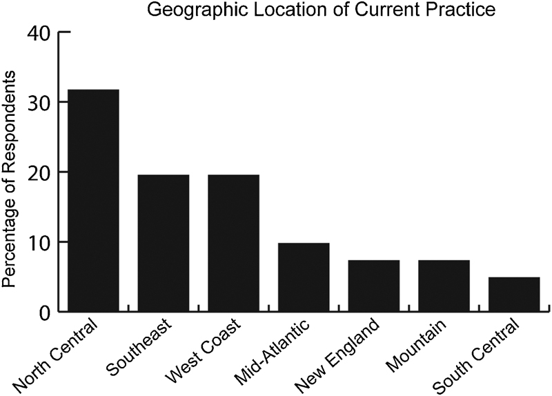
Geographic location of respondents in current practice.
Responses from neurotology-trained physicians comprised 48.8% of all responses, with rhinology-trained physicians and head and neck oncology–trained physicians making up 29.3 and 21.9% of responses, respectively. The main reasons that respondents decided to pursue their chosen fellowship were clinical interest and an important mentor, with salary being a lesser consideration (Fig. 2). The mean duration of practice was 2.1 years. Academic practice was more common than private practice (80.5 vs. 19.5%), and most respondents were affiliated with a dedicated skull base center (58.5 vs. 41.5%) (Table 1). Nearly all respondents reported working with residents (97.6%) and many also with fellows (43.9%).
Fig. 2.
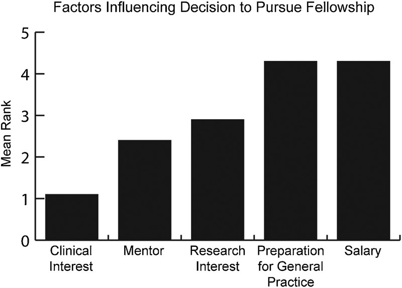
Ranking of factors influencing decision to pursue fellowship.
In residency, rhinology-trained physicians performed fewer than 20 skull base cases in 58.3% of respondents, 21 to 50 cases in 33.3% of respondents, and 76 to 100 cases in 8.3% of respondents (Fig. 3A). Head and neck oncology–trained physicians performed fewer cases in residency, with 88.9% of respondents performing fewer than 20 cases. Neurotology-trained physicians performed fewer than 20 cases in 50% of respondents, 21 to 50 cases in 25% of respondents, and more than 50 cases in 25% of respondents. Endoscopic ear surgery was rarely performed by neurotology-trained physicians during residency, with 100% of respondents performing fewer than 20 endoscopic ear surgery cases and 80% of these performing 0 cases (Fig. 4).
Fig. 3.
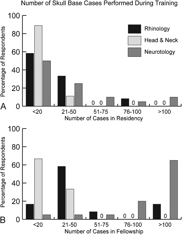
Number of skull base cases performed in residency (A) and fellowship (B) stratified by fellowship completed and reported as a percentage of respondents.
Fig. 4.
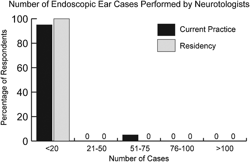
Number of endoscopic ear cases performed by neurotology-trained physicians in residency and annually in current practice.
During fellowship, rhinology-trained physicians performed fewer than 20 skull base cases in 16.7% of respondents, 21 to 50 cases in 58.3% of respondents, and more than 50 cases in 25% of respondents (Fig. 3B). Head and neck oncology–trained physicians performed fewer skull base cases with 66.7% of respondents performing fewer than 20 cases and 33.3% of respondents performing 21 to 50 cases. Neurotology-trained physicians performed more than 75 skull base cases in 85% of respondents during fellowship.
Among rhinology-trained physicians, 75% of respondents devoted less than 20% of their practice to skull base surgery, 16.7% of respondents devoted 21 to 50% of their practice, and 8.3% of respondents devoted greater than 50% of their practice to skull base surgery (Fig. 5). Head and neck oncology–trained physicians devoted less than 20% of their practice to skull base surgery in 100% of respondents. Neurotology-trained physicians devoted less than 20% of their practice to skull base surgery in 65% of respondents and 21 to 50% in 35% of respondents.
Fig. 5.
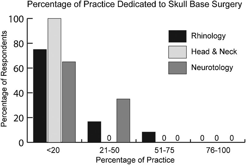
Percentage of current practice devoted to skull base surgery stratified by fellowship completed and reported as a percentage of respondents.
The total number of endoscopic skull base cases performed annually in current practice by rhinology-trained physicians was fewer than 20 in 50%, 21 to 50 in 33.3%, and more than 50 in 16.7% (Fig. 6A). Head and neck oncology–trained physicians performed fewer than 20 endoscopic skull base cases per year in 100% of respondents. The total number of open skull base cases performed annually in current practice by rhinology-trained physicians was fewer than 20 in 83.3% of respondents and more than 20 in 16.7% (Fig. 6B). Head and neck oncology–trained physicians performed less than 20 open skull base cases per year in 100% of respondents. Neurotology-trained physicians performed fewer than 20 skull base cases per year in current practice in 55% of respondents, 21 to 50 cases in 35% of respondents, and more than 50 cases in 10% of respondents (Fig. 7). Endoscopic ear cases were rarely performed in current practice among neurotology-trained physicians, with 95% of respondents performing fewer than 20 cases per year (Fig. 4).
Fig. 6.
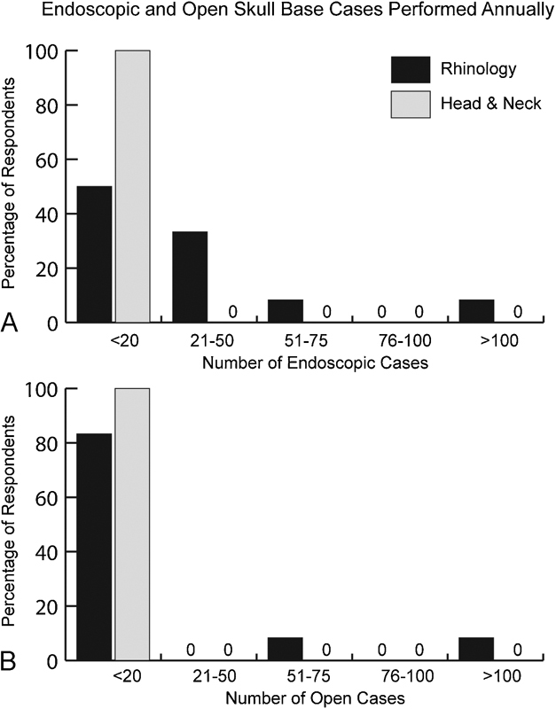
Number of endoscopic (A) and open (B) anterior skull base cases performed annually in current practice in rhinology and head and neck oncology–trained physicians reported as a percentage of respondents.
Fig. 7.
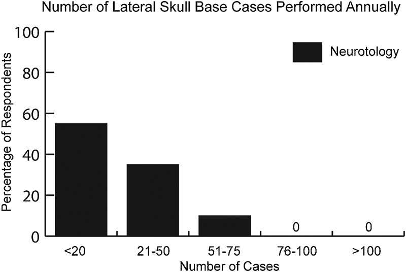
Number of lateral skull base cases performed annually in current practice among neurotology-trained physicians reported as a percentage of respondents.
Rhinology-trained and head and neck oncology–trained physicians were asked to report their comfort level while independently performing skull base procedures in general, as well as for a range of advanced endoscopic and open approaches to the anterior skull base. These confidence levels were stratified using a 5-point Likert scale ranging from 5 (extremely comfortable) to 1 (not comfortable at all), with the results presented in Fig. 8. Median comfort levels with skull base procedures in general were 4 among both rhinology-trained and head and neck oncology–trained physicians (p = 0.27). Overall median comfort levels with specific approaches ranged from 1 to 5. Rhinology-trained physicians reported a significantly higher comfort level, performing endoscopic CSF leak repair than head and neck oncology–trained physicians (5 vs. 3, p = 0.002), and had statistically significantly higher comfort levels with more advanced endoscopic approaches. Head and neck oncology–trained physicians reported a statistically significantly higher comfort level with open skull base approaches than rhinology-trained physicians (5 vs. 4, p = 0.02).
Fig. 8.
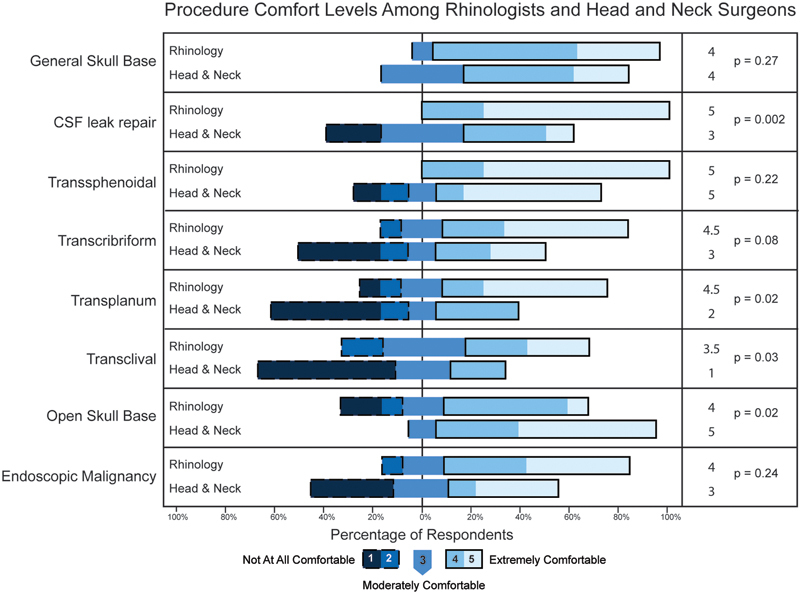
Comfort levels performing a range of skull base surgical approaches by rhinology and head and neck oncology–trained physicians reported as a distribution of responses ranging from 1 (not at all comfortable) to 5 (extremely comfortable). Median response scores and p values are shown in the right column.
Neurotology-trained physicians were asked to report their comfort level while independently performing skull base procedures in general, as well as for a variety of approaches to the lateral skull base and endoscopic ear surgery using the same scale as described above. The median comfort level for skull base surgery in general was 5, whereas the level for endoscopic ear surgery in general was 2 (Fig. 9). Median comfort levels for specific approaches to the lateral skull base ranged were 5 for all approaches, and the comfort level associated with endoscopic removal of middle ear cholesteatoma was 2.
Fig. 9.
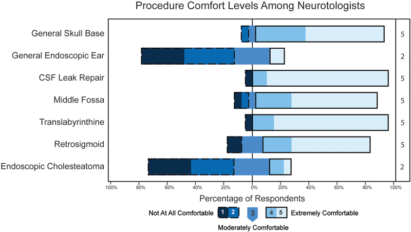
Comfort levels performing a range of lateral skull base surgical approaches and endoscopic ear surgery by neurotology-trained physicians reported as a distribution of responses ranging from 1 (not at all comfortable) to 5 (extremely comfortable). Median response scores are shown in the right column. CSF, cerebrospinal fluid.
Discussion
The field of skull base surgery has rapidly incorporated advanced endoscopic endonasal methods and instrumentation to complement traditional open craniofacial approaches. As part of a multidisciplinary team, otolaryngologists, working closely with neurosurgeons, play a key surgical role in these complex procedures.1 Recent surveys of the NASBS and ARS memberships revealed that endoscopic endonasal skull base techniques are now widely used.6 7 In light of the shift toward more endoscopic endonasal skull base procedures, we developed a survey to learn more about exposure and experience with skull base techniques during otolaryngology-head and neck surgery training, as well as formulate a snapshot of the skull base surgery practice patterns of young otolaryngologists.
The pathway to becoming a skull base surgeon for an otolaryngologist typically involves fellowship training in rhinology, head and neck oncology, neurotology, or a combined fellowship program. We therefore surveyed physicians who completed these fellowships within the past 5 years. Responses were received from a broad sample of young physicians from across the United States with a mean time in current practice of 2.1 years. Respondents were overwhelmingly located in academic settings with resident and fellow contact, and the majority worked in locations with a dedicated skull base program.
Exposure to skull base procedures during residency was similar across all respondents—61% were involved in fewer than 20 cases during residency, with rhinology- and neurotology-trained physicians reporting somewhat increased involvement. In fellowship, rhinology-trained physicians performed more skull base cases than their head and neck oncology–trained colleagues (Fig. 3B). Rhinologists almost exclusively use endoscopic endonasal surgical approaches, and the increased exposure to skull base surgery during rhinology fellowship training appears to parallel the shift toward endoscopic endonasal approaches to the cranial base. In addition, many endoscopic endonasal approaches are used for benign disease, and as such would not be routinely performed in head and neck oncology fellowship training programs resulting in lower overall case numbers. Rhinology-trained physicians also devoted a higher percentage of their practice to skull base surgery and performed more endoscopic skull base procedures in early practice after completion of fellowship training.
Regardless of fellowship training, respondents reported a high overall comfort level with skull base procedures in general. A lower comfort level was noted with more advanced endoscopic endonasal approaches (i.e., transplanum and transclival approaches), with statistically significantly higher comfort levels in rhinology-trained physicians compared with those who were head and neck oncology–trained. On the other hand, head and neck oncology–trained physicians reported comfort levels with open skull base approaches that were statistically significantly higher than those of rhinology-trained physicians. It is likely that advanced sinonasal malignancy cases requiring open surgical approaches are more frequently addressed in head and neck oncology practices, which would further increase exposure and comfort with open approaches among trainees.
Neurotology-trained physicians performed a large number of lateral skull base cases during fellowship, which is consistent with ACGME (Accreditation Council for Graduate Medical Education) accreditation requirements in the setting of a 2-year fellowship. In early practice, 65% of neurotology-trained physicians devoted less than 20% of their practice to skull base cases, although 45% of respondents performed more than 20 skull base cases in a typical year. Comfort levels with lateral skull base approaches were very high overall, whereas comfort levels with endoscopic ear surgery were quite low, consistent with little or no exposure to endoscopic ear techniques during training. Endoscopic techniques for ear surgery are not being widely used at this time, although proponents report promising outcomes for removal of middle ear cholesteatoma compared with traditional microscopic techniques.9 10 There are also reports of endoscopic techniques being used both exclusively and in combination with traditional approaches for lateral skull base lesions.11 12 It is unclear if endoscopic ear techniques will achieve widespread adoption similar to endoscopic methods in rhinology and endoscopic endonasal skull base surgery, and at this time neurotology training provides little or no exposure to endoscopic techniques.
This survey has important limitations that deserve mention. Though the response rate in this study was typical for surveys of this type, the overall response rate was low with disproportionate responses among each specialty. The experiences of those who did not respond are unknown, and all data were self-reported as an estimated range without verification. It is also important to note that this study is subject to recall bias, and true case numbers may be over- or underestimated. Respondents also may have had different interpretations of what constitutes a skull base case, which could alter the reported numbers. In addition, the reported comfort levels associated with performing procedures are subjective. The reported data apply only to the target population studied, and they cannot be generalized to otolaryngologists with differing characteristics (i.e., longer time in practice).
Conclusion
Skull base surgery is a rapidly advancing field, and the widespread adoption of endoscopic endonasal techniques is facilitating new approaches to address benign and malignant lesions. Otolaryngologists are key drivers of this transition, and this survey demonstrates that recent fellowship graduates are getting exposure to skull base procedures in training and incorporating these procedures into their practices at an early stage. Neurotology training affords excellent exposure to traditional lateral skull base procedures and recent graduates have a high level of confidence when performing these procedures independently. Exposure to burgeoning endoscopic ear methods is minimal in current training programs. Both rhinology and head and neck oncology training yield a high level of comfort performing skull base procedures in general after graduation. However, rhinology fellowship training is associated with a higher level of exposure to endoscopic anterior skull base surgery and may translate into improved expertise in advanced endoscopic endonasal approaches.
Acknowledgments
The authors thank rhinology, head and neck oncology, and neurotology program directors for providing contact information, as well as all survey participants. We thank the American Neurotology Society for disseminating the survey electronically.
Financial Disclosures The authors have no financial disclosures to report. Conflict of Interest None.
Note
These survey results were presented as a poster at the 2015 North American Skull Base Society Meeting.
References
- 1.Snyderman C, Carrau R, Kassam A. Who is the skull base surgeon of the future? Skull Base. 2007;17(6):353–355. doi: 10.1055/s-2007-986427. [DOI] [PMC free article] [PubMed] [Google Scholar]
- 2.Su S Y, Kupferman M E, DeMonte F, Levine N B, Raza S M, Hanna E Y. Endoscopic resection of sinonasal cancers. Curr Oncol Rep. 2014;16(2):369. doi: 10.1007/s11912-013-0369-6. [DOI] [PubMed] [Google Scholar]
- 3.Batra P S, Luong A, Kanowitz S J. et al. Outcomes of minimally invasive endoscopic resection of anterior skull base neoplasms. Laryngoscope. 2010;120(1):9–16. doi: 10.1002/lary.20680. [DOI] [PubMed] [Google Scholar]
- 4.Higgins T S, Thorp B, Rawlings B A, Han J K. Outcome results of endoscopic vs craniofacial resection of sinonasal malignancies: a systematic review and pooled-data analysis. Int Forum Allergy Rhinol. 2011;1(4):255–261. doi: 10.1002/alr.20051. [DOI] [PubMed] [Google Scholar]
- 5.Little R E, Taylor R J, Miller J D. et al. Endoscopic endonasal transclival approaches: case series and outcomes for different clival regions. J Neurol Surg B Skull Base. 2014;75(4):247–254. doi: 10.1055/s-0034-1371522. [DOI] [PMC free article] [PubMed] [Google Scholar]
- 6.Batra P S, Lee J, Barnett S L, Senior B A, Setzen M, Kraus D H. Endoscopic skull base surgery practice patterns: survey of the North American Skull Base Society. Int Forum Allergy Rhinol. 2013;3(8):659–663. doi: 10.1002/alr.21151. [DOI] [PubMed] [Google Scholar]
- 7.Lee J T, Kingdom T T, Smith T L, Setzen M, Brown S, Batra P S. Practice patterns in endoscopic skull base surgery: survey of the American Rhinologic Society. Int Forum Allergy Rhinol. 2014;4(2):124–131. doi: 10.1002/alr.21248. [DOI] [PubMed] [Google Scholar]
- 8.Snyderman C, Kassam A, Carrau R, Mintz A, Gardner P, Prevedello D M. Acquisition of surgical skills for endonasal skull base surgery: a training program. Laryngoscope. 2007;117(4):699–705. doi: 10.1097/MLG.0b013e318031c817. [DOI] [PubMed] [Google Scholar]
- 9.Presutti L, Gioacchini F M, Alicandri-Ciufelli M, Villari D, Marchioni D. Results of endoscopic middle ear surgery for cholesteatoma treatment: a systematic review. Acta Otorhinolaryngol Ital. 2014;34(3):153–157. [PMC free article] [PubMed] [Google Scholar]
- 10.Tarabichi M, Nogueira J F, Marchioni D, Presutti L, Pothier D D, Ayache S. Transcanal endoscopic management of cholesteatoma. Otolaryngol Clin North Am. 2013;46(2):107–130. doi: 10.1016/j.otc.2012.10.001. [DOI] [PubMed] [Google Scholar]
- 11.Presutti L, Alicandri-Ciufelli M, Rubini A, Gioacchini F M, Marchioni D. Combined lateral microscopic/endoscopic approaches to petrous apex lesions: pilot clinical experiences. Ann Otol Rhinol Laryngol. 2014;123(8):550–559. doi: 10.1177/0003489414525342. [DOI] [PubMed] [Google Scholar]
- 12.Presutti L, Nogueira J F, Alicandri-Ciufelli M, Marchioni D. Beyond the middle ear: endoscopic surgical anatomy and approaches to inner ear and lateral skull base. Otolaryngol Clin North Am. 2013;46(2):189–200. doi: 10.1016/j.otc.2012.12.001. [DOI] [PubMed] [Google Scholar]


