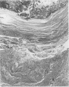
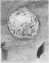
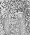
Abstract
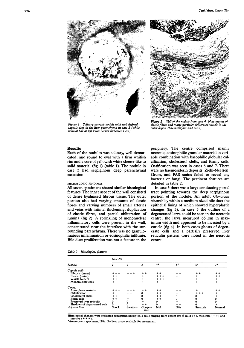
AIMS: To report further cases of solitary necrotic nodule of the liver and to study its nature. METHODS: Seven nodules were retrieved from 4000 necropsy and surgical liver specimens coming to light over the past five years. All of them satisfied the diagnostic criteria of solitary necrotic nodule: a solid lesion with a central necrotic core and a hyalinised fibrotic capsule containing elastic fibres. Their clinicopathological features were reviewed. RESULTS: The nodules were incidental findings at surgery or necropsy in four men and three women whose ages ranged from 48 to 79 years (mean 63.7 years). Four were found in the right lobe and three in the left. Six were subcapsular and only one deep in the parenchyma, with sizes ranging from 0.3-2.5 cm. Each of them was solitary, well demarcated, and round to oval with a firm, whitish rim and a core of yellowish white cheese-like to solid material. In addition to the basic architecture, there were a number of common and undescribed histological features: presence of varying numbers of small mural vessels with intimal fibrosis and obliteration, presence of cholesterol clefts and foamy cells among necrotic material, and sparsity of inflammatory cells. In the two cases where ghosts of degenerated cells and partially preserved liver reticulin pattern were noted, worms were identified, one being Clonorchis sinensis. CONCLUSIONS: The entity is believed to be a "burnt-out phase" of a variety of benign lesions. Parasitic infestation is another possible cause, and presence of ghosts of degenerate cells, partially preserved liver reticulin pattern, cholesterol clefts and foamy cells among necrotic material are auxiliary features pointing to such an aetiology. The variation in morphological fine details reflects both the lesion's diverse pathogenesis and the fact that it can be of varying duration.
Full text
PDF

Images in this article
Selected References
These references are in PubMed. This may not be the complete list of references from this article.
- Berry C. L. Liver lesions in an autopsy population. Hum Toxicol. 1987 May;6(3):209–214. doi: 10.1177/096032718700600306. [DOI] [PubMed] [Google Scholar]
- Berry C. L. Solitary "necrotic nodule" of the liver: a probable pathogenesis. J Clin Pathol. 1985 Nov;38(11):1278–1280. doi: 10.1136/jcp.38.11.1278. [DOI] [PMC free article] [PubMed] [Google Scholar]
- Mondou E. N., Gnepp D. R. Hepatic granuloma resulting from Enterobius vermicularis. Am J Clin Pathol. 1989 Jan;91(1):97–100. doi: 10.1093/ajcp/91.1.97. [DOI] [PubMed] [Google Scholar]
- Shepherd N. A., Lee G. Solitary necrotic nodules of the liver simulating hepatic metastases. J Clin Pathol. 1983 Oct;36(10):1181–1183. doi: 10.1136/jcp.36.10.1181. [DOI] [PMC free article] [PubMed] [Google Scholar]
- Shepherd N. A. Solitary necrotic nodule. J Clin Pathol. 1990 Apr;43(4):348–349. doi: 10.1136/jcp.43.4.348. [DOI] [PMC free article] [PubMed] [Google Scholar]
- Sundaresan M., Lyons B., Akosa A. B. 'Solitary' necrotic nodules of the liver: an aetiology reaffirmed. Gut. 1991 Nov;32(11):1378–1380. doi: 10.1136/gut.32.11.1378. [DOI] [PMC free article] [PubMed] [Google Scholar]


