Abstract
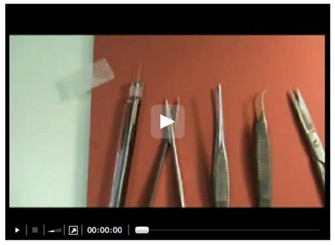
The MIND method involves intraductal injection of patient derived ductal carcinoma in situ (DCIS) cells and DCIS cell lines (MCF10DCIS.COM and SUM225) inside the mouse mammary ducts [Video 1 and Figure 1 in Behbod et al. (2009)]. This method mimics the normal environment of DCIS and facilitates study of the natural progression of human DCIS, i.e., their initial growth as carcinoma in situ within the ducts, followed by invasion into the stroma through the myoepithelial cell layer and basement membrane (Behbod et al., 2009; Valdez et al., 2011). In order to demonstrate that transplantation procedure is successful, the transplanted mammary glands may be excised as early as two weeks following intraductal injection of cells followed by Hematoxylin and Eosin (H&E) staining and/or immunofluorescence staining using human specific cytokeratin 5 and/or 19 [please see Figures 2–4 in Behbod et al. (2009)]. Additionally, the presence of trypan blue inside the mouse mammary ducts immediately following intraductal injection is the best indicator that the injection was successful (Video 1 starting at 4:33 sec).
Materials and Reagents
Hamilton syringe, 50 μl capacity, with a 30 gauge blunt-ended fixed 1/2-inch needle (Hamilton, catalog number: 80608)
1 ml Tuberculin (TB) syringe (Benton Dickenson, catalog number: 309625)
Wound clips (Becton Dickinson, catalog number: 427631)
-
Animals: 8–12 week old female immunocompromised mice
Note: NSG, NOD scid gamma [NOD.Cg-Prkdcscid Il2rgtm1Wjl/SzJ (HE JACKSON LABORATORY, catalog number: 005557)]. Alternatively, SCID/beige mice (Fox Chase SCID® Beige CB17.Cg-PrkdcscidLystbg-J/Crl) may be used for the injection of DCIS cell lines only. While a formal comparison has not been made, patient derived DCIS epithelial cells seem to grow more efficiently in the NSG mice.
Cells of interest [i.e. cell lines MCF10DCIS.COM and SUM225 (Behbod Lab, 2009 #329) or patient derived DCIS epithelial cells] in single cells
-
PBS (pH 7.4, 1x), sterile (Life technologies, catalog number: 10010023)
Note: Currently, it is “Thermo Fisher Scientific, catalog number: 10010023”.
Trypan blue 0.4%, sterile (Sigma-Aldrich, catalog number: T8154)
70% ethanol
Sterile water
Nembutal Sodium Solution (Pentobarbital sodium injection, USP) (Akorn, Oak Pharmaceuticals, catalog number: NDC 7647850120)
Ketofen™ (Ketoprofen) (Patterson Veterinary Supply, catalog number: 10004029)
Equipment
-
Scissors Small scale suitable for mice
Sharp-Blunt (Fine Science Tool, catalog number: 14028-10)
Castroviejo Spring Scissors (Fine Science Tool, catalog number: 15017-10)
Balled Scissors (Fine Science Tool, catalog number: 14086-09)
Clippers (Fine Science Tool, catalog number: 1501710)
Rounded end flat handle tweezers (ROBOZ, model: RS5095)
Spring scissor (Fine Science Tool, catalog number: 1501710)
Balled scissor (Fine Science Tool, catalog number: 1408609)
Rounded end tweezers (Fine Science Tool, catalog number: 1105210)
Rat tooth tweezers (Fine Science Tool, catalog number: 1105310)
Clip applier (Becton Dickinson, catalog number: 427630)
Heat pad (Sunbeam Products, model: PN126985900)
Clip remover (Becton Dickinson, catalog number: 427638)
Procedure
A. Prepare the cells for injection
Resuspend cells in PBS with 0.04% trypan blue. For cell lines, the final concentration should be 5,000–10,000 cells/μl. For primary cells the concentration should be 12,500–17,500 cells/μl. Trypan blue will indicate the presence of cells inside the duct after the injection procedure.
Be sure all instruments have been sterilized and the Hamilton syringe washed by alternating pulling up sterile water and 70% alcohol, ending with sterile water.
B. Prepare the mice for surgery
Weigh the mouse.
Using 1 ml TB syringe, administer Nembutal 40 μg/g body intraperitoneal (this will provide 1 h of sedation).
Wait for mouse to fall asleep; confirm by checking hind limb reflex by pinching foot.
Use clippers to shave the abdomen exposing the inguinal nipples and taking care not to cut them.
Using 1 ml TB syringe, administer Ketofen™ (ketoprofen) 5 mg/kg intraperitoneal for pain relief.
Place the mouse on surgical board and secure limbs with surgical tape.
Sterilize the abdomen with 70% alcohol.
C. Surgery
Cut off the tip of the nipples of the inguinal glands so that the syringe can be directly inserted through the nipple. Use the rounded end flat handle tweezers to hold the skin under the nipple and force it to protrude. Use the spring scissor to cut off about the top 0.5 mm of the nipple. Take care not to remove too much or the syringe will slide under the skin. A properly cut nipple will have a volcano cone shape.
Using the balled scissors, make an inverted Y-incision on the abdomen beginning on the midline slightly below the inguinal nipple and extending up about 1 inch. The other two cuts of the incision should extend between and fourth and fifth nipples on either side.
Using the rounded end tweezers and rat tooth tweezers, separate the skin covering the inguinal mammary fat pads to expose the inguinal gland.
Using the Hamilton syringe, draw up two microliters of PBS (with 0.04% trypan blue) containing cells at the proper concentration; holding the skin flap up, insert the tip of the syringe into the nipple at a 90 degree angle to the skin flap and carefully inject the cells. Trypan blue will make the injected liquid visible in the duct.
Reposition the skin flaps and suture with wound clips.
D. Post-surgery
Allow mice to recover on heat pad set on the lowest setting.
Administer Ketofen™ 5 mg/kg intraperitoneal 24 h later.
Remove clips after 7–10 days.
Video 1. Intraductal injection.

Note: The video contains material that can be disturbing to some viewers.
Acknowledgments
This protocol was developed in the Department of Molecular and Cellular Biology, Baylor College of Medicine and Department of Pathology and Laboratory Medicine, University of Kansas Medical School. We thank David Edwards for assistance in the production of the procedure video. The work was supported by NCI (1K99/R00 CA127462) to FB and NCI (1K22 CA160587-01A1) to KV.
References
- 1.Behbod F, Kittrell FS, LaMarca H, Edwards D, Kerbawy S, Heestand JC, Young E, Mukhopadhyay P, Yeh HW, Allred DC, Hu M, Polyak K, Rosen JM, Medina D. An intraductal human-in-mouse transplantation model mimics the subtypes of ductal carcinoma in situ. Breast Cancer Res. 2009;11(5):R66. doi: 10.1186/bcr2358. [DOI] [PMC free article] [PubMed] [Google Scholar]
- 2.Valdez KE, Fan F, Smith W, Allred DC, Medina D, Behbod F. Human primary ductal carcinoma in situ (DCIS) subtype-specific pathology is preserved in a mouse intraductal (MIND) xenograft model. J Pathol. 2011;225(4):565–573. doi: 10.1002/path.2969. [DOI] [PMC free article] [PubMed] [Google Scholar]


