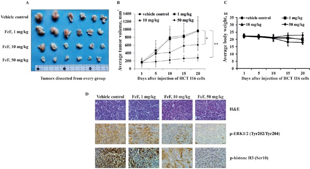
Figure 5. The effect of FeF on tumor growth and phosphorylation of TOPK downstream signaling targets in HCT 116 xenograft mouse model.

A. Tumors dissected from every group. B. FeF significantly inhibited colon tumor growth. The average tumor volume of vehicle-treated control mice and FeF-treated mice plotted over 20 days after HCT 116 cells injection. Data are shown as means ± standard deviation of measurements. The asterisk (*) indicates a significant inhibition of tumor growth by FeF (*p < 0.05, **p < 0.01). C. FeF does not affect mice body weight. Body weight from treated or untreated groups of mice was measured every other day. D. H&E staining and the immunohistochemical analysis of tumor tissues. Treated or untreated groups of mice were euthanized and tumors extracted. Colon cancer tissue slides were prepared with paraffin sections after fixation with formalin and then stained with H&E or p-ERK1/2 and p-histone H3. The magnification of representative photos for H&E and the immunohistochemical staining is ×40.
