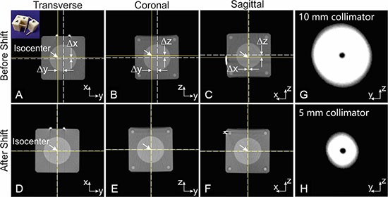
Figure 5. The mechanical precision test for the animal stage translation and collimator positioning.

(A–C): the orthogonal CBCT views of the mini-ball cube phantom before shift. The solid crosshair indicates the imaging/radiation isocenter, and dash crosshair the ball center. Δx, Δy, Δz are 0.39, 1.56 and 0.52 mm respectively. The ball center was aligned to the isocenter with only one stage adjustment. (D–F) confirmed the coincidence of isocenter and ball center after shift. The insert in the first panel is a photo of the mini-ball cube phantom. (G and H) are the x-ray exposures of a radiopaque ball centered at isocenter through 5 and 10 mm collimator respectively. The ball center and center of x-ray field are well aligned, indicating the coincidence of the isocenter and central axis of collimated radiation beams. The imperfect round shape of the collimated beam may be caused by some residual lead left on the inner edge of the collimator aperture.
