
Figure 6.
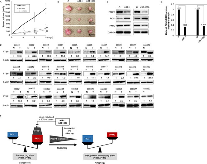
(A–D) MiR-1 and -133b have anti-tumor effects even in vivo. (A) Time course of tumor size in mice injected with control miRNA, miR-1 or -133b (n = 5). (B) Representative photograph of tumors. Left is the control. Middle is treatment with miR-1; and right, treatment with miR-133b. (C) PTBP1 and PTBP1-related protein expression in control, miR-1or -133b-treated tumor tissues as determined by Western blot analysis. (D) The ratio of PKM2/PKM1 protein was calculated based on densitometric value of PKM1 and PKM2 in “C”. Numbers represent the PKM2/PKM1 ratio of each sample, with the control taken as 1.000. (E) PTBP1 was overexpressed in clinical colorectal tumors. PTBP1 expression in 25 colorectal cancer (case 1–25) and 5 adenoma (case 26–30) samples as determined by Western blot analysis. Details of the characteristics of the samples are given in Supplementary Table 1. Densitometric values of PTBP1 were calculated. β-actin was used as the control. (F) Schematic diagram of the effects of miR-1 and -133b on colorectal tumors. MiR-1 and -133b directly bind to PTBP1, and PTBP1 is very frequently overexpressed in colorectal tumors and even in adenomas, with the overexpression induced by down-regulation of these miRs. Ectopic expression of these miRs induced switching of PKM isoform expression from PKM2 to PKM1 through down-regulation of PTBP1. These miRs induce autophagic cell death and the activation of oxidative stress through this pathway. These miRs and PTBP1 have great potential as therapeutic targets or as biomarkers in colorectal tumors.