Abstract
In the present study, in vitro interaction of nisin and perilla oil (PO) against 20 food-borne isolates of L. monocytogenes and S. aureus were assessed using a checkerboard microdilution method. Synergism was observed in tested strains with the fractional inhibitory concentration indexs (FICIs) ranges from 0.125–0.25 and 0.19–0.375, respectively. Scanning electron microscopy was carried out to investigate the effect of nisin and PO on the integrity of cell wall and membrane of L. monocytogenes and S. aureus. The results showed that nisin and PO were more effective in damaging cell wall and membrane in combination.
Keywords: Listeria monocytogenes, Staphylococcus aureus, Nisin, Perilla oil, Synergism, Milk
Introduction
Foodborne illnesses and food poisoning, a major concern for consumers, are cause for concern globally. The diseases are often caused by food contamination with pathogenic bacteria such as Listeria monocytogenes and Staphylococcus aureus due largely to poor sanitary habits or storage conditions. A majority of these outbreaks has been primarily due to the consumption of dairy and ready-to-eat meat products contaminated with L. monocytogenes (Mead et al. 2006). S. aureus often exists in food as a foodborne pathogen and is also associated with toxin mediate food poisoning (Rasooly and Do 2009). Milk and dairy products are often implicated in staphylococcal outbreaks. It drives us to find better ways to extend the shelf-life of foods.
There are a lot of strategies to prevent pathogenic and spoilage microorganisms in food, and using chemical preservative is one of the important ways. Over the past decade, there is a growing awareness that the continued widespread use of chemical preservatives might pose a serious health problem (Kito et al. 2002). Therefore, the application of natural antimicrobials has been of great interest to researchers and food manufacturers. Natural antimicrobial compounds which have been approved as a safe food additive can be applied directly to foods and food products to protect food quality and extend shelf life of food by inhibiting or slowing the growth of spoilage bacteria. Nisin, a bacteriocin or natural antibacterial peptide secreted from Lactococcus lactis, is active against a wide range of Gram-positive bacteria (Gálvez et al. 2007). However, some microorganisms may develop resistance to this compound and regrow under optimum conditions, such as L. monocytogenes (Gravesen et al. 2002), Clostridium botulinum (Mazzotta et al. 1997), and S. aureus (Peschel et al. 1999). The emergence of resistant strains to antimicrobial agents justifies the search for new antibacterial strategies. For this purpose, the combined utilization of antimicrobial compound could enhance the efficacy of antimicrobial compound, restore the sensitivity to the compound. It has also been demonstrated that the combined use of efflux pump inhibitors and antimicrobial compound delays the emergence of resistance to these drugs (Kim et al. 2011).
Perilla seeds (Perilla frutescens), a natural medicine found in eastern Asia, is primarily used as both a medicinal and culinary herb (Ito 2008). Its essential oil, perilla oil (PO), is used as cooking oil in Asian countries, and it is one of the naturally-flavored oils and obtained usually from roasted seeds to give good characteristic flavor. Furthermore, PO, an excellent source of plant-derived ω-3 fatty acid, is considered to be a healthy oil, nutritional studies have shown that perilla oil is beneficial for healthy aging and learning performance (Okuyama et al. 2007). Reportedly, PO has a variety of medicinal activities, such as antitumor, anti-inflammatory, hypotension, antiatherosclerosis, and antianaphylactic shock activities (Ezaki et al. 1999). Furthermore, the anti-microbial and anti-fungal properties of PO was reported, such as anti-trichophyton (Inouye et al. 2006), but no report has been published on the effect of PO against L. monocytogenes, and a few reports about its anti-S. aureus activity (Lv et al. 2011).
To the best of our knowledge there has been no research reported on the activity of nisin and PO incorporated together against L. monocytogenes or S. aureus. Therefore, the aim of this study was to evaluate the effectiveness of PO in combination with nisin to inhibit the growth of both L. monocytogenes and S. aureus, especially in pasteurized milk.
Materials and methods
Test bacteria, and natural antimicrobials
Twenty food-borne isolates of L. monocytogenes and S.aureus were obtained from Jilin Enrty-Exit Inspection and Quarantine Bureau. The quality control strains, ATCC 29213 and ATCC 19115, were obtained from the China Medical Culture Collection Center (CMCC). The strains were preserved as stabs in trypticase soy agar (TSA; BD Difco™), transferred to 10 ml of trypticase soy broth (TSB; BD Difco™) using an inoculation loop, and incubated overnight at 37 °C. Mueller-Hinton broth (MHB, BD Difco™) and brain-heart infusion (BHI) broth (BD Difco™) was used to determine the minimum inhibitory concentration (MIC) for S.aureus and L. monocytogenes, respectively. Nisin (Sigma-Aldrich) was resuspended in sterile 0.02 N HCl, and PO (National Institutes for Food and Drug Control) was dissolved in dimethyl sulfoxide (Sigma-Aldrich; DMSO). The final concentration of DMSO in all experimental groups was 0.1 %, including control groups.
MIC determination
The MICs of nisin and PO against S. aureus and L. monocytogenes were determined using the standard broth microdilution method, as described by the CLSI guidelines (Anonymous 2009, 2010). MIC was defined as the lowest concentration of antibiotic that produced the complete inhibition of visible growth.
Checkerboard dilution test
The synergistic antimicrobial effect of nisin and PO was studied using the checkerboard method against L. monocytogenes and S.aureus in nutrient broth (Pillai and Moellering 2005). Serial 2-fold dilutions of two different antimicrobial agents were mixed in MHB or BHI broth with final concentrations of the compounds ranged from 1/32 to 4 times the MIC for nisin and from 1/256 to 4 times the MIC for PO. The checkerboard plates were inoculated with 105 CFU/ml and incubated at 37 °C for 24 h. The concentration of the individual compound in the combination of nisin and PO in which the growth of organisms is completely inhibited is taken as the MIC of the individual compound in the combination. The combination for each reference strain was tested in triplicate. The data produced by the checkerboard assay were analyzed in terms of the fractional inhibitory concentration index (FICI) using the following equation:
where MICAalone and MICBalone are the MICs values of compound A and B when acting alone, and CAcomb and CBcomb are the concentrations of compound A and B at the isoeffective combinations. The results were interpreted as a synergistic effect if FICI ≤0.5; as an additive or indifferent effect if 0.5 < FICI ≤4 and as an antagonistic effect if FICI >4 (Oliveira et al. 2010).
Challenge tests in pasteurized milk
Commercial pasteurized milk was inoculated with 1 × 10⁶ CFU/ml of S. aureus and L. monocytogenes. Immediately after, nisin (16 μg/ml), PO (1 mg/ml) and a mixture of both, were also added. The milk was incubated at 37 °C without shaking and samples were taken at various times (0, 3, 6, 10 and 24 h). Survival of S. aureus and L. monocytogenes was determined by plating decimal dilutions on plates of Baird-Parker selective agar (Qingdao Hope Bio-Technology, China) and BD PALCAM Listeria agar which were incubated at 37 °C for 24 h and at 30 °C for 48 h, respectively. All experiments were conducted in triplicate, and the results are reported as the mean values. Synergism was defined as a decrease in the colony count of ≥2 log10 CFU/ml relative to the count that was obtained with the most active single compound. Antagonism was defined as a decrease of <2 log10 CFU/ml with respect to the least active compound (Jacqueline et al. 2005).
Agar disk diffusion assay
A 100-μl aliquot of a 106 CFU/ml suspension was spread uniformly on agar plates. Paper disks (6 mm diameter) impregnated with nisin and PO (5 μl of different concentration) alone or in combination were placed onto the agar surface, and the inhibition zones were measured after incubation at 37 °C for 24 h. Microbial inhibition was visually appraised as the diameter of the inhibition zones surrounding the disks (disk diameter included) and recorded in millimeter. The diameters of the inhibition zones were measured with a digital caliper. The agar disk diffusion tests were performed in triplicate. The antimicrobial activity of plant essential oils (EOs) can be classified into three levels (Rota et al. 2004): weak activity (inhibition zone ≤12 mm), moderate activity (12 mm < inhibition zone <20 mm) and strong activity (inhibition zone ≥20 mm).
Scanning electron microscopy (SEM)
Logarithmic phase bacteria were allowed to adhere to polylysine-coated coverslips for 10 h and were exposed to nisin (32 μg/ml) and PO (2 mg/ml) alone or in combination for 3 h. The cells were washed in PBS after incubation and were fixed for 2 h at 4 °C with 2.5 % glutaraldehyde. The cells were washed in the same buffer and were post-fixed for 30 min with osmium tetroxide in 0.1 M of cacodylate buffer (pH 7.2). The samples were dehydrated using sequential ethanol concentrations ranging from 30 to 100 %. The ethanol was replaced with tertiary butyl alcohol. Cells were freeze-dried with a vacuum freeze drier (Hitachi ES-2030), coated with an ion sputtering apparatus (Hitachi E-1010), and observed through SEM (Hitachi S-3400 N). The bacterial cells that were not exposed to antimicrobials were similarly processed and used as control. All tests were performed in triplicate.
Membrane damage
The damage to the bacterial membrane was assessed using A LIVE/DEAD BacLight kit containing SYTO-9 and propidium iodide dyes from Molecular Probes (Invitrogen). The organisms were grown overnight in Cation-adjusted Mueller-Hinton broth (CAMHB) at 37 °C under aeration. The culture was diluted 40 times with fresh CAMHB and grown to an OD600 of 0.5–0.6. The bacterial suspension was centrifuged at 10,000×g for 15 min, and the cell pellet was washed once in filter-sterilized distilled water. The cell pellet was resuspended to 1/10 of the original volume and then diluted 1:20 into either water or water containing test compounds at MIC. Bacteria and compounds were incubated at 37 °C on a tube rocker for 1 h. At the end of the incubation period, a sample was removed for the CFU determination, and the remaining suspension was centrifuged at 10,000×g for 10 min, washed once in water, and resuspended to an OD670 of 0.325. A volume of 100 μl of the bacterial suspension was removed and added to a 96-well microtiter plates. An equal volume of the BacLight reagent was then added to each well and incubated for 15 min in the dark at room temperature. At the end of the incubation period, green fluorescence was read at 530 nm, and red fluorescence was read at 645 nm at an excitation wavelength of 485 nm. The ratio of green to red fluorescence intensities was normalized to the control group and expressed as a percentage of the control (Hilliard et al. 1999).
Statistical analysis
All samples were analyzed in triplicate, and the data are presented as the mean values ± standard deviations (SD) (n = 3). Independent Student t tests were used to determine statistical differences, and p ≤ 0.05 was considered significant.
Results
Antibacterial activity
The in vitro activities of the studied nisin or PO alone and in combination against standard and food-borne isolates of L. monocytogenes and S.aureus are summarized in Table 1. In the present study, nisin and PO showed different antimicrobial activities against the tested strains based on the calculated MIC. The MIC values for nisin and PO against twenty isolates ranged from 32 to 64 μg/ml and 1 to 2 mg/ml, respectively. The MIC values of nisin and PO against the ATCC 29213, ATCC 19115 name the microorganisms used were 16 μg/ml and 2 mg/ml, 16 μg/ml and 1 mg/ml, respectively. As expected, the MIC values for nisin against food-borne isolates were higher than the standard strains ATCC 29213 and ATCC 19115. Nisin has been shown to inhibit L. monocytogenes and S. aureus, and its MIC ranges were determined to be 740-l05 IU/ml (Benkerroum and Sandine 1988) and 2–32 μg/ml (Dosler and Gerceker 2012), respectively, consistent with the value calculated in the present study. Based on our knowledge, little work has been carried out on the antibacterial activity of PO. For PO, the MIC values against S. aureus ATCC 6538 were 5 μl/ml (~4.5 mg/ml) (Lv et al. 2011), which were a little higher than those calculated in the present study. However, no report has been published on the effect of PO against L. monocytogenes. Some new studies about nisin combined with other natural compounds have been performed. Liu et al. (2015) have demonstrated the synergistic antibacterial effect of the combination of ε-Polylysine and nisin against Enterococcus faecalis. And Phongphakdee and Nitisinprasert (2015) have studied that the combination inhibition activity ofnisin and ethanol on the growth inhibition of pathogenic gram negative bacteria. The biological activity research of PO has gained great progress, such as antibacterial and antioxidant activity. Qiu et al. (2011) have reported that PO inhibited the growth of S. aureus, and the production of α-toxin, SEA, SEB, and TSST-1 in S. aureus was also decreased by PO. Lee et al. (2015) have reported that the protective effect against oxidative stress by PO was increased in a roasting temperature-dependent manner by attenuating intracellular ROS formation.
Table 1.
In vitro minimum inhibitory concentration (MIC) of Nisin and PO against L. monocytogenes and S.aureus
| Strains | Median MIC (range) of compound | |||
|---|---|---|---|---|
| Alone | In combination | |||
| Nisin (μg/ml) | PO (mg/ml) | Nisin (μg/ml) | PO (mg/ml) | |
| S.aureus JL-10011 | 32 (32–64) | 2 (2) | 4 (4) | 0.25 (0.25–0.5) |
| S.aureus JL-10012 | 32 (32) | 2 (2) | 4 (4) | 0.25 (0.25) |
| S.aureus JL-10013 | 32 (32) | 2 (2) | 4 (4–8) | 0.25 (0.25) |
| S.aureus JL-10014 | 32 (32–64) | 1 (1) | 4 (4) | 0.25 (0.25) |
| S.aureus JL-10015 | 64 (64) | 2 (2) | 4 (4) | 0.25 (0.25–0.5) |
| S.aureus JL-10016 | 32 (32) | 2 (2) | 2 (2–4) | 0.25 (0.25) |
| S.aureus JL-10017 | 32 (32) | 1 (1) | 4 (4) | 0.25 (0.25) |
| S.aureus JL-10018 | 64 (64) | 2 (1–2) | 8 (8) | 0.25 (0.25) |
| S.aureus JL-10019 | 64 (64) | 2 (2) | 8 (8) | 0.25 (0.25) |
| S.aureus JL-10020 | 64 (32–64) | 2 (2) | 4 (4) | 0.25 (0.25) |
| S.aureus ATCC 29213 | 32 (32) | 2 (2) | 2 (2–4) | 0.25 (0.25) |
| L. monocytogenes JL-2000 | 32 (32–64) | 2 (2) | 4 (4) | 0.125 (0.125) |
| L. monocytogenes JL-2001 | 32 (32) | 2 (2) | 2 (2–4) | 0.125 (0.125) |
| L. monocytogenes JL-2002 | 64 (64) | 1 (1) | 4(4) | 0.125 (0.0625–0.125) |
| L. monocytogenes JL-2003 | 64 (64) | 2 (2) | 4(4–8) | 0.125(0.125) |
| L. monocytogenes JL-2004 | 32 (32–64) | 2 (2) | 4(4) | 0.125 (0.125) |
| L. monocytogenes JL-2005 | 64 (64) | 2 (1–2) | 4(4) | 0.125 (0.125) |
| L. monocytogenes JL-2006 | 64 (64) | 2 (2) | 8(8) | 0.125 (0.125–0.25) |
| L. monocytogenes JL-2007 | 32 (32) | 1 (1) | 4(4) | 0.125(0.125) |
| L. monocytogenes JL-2008 | 32 (32–64) | 2 (2) | 4(4) | 0.25 (0.25) |
| L. monocytogenes JL-2009 | 32 (32) | 2 (2) | 4 (4) | 0.125 (0.125) |
| L. monocytogenes ATCC 19115 | 32 (32) | 2 (2) | 2 (2–4) | 0.125 (0.125) |
When both nisin and PO were combined in the checkerboard microtiter test, a synergistic effect was observed. In the checkerboard assay. The interaction between nisin and PO against the 11 L. monocytogenes strains and 11 S. aureus strains were synergistic with FICI values ranging from 0.125 to 0.25 and 0.19 to 0.375, respectively (Table 2). No indifference and antagonism was observed for any of the combinations evaluated. When in combinations, the MIC values of PO were listed as follows: 0.0625–0.25 mg/ml for L. monocytogenes and 0.25–0.5 mg/ml for S. aureus. The results revealed that in the presence of subinhibitory concentrations of nisin, a lower PO concentration was needed to fully inhibit L. monocytogenes and S. aureus growth.
Table 2.
In vitro interaction between Nisin and PO
| Strains | Nonparametric method | |
|---|---|---|
| FICI | ||
| Mean (range) | Interpretation | |
| S.aureus JL-10011 | 0.25 (0.19–0.375) | SYN |
| S.aureus JL-10012 | 0.25 (0.25) | SYN |
| S.aureus JL-10013 | 0.25 (0.25–0.375) | SYN |
| S.aureus JL-10014 | 0.375 (0.31–0.375) | SYN |
| S.aureus JL-10015 | 0.19 (0.19–0.31) | SYN |
| S.aureus JL-10016 | 0.19 (0.19–0.25) | SYN |
| S.aureus JL-10017 | 0.375 (0.375) | SYN |
| S.aureus JL-10018 | 0.25 (0.25–0.375) | SYN |
| S.aureus JL-10019 | 0.25 (0.25) | SYN |
| S.aureus JL-10020 | 0.28 (0.14–0.32) | SYN |
| S.aureus ATCC 29213 | 0.19(0.19–0.25) | SYN |
| L. monocytogenes JL-2000 | 0.19(0.125–0.19) | SYN |
| L. monocytogenes JL-2001 | 0.125(0.125–0.19) | SYN |
| L. monocytogenes JL-2002 | 0.19(0.125–0.19) | SYN |
| L. monocytogenes JL-2003 | 0.125(0.125–0.19) | SYN |
| L. monocytogenes JL-2004 | 0.19(0.125–0.19) | SYN |
| L. monocytogenes JL-2005 | 0.125(0.125–0.19) | SYN |
| L. monocytogenes JL-2006 | 0.19 (0.19–0.25) | SYN |
| L. monocytogenes JL-2007 | 0.25 (0.25) | SYN |
| L. monocytogenes JL-2008 | 0.25 (0.19–0.25) | SYN |
| L. monocytogenes JL-2009 | 0.19 (0.19) | SYN |
| L. monocytogenes ATCC 19115 | 0.125(0.125–0.19) | SYN |
SYN synergism, ANT antagonism, IND indifference; n number of interactions. For the FICI model, synergy was defined as an FICI of ≤0.5, antagonism was defined as an FICI of >4.0, and indifference was defined as an FICI of >0.5–4 (i.e., no interaction)
Enhanced inhibition of S. aureus and L. monocytogenes growth in milk
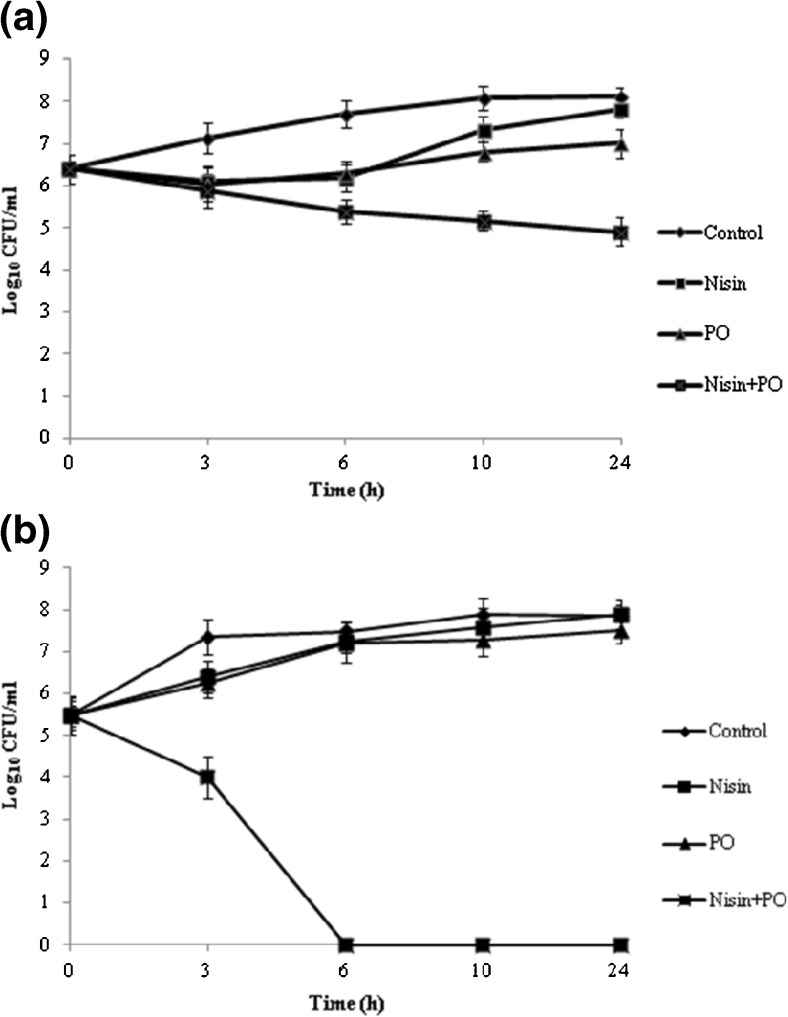
Combination studies were performed with both antimicrobials by challenge assay using S. aureus ATCC 29213 and L. monocytogenes ATCC 19115, the results were shown in Fig. 1. The compounds were assayed at their 1/2 × MIC and in the combinations 1/2 × MIC +1/2 × MIC. Addition of nisin and PO alone did not significantly affect the growth curve, especially over the 6 h of exposure. The combination of nisin and PO at sub-inhibitory concentrations yielded a ≥ 2 log10 CFU/ml decrease compared with nisin or PO alone of S. aureus and L. monocytogenes after 24 h and no recovery in viable count was noted in the remainder evaluated intervals. The results showed that a synergism was confirmed between nisin and PO against S. aureus ATCC 29213 and L. monocytogenes ATCC 19115. For L. monocytogenes, a 2.4-log10 CFU/ml decrease caused by the two-compound combination relative to nisin alone at 3 h, after 6 h, the decrease reached to more than 7-log10 CFU/ml and a complete bactericidal effect was observed for the combination. However, the maximum reduction of 2.9-log10 CFU/ml was observed at 24 h for S. aureus. The results revealed that a stronger bactericidal effect in milk might be exerted in nisin and PO combinations against L. monocytogenes than S. aureus.
Fig. 1.
Killing of S. aureus ATCC 29213 (a) and L. monocytogenes ATCC 19115 (b) with nisin and PO in pasteurized whole milk. The strains at a starting inoculum of 106 CFU/ml were exposed to in vivo-achievable concentrations of 16 μg/ml nisin; 1 mg/ml PO; 16 μg/ml nisin +1 mg/ml PO
Agar disk diffusion assay
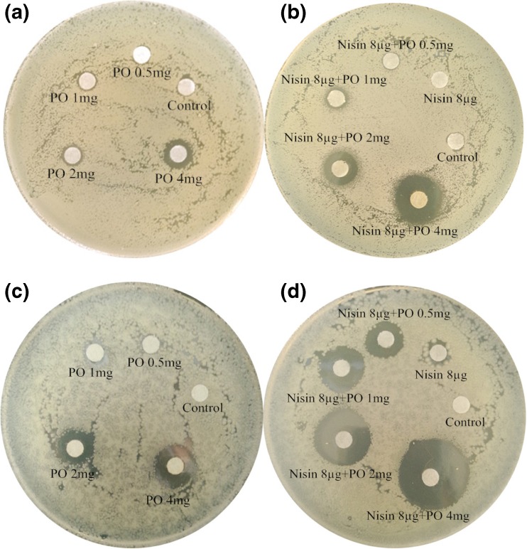
Agar disk diffusion tests helped to visualize the synergistic interaction between nisin and PO (Fig. 2). PO with different concentration displayed a variable degree of antibacterial activity against two tested strains. For S. aureus, treated with PO at 1, 2 and 4 mg, the inhibition zones were 7 mm, 8 mm and 11 mm, respectively. S. aureus was found to be least sensitive to PO. When treated with the same concentrations of PO in conbination with 8 μg nisin (showed no antibacterial activity here), the the zones of inhibition increased to 8 mm, 13 mm and 18 mm, the results showed that 2 and 4 mg of PO combined with nisin displayed moderate anti-S. aureus activity. Comparatively, PO has already showed moderate anti-L. monocytogenes activity when treated alone at 2 and 4 mg with the inhibition zones 13 and 15 mm, respectively. At the same concentrations, when combined with 8 μg nisin, L.monocytogenes showed maximum sensitivity to PO with inhibition zone of 20 and 25mm respectively. It was also worth noting that 0.5 and 1 mg PO combined nisin showed moderate anti-L. monocytogenes activity with inhibition zones of 13 and 17 mm, but PO showed no anti-L. monocytogenes activity at this two concentrations. Therefore, based on inhibition zone test results, the notable antibacterial effects of nisin at sub-inhibitory concentrations for L. monocytogenes and S. aureus had a different degree of improvement with the addition of PO at different concentrations.
Fig. 2.
Agar disk diffusion assay for nisin combined with PO against S.aureus ATCC 29213 (picture (a) and (b)) and L. monocytogenes ATCC 19115 (picture (c) and (d))
The effects of antimicrobials on the bacterial cell wall
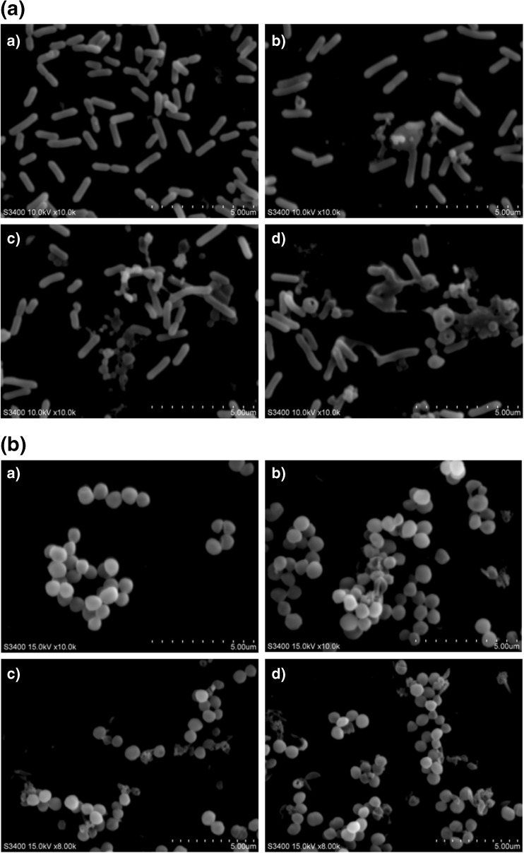
To investigate the morphology of bacteria in response to nisin, PO, and the combination of nisin with PO, the treated bacteria were observed using SEM (Fig. 3). Treatment of L. monocytogenes and S. aureus with nisin altered cell morphology such that it appeared to be pleomorphic or spiral shaped, as compared with the control experiment, which demonstrated that the cells were intact and smooth. In contrast, the damaging effect of PO to cell wall was stronger than that of nisin. Bacterial cells treated with the combination of PO and nisin were markedly damaged, such as splitting and a change in cell morphology due to deep wrinkles and distortion, and most of the outermost layer of the bacterial cells disappeared. These results show that the antibacterial activity of nisin and PO appears to be synergistic against the bacteria tested in this study, especially observed in L. monocytogenes.
Fig. 3.
SEMs of L. monocytogenes ATCC 19115 (a) and S. aureus ATCC 29213 (b) cells following various treatments after 3 h: a) untreated/control cells; b) cells after treatment with nisin; c) cells after treatment with PO; d) cells after treatment with a combination of nisin and PO
The effects of antimicrobials on cell membrane
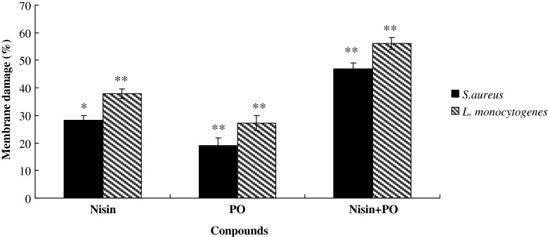
In this assay, the SYTO-9 and propidium iodide stains compete for binding to the bacterial nucleic acid. SYTO-9 labels cells with both damaged and intact membranes, whereas propidium iodide penetrates only cells with damaged membranes. Therefore, live cells appear green and dead ones appear red. The bacterial membrane damage caused by nisin and PO alone or in combination is represented as percentage of damage compared to that of control (Fig. 4). Nisin and PO were both less active against S. aureus ATCC 29213 with 28 % and 19 % membrane damage, whereas there were 38 % and 27 % damage in L. monocytogenes ATCC 19115. However, highest membrane damage was noted with nisin and PO in combination against the two micro-organisms, the combination were able to damage 47 % (p < 0.05) and 56 % (p < 0.05) of the S. aureus ATCC 29213 and L. monocytogenes ATCC 19115 membrane within 10 min, respectively. As expected, the combination of nisin and PO could cause higher membrane damage than the compounds used alone. It was worth noting that nisin and PO alone or in combination both displayed stronger membrane effects in L. monocytogenes than S. aureus, though these effects were just moderate membrane effects.
Fig. 4.
Effect of nisin (32 μg/ml) and PO (2 mg/ml) alone and in combination on bacterial membrane damage based on the LIVE/DEAD BacLight kit. Values are expressed as means ± standard deviation, *represents p < 0.05, ** represents p < 0.01
Discussion
Our results showed a bactericidal effect of nisin and PO combination on L. monocytogenes and S. aureus and demonstrated the potential of nisin and PO as a biopreservative in foods or food products when applied together, especially in pasteurized milk. Many approaches to enhancing the antibacterial activity of nisin and expand its range of application have been tried. Nisin has been found to act synergistically with various antimicrobials including chelators, small molecular weight substances from plants, reuterin (Arqués et al. 2004), proteins such as lysozyme and lactoferrin and milk-derived peptides (López-Expósito et al. 2008).
In our study, PO as one of the plant EOs was chosen to enhance the antimicrobial activity of nisin. The main constituents of PO are perillaldehyde (66.1 %), limonene (18.7 %) and β-caryophyllene (8.7 %) (Inouye et al. 2006). Some studies have concluded that whole EOs have a greater antibacterial activity than the major components mixed (Gill et al. 2002). Similarly, Burt (2004) also suggested that the minor components present in the EOs were more critical to the activity than EOs main components mixed, and the combination of major components with other minor components that have a weaker activity may achieve a synergistic effect. Because plant EOs exhibit a multi-component nature, it is more difficult for bacteria to develop resistance than many widely-used antibiotics, which have a single target site. Considering the above mentioned interaction of EO compositions, the EOs which consist of different biochemical components may increase the antimicrobial efficacy distinctly. Previous studies have reported that the combination of EOs and nisin was found to be an effective antimicrobial activity. Esmail et al. (2014) reported that the combination of nisin with sub-lethal doses of thyme oil in minced fish resulted in an increased reduction in the viable count of the foodborne pathogens than these compounds used alone. Furthermore, it has been confirmed that the antimicrobial activity of EOs constituents (e.g., carvacrol and thymol) are enhanced by the presence of nisin (Yoon et al. 2011). Our study indicated that the combination of nisin and PO can result in effective control of L. monocytogenes and S. aureus growth. The synergistic effects of PO and nisin would also mean that effective concentrations of nisin could be lowered considerably (up to 50 % in our studies) to achieve the desired antibacterial activity, thus making the cost of food preservation by nisin cheaper. Furthermore, the use of lower concentration of nisin may reduce the probability of the occurrence of nisin-resistant strains of bacteria.
Nisin has been reported to act on the cytoplasmic membranes of gram-positive bacteria to cause lesions and trigger autolysis of Staphylococcus simulans by activating Nacetylmuramoyl-L-alanine amidase (Bierbaum and Sahl 1987). Chemical analysis of EOs showed that the major active EO components are phenols, terpenes, aldehydes and ketones, and it is generally believed that EOs principally performed against the cell cytoplasmic membrane of microorganism. Leakage of intracellular constituents and impairment of microbial enzyme systems can then occur, and extensive loss of the cell contents will cause the death of cell (Moreira et al. 2005). In our study, we observed that nisin and PO both caused cell walls and cell membrane damage and the damage of combination improved indeed.
Although nisin is an antimicrobial agent that inhibits the growth of L. monocytogenes and S. aureus, exposure to this agent has also been associated with the development of nisin resistance. Our studies demonstrated that a combination of PO with nisin effectively inhibited both L. monocytogenes and S. aureus, especially L. monocytogenes according to the results of FICI values, challenge tests in pasteurized milk and agar disk diffusion assays. Additionally, the following synergistic antibacterial mechanism of PO and nisin has been proposed. The compounds in combination resulted in stronger damage to both of cell walls and cell membrane than nisin or PO alone in L. monocytogenes and S. aureus. The combination of PO with nisin and resulting damage suggests that the cell walls and membrane is the main target of this antimicrobial combination.
Nisin, the most well-studied bacteriocin, is used in over 50 countries as a natural food preservative agent. In addition, nisin is Generally Recognised As Safe (GRAS), and, is the only bacteriocin that has found practical application as a natural food preservative in processed cheese, milk, dairy products, canned foods, hot baked flour products (crumpets) and pasteurised liquid egg (Periago and Moezelaar 2001). Perilla is widely cultivated in East Asia and perilla oil is used in Asian countries as an ingredient in foods such as salad dressings, seasonings, and dipping sauces (Nitta et al. 2006). PO is one of the naturally-flavored oils and obtained usually from roasted seeds to give good characteristic flavor. Yang et al. (2012) reported the consumer acceptability of perilla oil porridges, Moritz et al. (2012) have demonstrated that the consumer acceptability of essential oils used in fermented milk. In this research, nisin and PO synergistically and significantly inhibited the growth of S. aureus and L. monoctogenes in milk samples. In conclusion, combinations of nisin and PO have the significant antibacterial activities, which might be used in food industry to control the growth of pathogens.
Acknowledgments
Financial support for this work came from the following sources: the National Nature Science Foundation of China (No. 31271951 and No. 81573448), China Postdoctoral Science Foundation (2013 M530142), the Program for New Century Excellent Talents in University (NCET-13-024), the Important National Science and Technology Specific Projects (2012ZX10003002).
Footnotes
Xingchen Zhao and Ce Shi contributed equally to this work.
References
- Anonymous (2009) Clinical and laboratory standards institute (CLSI). Methods for dilution antimicrobial susceptibility tests for bacteria that grow aerobically, (8th ed.) edn. CLSI, Wayne Approved Standard M7-A8
- Anonymous . Clinical and laboratory standards institute (CLSI). Methods for antimicrobial dilution and disk susceptibility testing of infrequently isolated or fastidious bacteria-second edition. Wayne: CLSI; 2010. [DOI] [PubMed] [Google Scholar]
- Arqués JL, Fernández J, Gaya P, Nuñez M, Rodríguez E, Medina M. Antimicrobial activity of reuterin in combination with nisin against food-borne pathogens. Int J Food Microbiol. 2004;95:225–229. doi: 10.1016/j.ijfoodmicro.2004.03.009. [DOI] [PubMed] [Google Scholar]
- Benkerroum N, Sandine WE. Inhibitory action of nisin against Listeria monocytogenes. J Dairy Sci. 1988;71:3237–3245. doi: 10.3168/jds.S0022-0302(88)79929-4. [DOI] [PubMed] [Google Scholar]
- Bierbaum G, Sahl HG. Autolytic system of Staphylococcus aimulans 22: influence of cationic peptides on activity of N-acetylmuramoyl-L-alanine amidase. J Bacteriol. 1987;169:5452–5458. doi: 10.1128/jb.169.12.5452-5458.1987. [DOI] [PMC free article] [PubMed] [Google Scholar]
- Burt S. Essential oils: their antibacterial properties and potential applications in foods-a review. Int J Food Microbiol. 2004;94:223–253. doi: 10.1016/j.ijfoodmicro.2004.03.022. [DOI] [PubMed] [Google Scholar]
- Dosler S, Gerceker AA. In vitro activities of antimicrobial cationic peptides; melittin and nisin, alone or in combination with antibiotics against gram-positive bacteria. J Chemother. 2012;24:137–143. doi: 10.1179/1973947812Y.0000000007. [DOI] [PubMed] [Google Scholar]
- Esmail A, Masoud R, Hedayat H. Antibacterial activity of plant essential oils and extracts: the role of thyme essential oil, nisin, and their combination to control Listeria monocytogenes inoculated in minced fish meat. Food Control. 2014;35:177–183. doi: 10.1016/j.foodcont.2013.07.004. [DOI] [Google Scholar]
- Ezaki O, Takahashi M, Shigematsu T, et al. Longterm effects of dietary alpha-linolenic acid from perilla oil on serum fatty acids composition and on the risk factors of coronary heart disease in Japanese elderly subjects. J Nutr Sci Vitaminol. 1999;45:759–772. doi: 10.3177/jnsv.45.759. [DOI] [PubMed] [Google Scholar]
- Gálvez A, Abriouel H, López RL, Omar NB. Bacteri ocin-based strategies for food biopreserv ation. Int J Food Microbiol. 2007;120:51–70. doi: 10.1016/j.ijfoodmicro.2007.06.001. [DOI] [PubMed] [Google Scholar]
- Gill AO, Delaquis P, Russo P, Holley RA. Evaluation of antilisterial action of cilantro oil on vacuum packed ham. Int J Food Microbiol. 2002;73:83–92. doi: 10.1016/S0168-1605(01)00712-7. [DOI] [PubMed] [Google Scholar]
- Gravesen A, Jydegaard Axelsen AM, Mendes da Silva J, Hansen TB, Knøchel S. Frequency of bacteriocin resistance development and associated fitness costs in Listeria monocytogenes. Appl Environ Microbiol. 2002;68:756–764. doi: 10.1128/AEM.68.2.756-764.2002. [DOI] [PMC free article] [PubMed] [Google Scholar]
- Hilliard JJ, Goldschmidt RM, Licata L, Baum EZ, Bush K. Multiple mechanisms of action for inhibitors of histidine protein kinases from bacterial two-component systems. Antimicrob Agents Chemother. 1999;43:1693–1699. doi: 10.1128/aac.43.7.1693. [DOI] [PMC free article] [PubMed] [Google Scholar]
- Inouye S, Nishiyama Y, Uchida K, Hasumi Y, Yamaguchi H, Abe S. The vapor activity of oregano, perilla, tea tree, lavender, clove, and geranium oils against a Trichophyton mentagrophytes in a closed box. J Infect Chemother. 2006;12:349–354. doi: 10.1007/s10156-006-0474-7. [DOI] [PubMed] [Google Scholar]
- Ito M. Studies on perilla, agarwood, and cinnamon through a combination of fieldwork and laboratory work. J Nat Med. 2008;62:387–395. doi: 10.1007/s11418-008-0262-z. [DOI] [PubMed] [Google Scholar]
- Jacqueline C, Navas D, Batard E, et al. In vitro and In vivo synergistic activities of linezolid combined with subinhibitory concentrations of imipenem against methicillin-resistant Staphylococcus aureus. Antimicrob Agents Chemother. 2005;49:45–51. doi: 10.1128/AAC.49.1.45-51.2005. [DOI] [PMC free article] [PubMed] [Google Scholar]
- Kim KY, Park JH, Kwak HS, Woo GJ. Characterization of the quinolone resistance mechanism in foodborne Salmonella isolates with high nalidixic acid resistance. Int J Food Microbiol. 2011;146:52–56. doi: 10.1016/j.ijfoodmicro.2011.01.037. [DOI] [PubMed] [Google Scholar]
- Kito M, Onji Y, Yoshida T, Nagasawa T. Occurrence of ε-poly-L-lysine degrading enzyme in ε-poly-L-lysine-tolerant Sphingobacterium multivorum OJ10: purification and characterization. FEMS Microbiol Lett. 2002;207:147–151. doi: 10.1111/j.1574-6968.2002.tb11043.x. [DOI] [PubMed] [Google Scholar]
- Lee S, Lee Y, Sung JS, et al. Influence of roasting conditions on the chemical properties and antioxidant activity of perilla oils. J Korean Soc Appl Biol Chem. 2015;58:325–334. doi: 10.1007/s13765-015-0046-6. [DOI] [Google Scholar]
- Liu F, Liu M, Du L, Wang D, Geng Z, Zhang M, Sun C, Xu X, Zhu Y, Xu W. Synergistic antibacterial effect of the combination of ε-polylysine and nisin against Enterococcus faecalis. J Food Prot. 2015;78:2200–2206. doi: 10.4315/0362-028X.JFP-15-220. [DOI] [PubMed] [Google Scholar]
- López-Expósito I, Pellegrini A, Amigo L, Recio I. Synergistic effect between different milk-derived peptides and proteins. J Dairy Sci. 2008;91:2184–2189. doi: 10.3168/jds.2007-0037. [DOI] [PubMed] [Google Scholar]
- Lv F, Liang H, Yuan Q, Li C. In vitro antimicrobial effects and mechanism of action of selected plant essential oil combinations against four food-related microorganisms. Food Res Int. 2011;44:3057–3064. doi: 10.1016/j.foodres.2011.07.030. [DOI] [Google Scholar]
- Mazzotta AS, Crandall AD, Montville TJ. Nisin resistance in Clostridium botulinum spores and vegetative cells. Appl Environ Microbiol. 1997;6:2654–2659. doi: 10.1128/aem.63.7.2654-2659.1997. [DOI] [PMC free article] [PubMed] [Google Scholar]
- Mead PS, Dunne EF, Graves L, et al. Nationwide outbreak of listeriosis due to contaminated meat. Epidemiol Infect. 2006;134:744–751. doi: 10.1017/S0950268805005376. [DOI] [PMC free article] [PubMed] [Google Scholar]
- Moreira MR, Ponce AG, Del Valle CE, Roura SI. Inhibitory parameters of essential oils to reduce a foodborne pathogen. LWT-Food Sci Technol. 2005;38:565–570. doi: 10.1016/j.lwt.2004.07.012. [DOI] [Google Scholar]
- Moritz CM, Rall VL, Saeki MJ, Júnior AF. Inhibitory effect of essential oils against lactobacillus rhamnosus and starter culture in fermented milk during its shelf-life period. Braz J Microbiol. 2012;43:1147–1156. doi: 10.1590/S1517-83822012000300042. [DOI] [PMC free article] [PubMed] [Google Scholar]
- Nitta M, Kobayashi H, Ohnishi KM, Nagamine T, Yoshida M (2006) Essential oil variation of cultivated and wild perilla analyzed by GC/MS. Biochem Syst Ecol 34:25–37.
- Okuyama H, Yamada K, Miyazawa D, Yasui Y, Ohara N. Dietary lipids impacts on healthy ageing. Lipids. 2007;42:821–825. doi: 10.1007/s11745-007-3073-1. [DOI] [PubMed] [Google Scholar]
- Oliveira CEV, Stamford TLM, Gomes Neto NJ, Souza EL. Inhibition of Staphylococcus aureus in broth and meat broth using synergies of phenolics and organic acids. Int J Food Microbiol. 2010;137:312–316. doi: 10.1016/j.ijfoodmicro.2009.11.019. [DOI] [PubMed] [Google Scholar]
- Periago PM, Moezelaar R. Combined effect of nisin and carvacrol at different pH and temperature levels on the viability of different strains of Bacillus cereus. Int J Food Microbiol. 2001;68:141–148. doi: 10.1016/S0168-1605(01)00461-5. [DOI] [PubMed] [Google Scholar]
- Peschel A, Otto M, Jack RW, Kalbacher H, Jung G, Götz F. Inactivation of the dlt operon in Staphylococcus aureus confers sensitivity to defensins, protegrins and other antimicrobial peptides. J Biol Chem. 1999;274:8405–8410. doi: 10.1074/jbc.274.13.8405. [DOI] [PubMed] [Google Scholar]
- Phongphakdee K, Nitisinprasert S. Combination inhibition activity of nisin and ethanol on the growth inhibition of pathogenic gram negative bacteria and their application as disinfectant solution. J Food Sci. 2015;80:M2241–M2246. doi: 10.1111/1750-3841.13015. [DOI] [PubMed] [Google Scholar]
- Pillai SK, Moellering RC. Antimicrobial combinations. Antibiotics in laboratory medicine. New York: Lippincott Williams and Wilkins; 2005. pp. 365–400. [Google Scholar]
- Qiu J, Zhang X, Luo M, Li H, Dong J, Wang J, Leng B, Wang X, Feng H, Ren W, Deng X. Subinhibitory concentrations of perilla oil affect the expression of secreted virulence factor genes in Staphylococcus aureus. PLoS One. 2011;6 doi: 10.1371/journal.pone.0016160. [DOI] [PMC free article] [PubMed] [Google Scholar]
- Rasooly R, Do PM. In vitro cell-based assay for activity analysis of staphylococcal enterotoxin a in food. FEMS Immunol Med Microbiol. 2009;56:172–178. doi: 10.1111/j.1574-695X.2009.00561.x. [DOI] [PubMed] [Google Scholar]
- Rota MC, Carraminana JJ, Burillo J, Herrera A. In vitro antimicrobial activity of essential oils from aromatic plants against selected foodborne pathogens. J Food Prot. 2004;67:1252–1256. doi: 10.4315/0362-028x-67.6.1252. [DOI] [PubMed] [Google Scholar]
- Yang JE, Kim HJ, Chun L (2012) Sensory characteristics and consumer acceptability of perilla porridges. Food Sci Biotechnol 21:785–797.
- Yoon JI, Bajpai VK, Kang SC (2011) Synergistic effect of nisin and cone essential oil of Metasequoia glyptostroboides Miki ex Hu against Listeria monocytogenes in milk samples. Food Chem Toxicol 49:109–114. [DOI] [PubMed]


