

Abstract
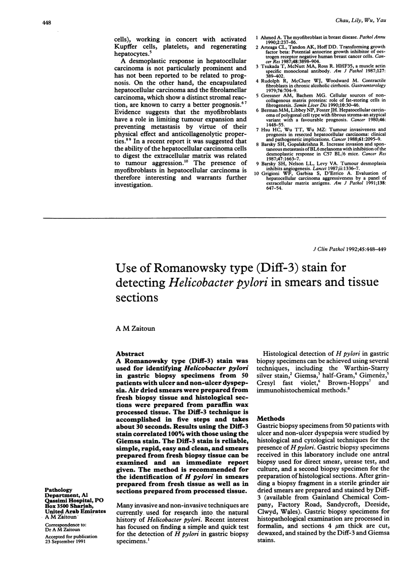
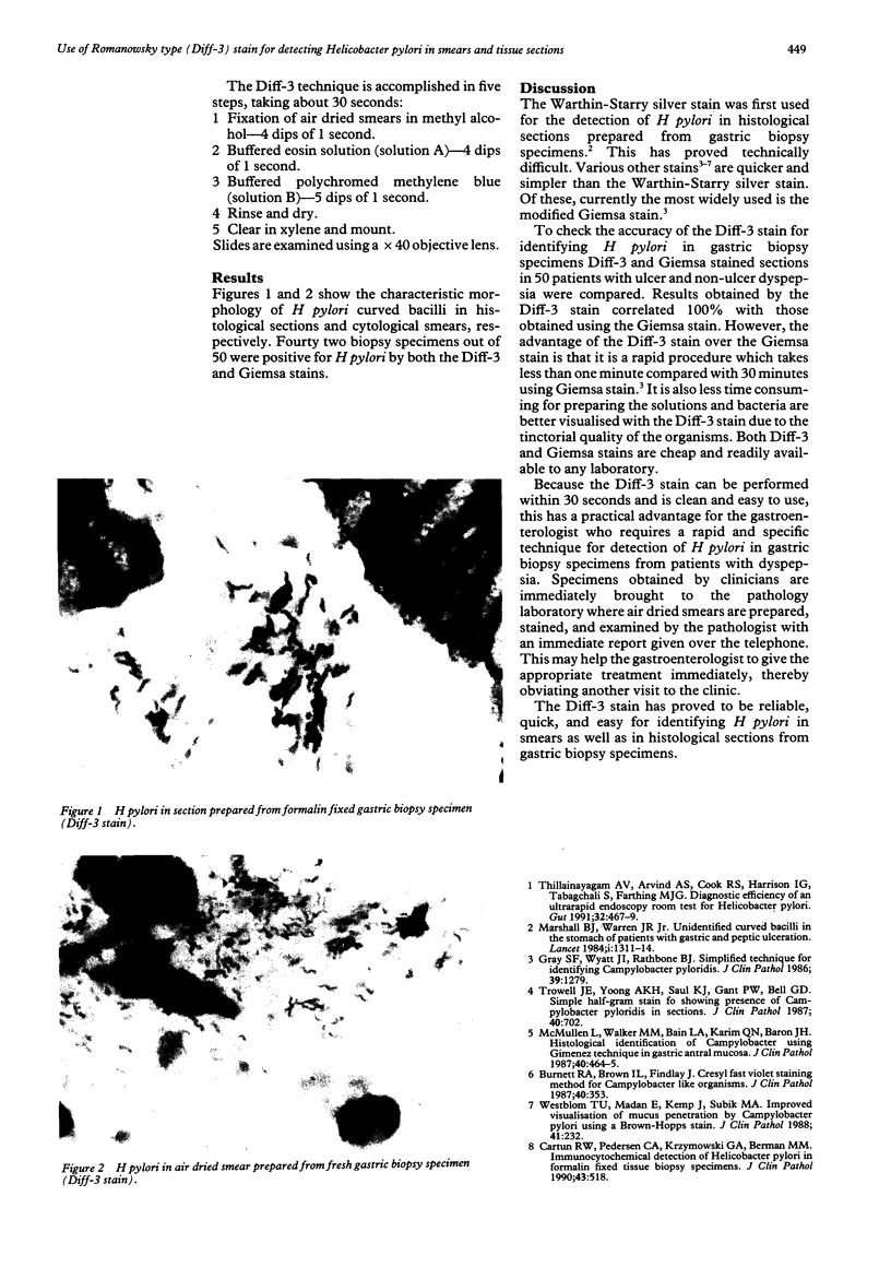
A Romanowsky type (Diff-3) stain was used for identifying Helicobacter pylori in gastric biopsy specimens from 50 patients with ulcer and non-ulcer dyspepsia. Air dried smears were prepared from fresh biopsy tissue and histological sections were prepared from paraffin wax processed tissue. The Diff-3 technique is accomplished in five steps and takes about 30 seconds. Results using the Diff-3 stain correlated 100% with those using the Giemsa stain. The Diff-3 stain is reliable, simple, rapid, easy and clean, and smears prepared from fresh biopsy tissue can be examined and an immediate report given. The method is recommended for the identification of H pylori in smears prepared from fresh tissue as well as in sections prepared from processed tissue.
Full text
PDF
Images in this article
Selected References
These references are in PubMed. This may not be the complete list of references from this article.
- Burnett R. A., Brown I. L., Findlay J. Cresyl fast violet staining method for Campylobacter like organisms. J Clin Pathol. 1987 Mar;40(3):353–353. doi: 10.1136/jcp.40.3.353-b. [DOI] [PMC free article] [PubMed] [Google Scholar]
- Cartun R. W., Pedersen C. A., Krzymowski G. A., Berman M. M. Immunocytochemical detection of Helicobacter pylori in formalin fixed tissue biopsy specimens. J Clin Pathol. 1990 Jun;43(6):518–518. doi: 10.1136/jcp.43.6.518-a. [DOI] [PMC free article] [PubMed] [Google Scholar]
- Gray S. F., Wyatt J. I., Rathbone B. J. Simplified techniques for identifying Campylobacter pyloridis. J Clin Pathol. 1986 Nov;39(11):1279–1279. doi: 10.1136/jcp.39.11.1279-a. [DOI] [PMC free article] [PubMed] [Google Scholar]
- Marshall B. J., Warren J. R. Unidentified curved bacilli in the stomach of patients with gastritis and peptic ulceration. Lancet. 1984 Jun 16;1(8390):1311–1315. doi: 10.1016/s0140-6736(84)91816-6. [DOI] [PubMed] [Google Scholar]
- McMullen L., Walker M. M., Bain L. A., Karim Q. N., Baron J. H. Histological identification of Campylobacter using Gimenez technique in gastric antral mucosa. J Clin Pathol. 1987 Apr;40(4):464–465. doi: 10.1136/jcp.40.4.464. [DOI] [PMC free article] [PubMed] [Google Scholar]
- Thillainayagam A. V., Arvind A. S., Cook R. S., Harrison I. G., Tabaqchali S., Farthing M. J. Diagnostic efficiency of an ultrarapid endoscopy room test for Helicobacter pylori. Gut. 1991 May;32(5):467–469. doi: 10.1136/gut.32.5.467. [DOI] [PMC free article] [PubMed] [Google Scholar]
- Trowell J. E., Yoong A., Saul K. J., Gant P. W., Bell G. D. Simple half-gram stain for showing presence of Campylobacter pyloridis in sections. J Clin Pathol. 1987 Jun;40(6):702–702. doi: 10.1136/jcp.40.6.702-a. [DOI] [PMC free article] [PubMed] [Google Scholar]
- Westblom T. U., Madan E., Kemp J., Subik M. A., Tseng J. Improved visualisation of mucus penetration by Campylobacter pylori using a Brown-Hopps stain. J Clin Pathol. 1988 Feb;41(2):232–232. doi: 10.1136/jcp.41.2.232-b. [DOI] [PMC free article] [PubMed] [Google Scholar]