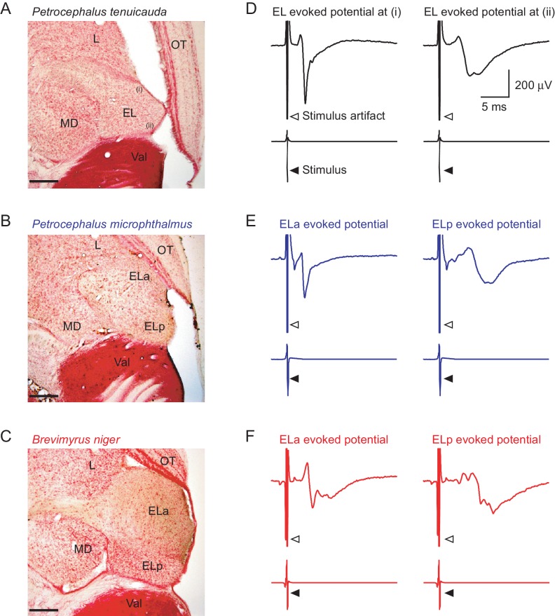
Figure 1. The central electrosensory system detects transient synchrony among oscillating receptors.
50 µm horizontal sections of the midbrain of P. tenuicauda (A), P. microphthalmus (B), and B. niger (C). The midbrain exterolateral nucleus (EL) in P. tenuicauda is small and undifferentiated; in the other two species, EL is enlarged and subdivided into separate anterior (ELa) and posterior (ELp) nuclei. (D) Representative mean evoked potentials (n = 10 traces) from the EL of P. tenuicauda obtained from relatively more anterior (left) and posterior (right) regions. Representative mean evoked potentials (n = 10 traces) obtained from ELa (left) and ELp (right) in P. microphthalmus (E) and B. niger (F). Scale bars in (A), (B), and (C) represent 200 µm. L, lateral nucleus; OT, optic tectum; MD, mediodorsal nucleus; Val, valvula cerebellum.

