Abstract
Traumatic right diaphragmatic hernia is rare in children. Its diagnosis can be difficult in the acute phase of trauma because its signs are not specific, especially in a poly trauma context. We report two cases of traumatic right diaphragmatic hernia following a blunt thoraco-abdominal trauma, highlighting some difficulties in establishing an early diagnosis and the need for a high index of suspicion.
Keywords: Children, diagnosis, right diaphragmatic hernia, trauma
INTRODUCTION
Diaphragmatic hernia occurs when abdominal viscera pass into the thoracic cavity through an opening in the diaphragm. It is usually a congenital, but many cases of traumatic origin have been described in children.[1,2] Traditionally, diaphragmatic ruptures were considered to be more frequent on the left than the right. Previously, it was the belief that the buffering effect played by the liver and right kidney is responsible for the relative lack of involvement of the right hemi-diaphragm.[1] However, recent review of the literature showed that traumatic diaphragmatic ruptures does occur on the right as often as on the left, but less obvious clinically, because most are often overlooked,[3,4] due to non-specific features. Despite modern imaging, diagnosis is not always easy in the emergency setting. We present the report two cases of traumatic right diaphragmatic hernia following a blunt thoraco-abdominal trauma, highlighting some difficulties in establishing an early diagnosis and the need for a high index of suspicion.
CASE 1
A 6-year-old boy referred from a regional hospital for the management of blunt chest trauma and head injury following a road traffic accident. He was admitted at the regional hospital and after a clinical and laboratory evaluation, a diagnosis of a blunt right hemothorax made. He had a chest tube thoracostomy, which drained 1,500 cc of altered blood but without clinical or radiographic improvement. A second drainage was immediately inserted, again without improvement. This necessitated his referral to our centre 7 days post-trauma. Further, radiology of the chest showed a dense homogeneous opacity occupying the entire lung field with a displacement of the mediastinum [Figures 1 and 2]. His haemoglobin level was 6 g/dl. Thoraco-abdominal computed tomography (CT)-scan showed a diaphragmatic rupture with an intrathoracic herniation. Surgical exploration revealed some haemoperitoneum, a transverse rupture of the right hemi diaphragm about 10 cm with irregular edges, liver in the chest with fracture of lower pole segment V and a laceration of about 0.5 cm on the upper surface caused by the chest tube drain [Figures 3 and 4]. He also had contusion of the base of the right lung and the right hepatic flexure. The liver was reduced from the chest and the diaphragmatic rent was closed in two layers after placing a chest tube. The chest tube was removed on the third post-operative day. The child was discharge on post-operatively day ten in satisfactory condition. Two years on follow-up, the child remained symptom free with a normal chest radiograph [Figure 5].
Figure 1.
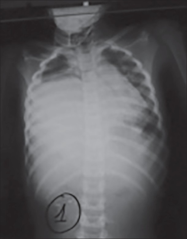
Chest radiography showing right lung opacity with left Mediastinal shift (observation 1) (Up left)
Figure 2.
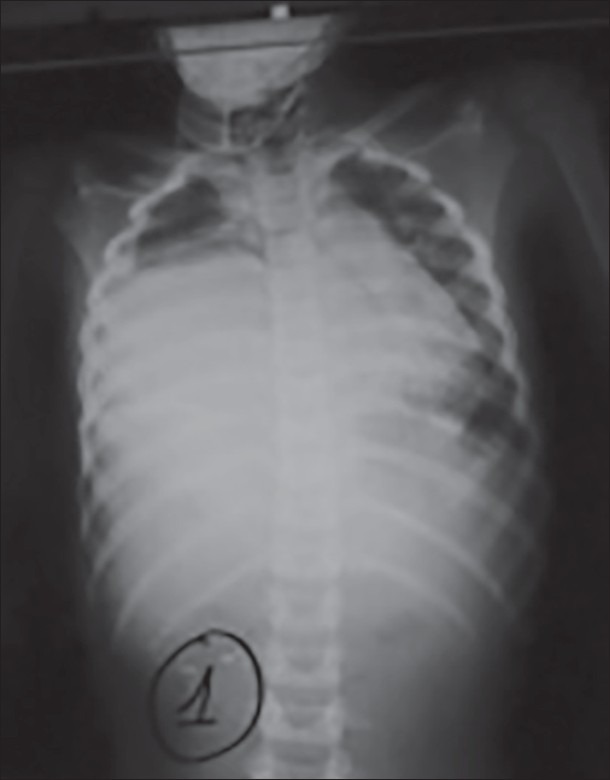
Image after drainage (observation 1) (Up left)
Figure 3.
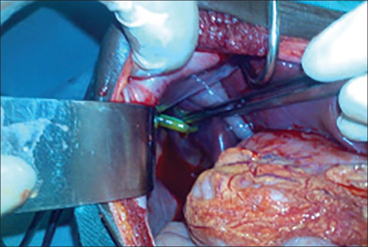
Intrathoracic chest tube in the liver (observation 1) (Up left)
Figure 4.
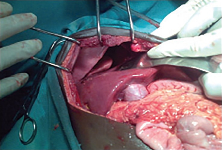
Right diaphragmatic rupture after reduction of the liver (observation 1) (Up left)
Figure 5.
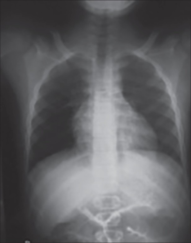
Control chest radiography on 24 months post-operative (observation 1) (Up left)
CASE 2
A 2-year-old girl with unremarkable medical history was admitted our emergency department on account of a blunt chest trauma and brain injury following involvement in a car accident. On admission 2 h after the accident, examination revealed an obsessed child with a mild pallor of conjunctiva and difficulty in breathing with abdominal breathing type. The general condition was fair with a blood pressure 110/80 mmHg, a respiratory rate of 52 cycles/min, pulse 124 beats/min and a normal temperature to 37.2°C. There were multiple abrasions, but abdomen was soft with neither a mass nor depression on palpation.
An antero-posterior chest radiograph showed a fracture the anterior arcs the 6th, 7th, 8th, and 9th left ribs, right lung opacity with right apical pneumothorax [Figure 6]. Abdominal ultrasound confirmed the absence of haemoperitoneum and visceral lesions. Due to the worsening of respiratory distress, the child was admitted into the intensive-care unit for better support.
Figure 6.
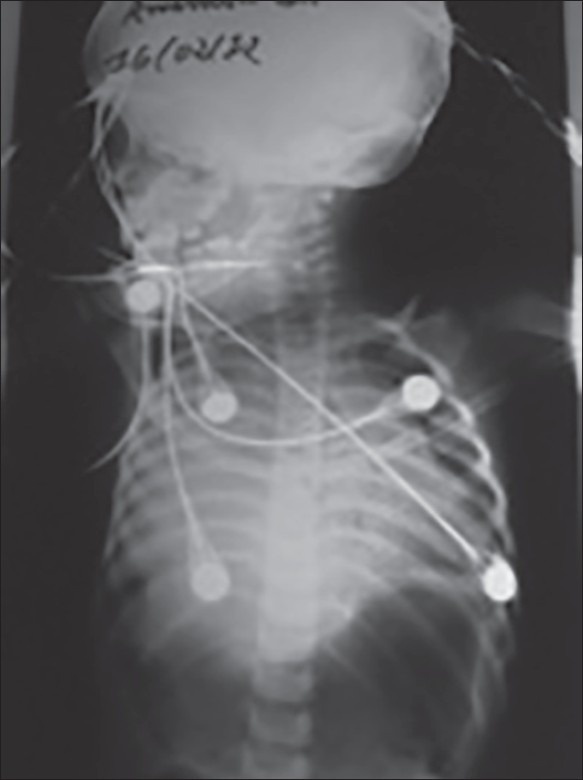
Chest radiograph (AP) showing right lung opacity (observation 2) (Up left)
Despite the oxygenation, breathing difficulties worsened. Right pleural tap yielded altered blood, suggesting a haemothorax. Chest tube thoracostomy performed confirmed the haemothorax. The radiograph showed a lack of improvement in radiological images [Figure 7]. Based on diagnostic uncertainty, thoraco-abdominal CT scan performed on day 2 of hospitalization showed a liver with intrathoracic drain within it [Figure 8].
Figure 7.
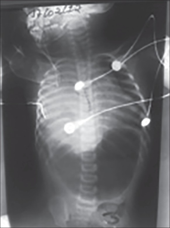
Image after drainage (observation 2) (Up left)
Figure 8.
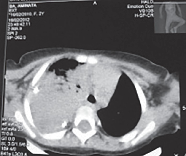
Chest computed tomography-scan objectivising an intrahepatic drain (arrow) with the liver in intrathoracic position (observation 2) (Fronf left)
At surgery, the liver had herniated into the thoracic cavity through a gap of about 10 cm in diameter in the right hemi diaphragm [Figure 9]. The base of the right lung was also contused. After reduction of the liver and insertion of a chest drain, the diaphragmatic rent was repaired with simple suture.
Figure 9.
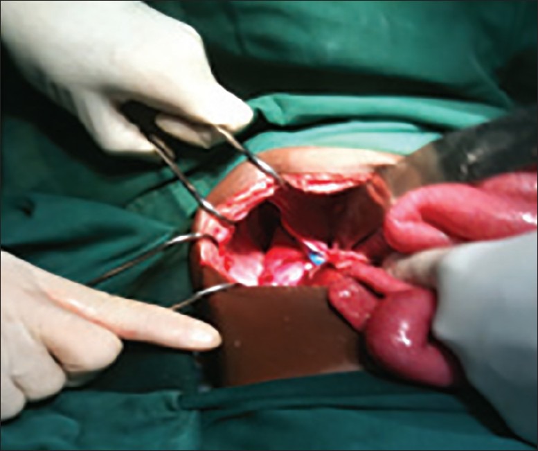
diaphragmatic rupture after reduction of liver and visualization of chest tube (observation 2) (Up left)
Clinically, the child did well and the chest radiograph on the first post-operative showed a remarked improvement. The chest drain was removed on the second post-operative day and the child was discharged 10 days after the operation. Sixteen months later, the child has symptom free with a normal radiograph [Figure 10].
Figure 10.
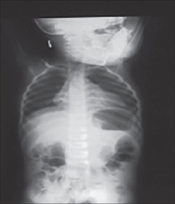
Control post-operative chest X-ray M 16 (observation 2) (Up left)
DISCUSSION
Traumatic diaphragmatic hernia is rare in children.[5] Its pathophysiology is poorly known, however, probably it results from the transmission to the diaphragm a pressure induced by the abdominal viscera. Road traffic accidents are the cause in 90% of cases.[6] The left hemi diaphragm is usually affected,[7,8] however, recent literature shows an increased incidence on the right side. Right diaphragmatic ruptures are often more difficult to diagnose in children. Their diagnostic delay contributes to mortality and morbidity.[9] In fact, they are usually associated with other severe trauma, which very often attracts more attention as was our experience in this report.
The clinical signs of right diaphragmatic rupture are not usually specific, although respiratory distress is often at the forefront. Therefore, the diagnosis is usually made by radiological examinations and should be performed early in suspected cases. Radiological signs, which point towards a diaphragmatic rupture include the inability to see clearly the hemi diaphragm, an abnormal elevation of the latter and any abnormal intrathoracic air image.[2] Since, these results are often not correlated with the probability of diaphragmatic rupture, the sensitivity of chest radiography in the diagnosis of right diaphragmatic rupture remains low and is also the order of 17%.[10] Besides, many series in the literature report that 30-50% of initial chest radiographs were normal in patients with diaphragmatic rupture, thus, emphasizing the importance of repeated radiographs.[4,11] The absence of intrathoracic air images makes the diagnosis difficult. In fact, the liver that is often herniated contributes greatly to the difficulty of early diagnosis.
Plain X-ray image in such a circumstance simulates a pulmonary opacity and it is difficult to differentiate a pulmonary atelectasis, hemothorax, and pulmonary contusion.
In any child who develops respiratory distress following trauma, the first differential diagnosis should be haemothorax. Although pleural tap may yield blood, it is possible for the blood to have come from the blood-congested liver. In such a dilemma, lack of clinical improvement despite a chest tube insertion for a suspected haemothorax should raise the suspicion of a herniated solid organ like the liver into the chest. CT-Scan can be very useful in establishing the diagnosis. Thoracoabdominal CT scan in thin cuts, allowing coronal and sagittal reconstructions is the gold standard for recent and old fractures. It recognizes 80% of ruptures to the left, and 50% of ruptures to the right.[12] It also allows users to review the associated lesions. The use of spiral CT has improved the accuracy in the diagnosis of diaphragmatic rupture with an overall sensitivity of 71% and a specificity of 100%.[10,12] In our study, CT-Scan has yet to direct the diagnosis by showing the liver in an intrathoracic position and hence, the indication for surgery in both cases after a period of 8 and 2 days respectively.
The use of magnetic resonance imaging for the diagnosis of diaphragmatic rupture in the context of emergency is limited due to problems of monitoring in a potentially unstable patient, the need for general anaesthesia in children and duration long examination.[10] Ultrasound can show a ruptured diaphragm, the absence of diaphragm, a floating diaphragm or the liver in intrathoracic position.[8] It is especially, useful when the patient is unstable and cannot be moved. Once confirmed, a diaphragmatic rupture requires surgical repair, because there is no spontaneous healing. Surgical exploration allowed us to confirm the traumatic hernia and its repair. Repair is usually done abdominally, unless there is an intrathoracic injury requiring thoracotomy.[6]
CONCLUSION
Although right traumatic diaphragmatic hernia is rare in children, it is often overlooked because the clinical signs are not specific. Un-resolving respiratory distress, despite chest tube insertion, in any child following trauma should suspicion of possible right traumatic diaphragmatic hernia should thought about. In our experience, CT-Scan can guide the diagnosis, to review the associated injuries and deal with them quickly, thus, avoiding the morbidity and mortality associated with delayed diagnosis.
Footnotes
Source of Support: Nil.
Conflict of Interest: None Conflict of Interest.
REFERENCES
- 1.Estrera AS, Landay MJ, McClelland RN. Blunt traumatic rupture of the right hemidiaphragm: Experience in 12 patients. Ann Thorac Surg. 1985;39:525–30. doi: 10.1016/s0003-4975(10)61992-3. [DOI] [PubMed] [Google Scholar]
- 2.Sharma AK, Kothari SK, Gupta C, Menon P, Sharma A. Rupture of the right hemidiaphragm due to blunt trauma in children: A diagnostic dilemma. Pediatr Surg Int. 2002;18:173–4. doi: 10.1007/s003830100686. [DOI] [PubMed] [Google Scholar]
- 3.Barsness KA, Bensard DD, Ciesla D, Partrick DA, Hendrickson R, Karrer FM. Blunt diaphragmatic rupture in children. J Trauma. 2004;56:80–2. doi: 10.1097/01.TA.0000103989.78049.46. [DOI] [PubMed] [Google Scholar]
- 4.Soundappan SV, Holland AJ, Cass DT, Farrow GB. Blunt traumatic diaphragmatic injuries in children. Injury. 2005;36:51–4. doi: 10.1016/j.injury.2003.11.019. [DOI] [PubMed] [Google Scholar]
- 5.Al-Salem AH, Qaisaruddin S. Thoracic and abdominal trauma in children. Ann Saudi Med. 1999;19:58–61. doi: 10.5144/0256-4947.1999.58. [DOI] [PubMed] [Google Scholar]
- 6.Favre J-P, Cheynel N, Benoit L, Favoulet P. Traitement chirurgical des ruptures traumatiques du diaphragme. EMC-Chir. 2005;2:242–51. [Google Scholar]
- 7.Ramos CT, Koplewitz BZ, Babyn PS, Manson PS, Ein SH. What have we learned about traumatic diaphragmatic hernias in children? J Pediatr Surg. 2000;35:601–4. doi: 10.1053/jpsu.2000.0350601. [DOI] [PubMed] [Google Scholar]
- 8.Chaudhary IA, Masood RS, Muftee M, Jilani R. Traumatic rupture of diaphragm: Not an Easy diagnosis. Rawal Med J. 2007;32:85–6. [Google Scholar]
- 9.Ninan G, Puri P. Late presentation of traumatic rupture of the diaphragm in a child. BMJ. 1993;306:643–4. doi: 10.1136/bmj.306.6878.643. [DOI] [PMC free article] [PubMed] [Google Scholar]
- 10.Iochum S, Ludig T, Walter F, Sebbag H, Grosdidier G, Blum AG. Imaging of diaphragmatic injury: A diagnostic challenge? Radiographics. 2002;22:S103–116. doi: 10.1148/radiographics.22.suppl_1.g02oc14s103. [DOI] [PubMed] [Google Scholar]
- 11.Jain P, Kushwaha AS, Pant N, Debnath PR, Chadha R, Choudhury SR, et al. Isolated post-traumatic right-sided diaphragmatic hernia. Indian J Pediatr. 2009;76:1167–8. doi: 10.1007/s12098-009-0282-z. [DOI] [PubMed] [Google Scholar]
- 12.William RR, Sankhla D, Al-Qassabi B, Al Ramadani K. Traumatic Rupture of the Right Hemidiaphragm: Diagnosis aided by Computerized Tomography and Image Reformation: A Case Report. Sultan Qaboos Univ Med J. 2008;8:219–22. [PMC free article] [PubMed] [Google Scholar]


