Summary
Aim
The goal of the study was to compare the ability of two different carrier based obturation (CBO) techniques to reach working length and fill in three-dimensions root canal systems, by using CBCT.
Materials and Methods
Twenty-six extracted molars were scanned with CBCT and 40 curved canals were selected (between 30° and 90°) and divided in two similar groups (n=20). All canals were prepared up to size 25 taper .06 using nickel-titanium instrumentation. The canals in the Group SC were obturated using Soft-Core obturators (Kerr, Romulus, Mi, USA), while Group TH canals (n= 20) were obturated using Thermafil Endodontic Obturators (Tulsa Dental Products, Tulsa, OK, USA), strictly following manufacturers’ instructions for use. The obturations were analyzed by means of CBCT to measure the distance from the apical limit of obturation to the apical foramen and the presence of voids inside root canals.
Results
There was no significant difference between the two groups in the mean distance of the apical extent of the obturation (t test, p>0.05). Overfilling occurred in only 3 cases (2 in Group TH and 1 in Group SC). The percentages of voids in both groups were very low with no significant difference (Z test, p>0.05).
Conclusions
The two tested CBO techniques showed similar positive results in terms of performance, even if, after checking with verifiers, in most cases the size of the selected Soft-Core obturator was one size smaller than Thermafil.
Keywords: endodontic obturation, Thermafil, Soft-Core, Cone-beam Computed Tomography
Introduction
The success of endodontic therapy is directly related to the sealing of the root canal system, by means of a three-dimensional hermetic endodontic filling (1, 2). The primary objective of the endodontic obturation in root canal treatment is to prevent the ingress of microorganisms or fluids, from both oral cavity or periapical area, and also refrain the growth of any residual bacteria left in the canal system (1–4). An inadequate seal might result in contamination of the canal spaces, thus leading to periapical disease (1, 2, 4).
The combination of gutta-percha with a layer of endodontic sealer is the most commonly used material for root canal obturations. A variety of techniques have been developed to provide the proper adaptation of the gutta-percha root canal walls, aiming the complete filling of the root canal system (2, 4). Johnson (5) developed an obturation technique using flexible metal/plastic carriers coated with α-phase gutta-percha (Thermafil Endodontic Obturator, Tulsa Dental Products, Tulsa, OK, USA). The main goal of this carrier based obturation (CBO) technique is to obtain a predictable thermo-plasticization and the consequent flow of the gutta-percha, by using a specific oven with a precise temperature control.
Previous studies have reported a good sealing ability of Thermafil obturations (6–10). The advantages of the carrier-based obturation (CBO) are related to the flow of the gutta-percha inside the root canal space, which is achieved by the combination of the heating process and the fact that the carrier facilitates the insertion of filling material and also slightly pressure it alongside the canal walls. Moreover, the simplicity of the CBO prevents the risks and problems related to the use of spreaders, pluggers, heaters and compactors inside complex root canals. Therefore, different types of CBO have been developed, such as the Soft-Core™ obturators (Kerr, Romulus, MI, USA). According to the manufacturer, the Soft-Core obturators present one universal taper size and a thinner core in apical part to allow greater flexibility to navigate curved canals.
On the other hand, the disadvantages of CBO techniques include the possible risk of overfilling (apical extrusion of the sealer, gutta-percha, and/or carrier) and underfilling, if the obturators do not reach the full working length (WL) due to the complex canal curvatures or less tapered preparation (11, 12). Another potential disadvantage of CBO, especially in severely curved canals, is the gutta-percha separating from the carrier, thus leading to the formation of gaps (voids) between the root canal filling and the canal walls. Moreover, CBO might be influenced by many factors (e.g., different ovens with different temperatures, type/amount of gutta-percha, design/dimensions of carrier, flexibility and material of the carrier, etc.), which might impair the apical sealing, mostly in curved canals (11, 13, 14).
Therefore, the aim of the present study was to compare the two different CBO brands, Thermafil and Soft-Core, in the ability to fill curved root canal systems, by using Cone-beam computed tomographic (CBCT) evaluation.
Materials and methods
A total of 26 molar was obtained from a pool of extracted teeth. A pre-operative CBCT scan evaluation was conducted to analyze the anatomical parameters and 40 canals presenting curvatures between 30° and 90° were selected. The canals were divided into two groups (n=20), with similar curvatures.
After access cavity in the teeth, each selected canal was checked for patency and the working length was established 0.5 mm short to the apical foramen. A mechanical glide path was performed with manual stainless steel K-files attached to the M4 handpiece (SybronEndo, Glendora, CA, USA), up to a size 15. Then, each canal was prepared using TFA nickel-titanium instruments (SybronEndo, Glendora, CA, USA) sizes 04.20 and 06.25. Irrigation was performed after each instrument using NAOCl 5% and final rinse with 3 mL of 17% EDTA and saline. After shaping procedures were completed, all canals were checked for patency with a 10 K-file and dried by paper points.
The Group SC canals (n=20) were obturated using Soft-Core, while Thermafill Obturators were used for the Group TH (n= 20). For each group the correspondent oven and verifiers were used, strictly following manufacturers’ instructions for use. Briefly, first it was inserted a verifier up to the WL to check the correct obturator size. Then, the root canal was coated with a small layer of endodontic sealer (Tubliseal Xpress, Kerr, Romulus, MI, USA) with the aid of a K-10 file. The obturator was calibrated at the WL with a rubber stop, heated in the oven and then inserted into the root canal. All the procedures were conducted by the same operator.
The teeth of both groups were radiographed in mesiodistal and buccolingual directions and then kept at 100% humidity and 37° C for 1 week, for the setting of the sealer. The post-operative CBCT was taken by using the i-Cat 500 (0,12 mm resolution). The quality of obturation was assessed by an experienced radiologist, using the I-Cat Vision software (Kavo, Biberach an der Riss, Germany).
Images were analyzed in the three planes (coronal, transversal and sagittal) to detect visible voids and to check the apical limit of obturation. The number of visible voids were calculated for each canal, while the underfilling was measured by determining the distance (in mm) from the apex to the end of the root canal obturation. The CBCT images were submitted to a rendering process to visualize more clearly any defects of the obturation (voids or underfilling) in the three-dimensional reconstructions. Data was submitted to statistical analysis with significance set at p<0.05.
Results
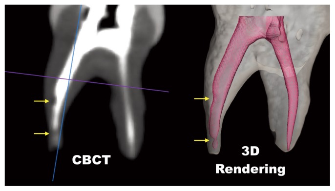
In three cases a slight overfilling of gutta-percha was observed of the root canals (2 in the Group TH and 1 in for the SC). These cases were excluded for the measurement of the distance between the limit of obturation and the apical foramen (Tab. 1). No significant differences were noted between the mean distances of the groups (t test, p>0.05). Visible voids were detected only in three canals (2 of Group TH and 1 of Group SC) and in only one case more than one void was present inside the canal (Fig. 1). The proportions Z test showed no difference in the percentage of canals with voids between the two groups (p >0.05).
Table 1.
Distance between the apical limit of the obturation to the apical foramen (mm) and percentage of the cases with voids.
| Distance | Voids | |||
|---|---|---|---|---|
|
|
|
|||
| Group | n | Mean (SD) | n | % |
| Thermafil | 18 | 0.32 a (0.24) | 2 | 10%a |
| Soft-Core | 19 | 0.27 a (0.18) | 1 | 5%a |
Different superscript letters indicate statistically significant difference within column.
Figure 1.
The CBCT (sagittal plane) and 3D rendering of the same obturated tooth showing the presence of voids (arrows).
Discussion
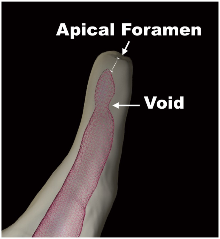
Several methods have been proposed to investigate the quality of root canal fillings technique (1, 3, 8, 9, 11–13, 15–17), such as radiographs, bioluminescence, histological sections, dye leakage, microleakage models and clearing techniques. The use of CT or CBCT is a quite recent methodology, which has many advantages: convenience, decreased examination time, lack of removal dentinal tissue, lack of damage of samples, possibility of different plane of examination and different sections of canals, and repeatability. CT is more costly and time consuming, but more precise. CBCT is easier and quicker to perform, and is more similar to clinical imaging. Moreover, the CBCT analysis allowed not only the evaluation of voids, but also the precise measurement of the apical length of the obturation. Although the CBCT images can be more easily visualized by three-dimensional rendering using specific softwares (Fig. 1), this image processing was made as an experimental model to confirm the CBCT findings (Fig. 2).
Figure 2.
Detection of voids (arrow) and measurement of distance between apical foramen and obturation (dashed line), using 3D rendering.
Results of the present study showed that there was no significant difference between the two tested CBO techniques in the apical extent of the root canal filling. Differently from some previous studies (11, 12), we found that despite the canal curvature, the apical limit of obturation was very satisfactory. Most of canals were sealed very close to the established working length, and a slight overfilling of gutta-percha was recorded in only two Thermafill cases and in a Soft-Core one, but there was no carrier protrusion beyond the apex.
These positive results are clearly related to a proper use of verifiers. Although the root canals were shaped up to a taper .06 and size 25, for the SC group the majority of canals (n=16) were filled with obturator size 20. It was observed that the Soft-Core verifier size 20 was correctly reaching WL while the size 25 verifier was short in thoses cases. For TH group, 18 all canals were obturated with size 25. This difference between the brands are probably related to their features: the Thermafil obturators present a smaller coronal dimensions and a bigger plastic core, while Soft-Core obturators have a thinner core and a greater amount of gutta-percha. A thinner core could also be related to a denser filling, since a greater gutta-percha ratio is considered preferable in maintaining the long-term apical seal (8, 9).
The apparently mismatch of sizes among the CBO brands proved not to be clinically relevant because the results of both were similar and satisfactory. This might be explained by the fact that in a CBO technique, the obturator is always well plasticized along its entire length. Accordingly, Marciano et al. showed that both Thermafil and System B were more effective than cold condensation techniques in the obturation of roots with isthmuses (9). Therefore, the gutta-percha flows not only apically but also laterally, filling a slightly wider canal space adequately, especially if we have more mass of gutta-percha, likewise in the Soft-Core obturators. Nevertheless, CBO are less effective than other warm obturation techniques in the filling of internal resorptive cavities (15).
Moreover a more flexible and smaller size carrier will reach working length with less risk of striping of the gutta-percha (11). Clinicians need only to pay attention at not pushing the carrier beyond the apex, which can be easily achieved by correctly placing the rubber stop on the obturator at the proper working length. Overall, when size 20 was used, Soft-Core obturators reached working length in severely curved canals more easily and predictably than Thermafil size 25. Table 1 shows the mean data concerning the distance between the apical foramen and root canal filling for both techniques.
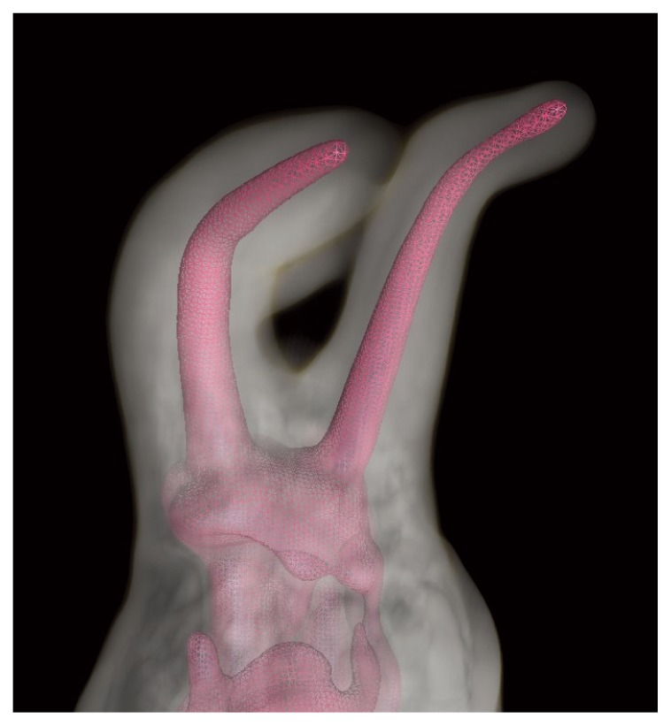
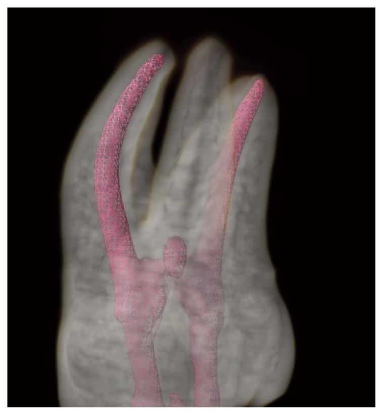
Likewise, no significant difference in terms of voids and gaps was found between the two techniques, showing the excellent performance of CBO in severe curvatures. The 3D rendering images clearly shows how canals were three-dimensionally filled (Figs. 3, 4). Present results are in agreement with previous researches that also showed no difference in the percentage of canal area filled and voids between Thermafil and Soft-Core (15).
Figure 3.
3D rendering of a tooth obturated with Soft-Core.
Figure 4.
3D rendering of a tooth obturated with Thermafil.
There have been many studies comparing obturation methods in vitro showing the effectiveness and simplicity of CBO techniques (6–10, 15). The findings of the present study confirmed these positive results, with a minimal amount of voids and underfilling. De Moor and Hommez compared different obturation materials with a dye leakage study and found that there were no differences in the long-term sealing ability between Thermafil and Soft-Core (18). A prospective clinical study (19) found no difference in success rates when filling the root canals with Soft-Core or lateral condensation. Another clinical study (20) did not find any difference in clinical outcomes between the lateral condensation and CBO technique performed with Thermafil obturator.
Hence we may conclude that the two tested CBO showed similar positive results. Both filling systems were quick and easy to perform even when severe curvatures were present. The present study emphatized the need to precisely verify the matching between preparation and obturators with verifiers.
References
- 1.Gillen BM, Looney SW, Gu L-S, Loushine BA, Weller RN, Loushine RJ, et al. Impact of the Quality of Coronal Restoration versus the Quality of Root Canal Fillings on Success of Root Canal Treatment: A Systematic Review and Meta-analysis. J Endod. 2011 Jul;37(7):895–902. doi: 10.1016/j.joen.2011.04.002. [DOI] [PMC free article] [PubMed] [Google Scholar]
- 2.Johnson WT, Kulild JC. Obturation of the cleaned and shaped root canal system. In: Hargreaves KM, Cohen S, editors. Cohen’s Pathways of the Pulp. 10th edition. Mosby; St. Louis, Mo, USA: 2010. pp. 349–351. [Google Scholar]
- 3.Hale R, Gatti R, Glickman GN, Opperman LA. Comparative analysis of carrier-based obturation and lateral compaction: A retrospective clinical outcomes study. Int J Dent. 2012;2012 doi: 10.1155/2012/954675. [DOI] [PMC free article] [PubMed] [Google Scholar]
- 4.Schilder H. Filling root canals in three dimensions. Dental Clinics of North America. 1967;11:723–744. [PubMed] [Google Scholar]
- 5.Endodontics JOB, Report W. A New Gutta-Percha Technique. 1978;4(6) doi: 10.1016/S0099-2399(78)80173-3. [DOI] [PubMed] [Google Scholar]
- 6.Gutmann JL, Saunders WP, Saunders EM, Nguyen L. An assessment of the plastic Thermafil obturation technique. Part 2. Material adaptation and sealability. Int Endod J. 1993;26:179–183. doi: 10.1111/j.1365-2591.1993.tb00790.x. [DOI] [PubMed] [Google Scholar]
- 7.Abarca AM, Bustos A, Navia M. A comparison of apical sealing and extrusion between Thermafil and lateral condensation techniques. J Endod. 2001;27:670–672. doi: 10.1097/00004770-200111000-00004. [DOI] [PubMed] [Google Scholar]
- 8.De-Deus G, Gurgel-Filho ED, Magalhães KM, Coutinho-Filho T. A laboratory analysis of gutta-percha-filled area obtained using Thermafil, System B and lateral condensation. Int Endod J. 2006;39(5):378–383. doi: 10.1111/j.1365-2591.2006.01082.x. [DOI] [PubMed] [Google Scholar]
- 9.Marciano M, Ordinola-Zapata R, Cunha TVRN, Duarte MAH, Cavenago BC, Garcia RB, et al. Analysis of four gutta-percha techniques used to fill mesial root canals of mandibular molars. Int Endod J. 2011;44(4):321–329. doi: 10.1111/j.1365-2591.2010.01832.x. [DOI] [PubMed] [Google Scholar]
- 10.Gencoglu N, Garip Y, Bas M, Samani S. Comparison of different gutta-percha root filling techniques: thermafil, Quickfill, System B, and lateral condensation. Oral Surg Oral Med Oral Pathol Oral Rad and Endodontics. 2002;93:333–336. doi: 10.1067/moe.2002.120253. [DOI] [PubMed] [Google Scholar]
- 11.Juhlin JJ, Walton RE, Dovgan JS. Adaptation of thermafil components to canal walls. J Endod. 1993;19(3):130–135. doi: 10.1016/S0099-2399(06)80507-8. [DOI] [PubMed] [Google Scholar]
- 12.Scott AC, Vire DE. An evaluation of the ability of a dentin plug to control extrusion of thermoplasticized gutta-percha. J Endod. 1992;18(2):52–57. doi: 10.1016/S0099-2399(06)81370-1. [DOI] [PubMed] [Google Scholar]
- 13.De Moor RJ, De Boever JG. The sealing ability of an epoxy resin root canal sealer used with five gutta-percha obturation techniques. Endod Dent Traumatol. 2000;16(6):291–297. doi: 10.1034/j.1600-9657.2000.016006291.x. [DOI] [PubMed] [Google Scholar]
- 14.Alhashimi RA, Foxton R, Romeed S, Deb S. Sci World J. Vol. 2014. Hindawi Publishing Corporation; 2014. An in vitro assessment of gutta-percha coating of new carrier-based root canal fillings. [DOI] [PMC free article] [PubMed] [Google Scholar]
- 15.Gencoglu N, Yildirim T, Garip Y, Karagenc B, Yilmaz H. Effectiveness of different gutta-percha techniques when filling experimental internal resorptive cavities. Int Endod J. 2008;41(10):836–842. doi: 10.1111/j.1365-2591.2008.01434.x. [DOI] [PubMed] [Google Scholar]
- 16.Dummer MH, Lyle L, Kennedy JK. A laboratory study of root fillings in teeth obturated by lateral condensation of gutta-percha or Thermafil obturators. 1994:32–38. doi: 10.1111/j.1365-2591.1994.tb00226.x. [DOI] [PubMed] [Google Scholar]
- 17.Kontakiotis EG, Chaniotis A, Georgopoulou M. Fluid filtration evaluation of 3 obturation techniques. Quintes Int. 2007;38:410–441. [PubMed] [Google Scholar]
- 18.De Moor RJG, Hommez GMG. The long-term sealing ability of an epoxy resin root canal sealer used with five gutta-percha obturation techniques. Int Endod J. 2002;35:275–282. doi: 10.1046/j.1365-2591.2002.00481.x. [DOI] [PubMed] [Google Scholar]
- 19.Özer SY, Aktener BO. Outcome of root canal treatment using soft-coreTM and cold lateral compaction filling techniques: A randomized clinical trial. J Contemp Dent Pract. 2009;10:74–81. [PubMed] [Google Scholar]
- 20.Chu CH, Lo ECM, Cheung GSP. Outcome of root canal treatment using Thermafil and cold lateral condensation filling techniques. Int Endod J. 2005;38:179–185. doi: 10.1111/j.1365-2591.2004.00929.x. [DOI] [PubMed] [Google Scholar]


