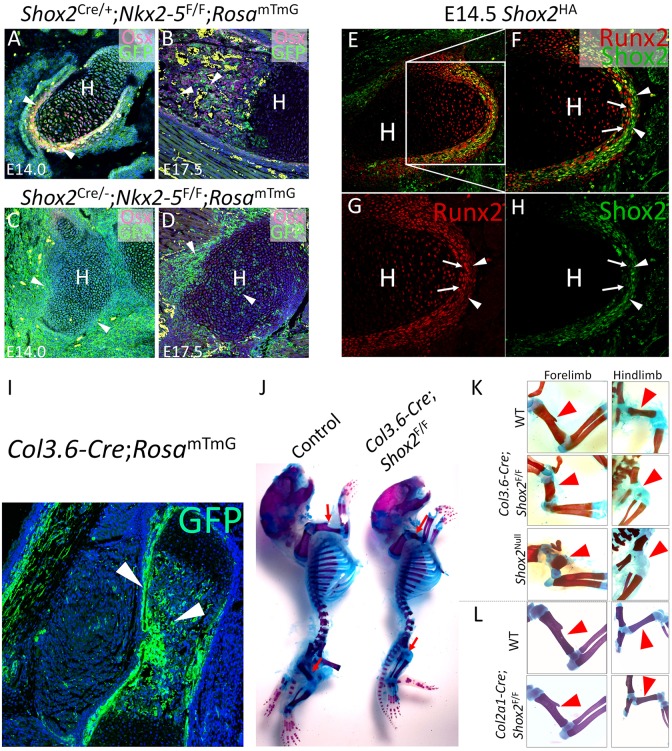
Fig. 3.
Shox2 inactivation in osteogenic lineage recapitulates limb defects in Shox2−/− mice. (A-D) Co-immunofluorescence of Osx and GFP shows the absence of osteogenic cells (arrowheads) in the humerus of Shox2Cre/−;Nkx2-5F/F;RosamTmG mice at E14.0 (C) and E17.5 (D) in comparison to littermate control Shox2Cre/+;Nkx2-5F/F;RosamTmG mice (A,B). (E-H) Co-immunofluorescence on E14.5 Shox2HA/+ embryo shows restricted expression of Shox2 in the outer layer (arrowheads) of the perichondrium and strong Runx2 expression in the inner layer (arrows). (I) GFP staining shows specific osteogenic activation (arrowheads) by Col3.6-Cre in a Col3.6-Cre;RosamTmG embryo at E17.5. (J) Alcian Blue/Alizarin Red staining on a representative Col3.6-Cre;RosamTmG mouse in comparison to its littermate control at P0. Red arrows indicate stylopodial skeleton. (K,L) Alcian Blue/Alizarin Red staining shows specific stylopodial defect (arrowheads) in Col3.6-Cre;Shox2F/F mice similar to that of Shox2−/− mice, compared with the Col2a1-Cre;Shox2F/F mice (L) and WT mice at P0 (the littermate control for Col2a1-Cre;Shox2F/F mice at P0 is shown in L). H, humerus.

