Abstract
Bacillus anthracis, the causative agent of anthrax, was utilized as a bioterrorism agent in 2001 when spores were distributed via the U.S. postal system. In responding to this event, the Federal Bureau of Investigation used traditional bacterial culture viability assays to ascertain the extent of contamination of the postal facilities within 24 to 48 h of environmental sample acquisition. Here, we describe a low-complexity, second-generation reporter phage assay for the rapid detection of viable B. anthracis spores in environmental samples. The assay uses an engineered B. anthracis reporter phage (Wβ::luxAB-2) which transduces bioluminescence to infected cells. To facilitate low-level environmental detection and maximize the signal response, expression of luxAB in an earlier version of the reporter phage (Wβ::luxAB-1) was optimized. These alterations prolonged signal kinetics, increased light output, and improved assay sensitivity. Using Wβ::luxAB-2, detection of B. anthracis spores was 1 CFU in 8 h from pure cultures and as low as 10 CFU/g in sterile soil but increased to 105 CFU/g in unprocessed soil due to an unstable signal and the presence of competing bacteria. Inclusion of semiselective medium, mediated by a phage-expressed antibiotic resistance gene, maintained signal stability and enabled the detection of 104 CFU/g in 6 h. The assay does not require spore extraction and relies on the phage infecting germinating cells directly in the soil sample. This reporter phage displays promise for the rapid detection of low levels of spores on clean surfaces and also in grossly contaminated environmental samples from complex matrices such as soils.
INTRODUCTION
Anthrax can be a fatal bacterial infection that occurs when Bacillus anthracis endospores enter the body through inhalation, ingestion, injection, or cutaneous exposure due to abrasions in the skin (1–3). Although anthrax infections occur infrequently in the natural environment, B. anthracis constitutes a biological threat as a military and/or terrorist weapon due to the longevity of its spores and the relative ease with which large quantities can be produced and stockpiled (2). The World Health Organization (WHO) estimated that 50 kg of dried spore powder dispersed over an urban population would result in ∼100,000 deaths and a concomitant breakdown of medical resources and civilian infrastructure (2, 4).
The first use of B. anthracis during a bioterrorism event in the United States was in 2001, when envelopes containing B. anthracis spores were distributed via the U.S. postal system to political and media targets. In addition to causing five fatalities and extensive social disruption, the event incurred a substantial emergency response and remediation costs; testing and remediation for the 42 contaminated buildings cost ∼$320 million (5). Given that these costs were the result of the distribution of only seven letters, the cost and scope of remediation and associated viability testing in the wake of a wide-area bioterrorism scenario would be extensive. The issues associated with remediation and the longevity of spores in soil are exemplified in the case of the Scottish island of Gruinard, which was deliberately contaminated with spores during biological weapon trials in World War II. Soil sampling 30 years after initial release indicated that although the numbers of spores were gradually declining, significant contamination remained and was likely to persist well into the next century (6). Cost-effective methodologies that expedite the large-scale and high-throughput sampling requirements for environmental detection and postclearance testing for viable B. anthracis bacteria would be of value.
The use of traditional culturing onto blood agar to isolate and identify B. anthracis from environmental samples is limited due to several complicating factors. For samples from relatively clean environments, such as solid indoor or outdoor surfaces, processing a single sample through elution, dilution, plating, and incubation can take days. The sampling and viability analysis steps are further complicated with a more complex environment, such as soil. These additional complications include the findings that up to 2 × 10⁹ background bacteria/g is found in the top 1 m of soil (7) and that closely related species within the Bacillus cereus group (e.g., B. cereus, B. anthracis, Bacillus thuringiensis, Bacillus mycoides, and Bacillus weihenstephanensis) are present as natural inhabitants of soil. For example, environmental isolates of B. anthracis have been shown to be beta-hemolytic, and B. cereus isolates displaying B. anthracis-like characteristics (such as lack of hemolysis, nonmotility, penicillin sensitivity, and/or phage susceptibility) have been identified (8, 9). A further complication is that a small number of B. anthracis spores may be present among a large number of naturally occurring spores of other Bacillus spp. (8). A semiselective agar consisting of polymyxin, lysozyme, disodium EDTA, and thallium acetate (PLET) was previously developed for differential selection of B. anthracis (10–12). However, the use of PLET with soil samples is limited for the following reasons: (i) multiple species within the B. cereus group are capable of growing on PLET, (ii) high numbers of B. cereus and B. thuringiensis bacteria naturally present in soil samples may outcompete growth of low numbers of B. anthracis, and (iii) polymyxin B is an ineffective inhibitor of Gram-negative bacteria (13).
Due to the limitations of traditional culture methods, the gold standard for quantification and detection of B. anthracis in environmental samples is nucleic acid-based detection (endpoint and real-time PCR) (8, 14). However, due to genetic similarity within the B. cereus group, cross-reactivity has often been observed (8, 15). With the advent of more rapid and less expensive whole-genome sequencing approaches, discrimination between closely related species and strains is readily attainable (16, 17). Nevertheless, chemical constituents of soil (such as organics, humic acids, and/or heavy metals) often interfere with nucleic acid-based chemistry and make direct detection of B. anthracis extremely difficult (18–21). Consequently, the majority of detection assays incorporate sample processing methods to separate, concentrate, and purify B. anthracis spores from soil prior to DNA extraction (21). Further complications arise during clearance monitoring as small numbers of viable spores must be recognized amid a landscape of nonviable spores. Currently, rapid viability PCR (RV-PCR) is the only diagnostic validated by the Environmental Protection Agency (EPA) for the detection of live B. anthracis spores in environmental samples (22, 23). This diagnostic expands the current capabilities of real-time PCR by measuring changes in cycle threshold (CT) values pre- and postincubation of sample. As only viable B. anthracis spores/cells can germinate and/or grow during the incubation period, reduced CT values postincubation are indicative of the presence of viable cells in the original environmental sample. RV-PCR can detect 10 live spores in the presence of 106 autoclaved spores and can process ∼96 samples in 24 h with a single robot and with personnel working in successive 8-h shifts. Although this technique has been validated for use with air filters and water and surface samples (24), it has yet to be validated for use in soil samples.
Bacteriophages (phages) are viruses which exclusively infect bacteria. This host tropism led to the development of phage typing schemes that have been used for decades to identify pathogens and that are now utilized in a number of different applications for bacterial detection (25, 26). A temperate B. anthracis phage (Wβ) identified in the 1950s was shown to display species specificity for B. anthracis by its ability to infect all B. anthracis strains tested (n = 171) (27) and an inability to lyse 242 out of the 244 strains (99% specificity) analyzed from 17 different non-anthracis Bacillus species (28). However, a few unusual B. cereus strains that manifest phenotypes of both B. cereus and B. anthracis have been identified which are phage susceptible (29). Using Wβ, we previously generated a light-tagged reporter phage by integrating the genes encoding bacterial luciferase (luxA and luxB) into the Wβ genome (30). Wβ::luxAB-1 transduces bioluminescence to cultured cells within 20 min. Although this reporter phage was able to detect 104 spores/ml in pure cultures within 3 h, improvements were required in order to facilitate detection from complex samples. Inclusivity experiments against wild-type B. anthracis isolates indicated that the reporter was able to confer bioluminescence to all B. anthracis strains tested (n = 38) (31). Specificity experiments with members of the closely related B. cereus group (B. cereus, B. thuringiensis, B. weihenstephanensis, and B. mycoides) indicated that 6 strains out of 119 analyzed displayed bioluminescence signals above those of background controls (95% specificity). Of these, five of the six positive strains elicited signals that were 10- to 100-fold lower than the B. anthracis signal. Fifteen other species of Bacillus and non-Bacillus members (comprising 47 strains) did not elicit a response with the reporter phage (31). We reconstructed Wβ::luxAB (Wβ::luxAB-2) to generate a brighter reporter phage with improved sensitivity, and here we demonstrate its utility in detecting viable B. anthracis spores from soils.
MATERIALS AND METHODS
Construction and propagation of Wβ::luxAB-2.
The Wβ::luxAB-2 reporter phage was constructed by targeted homologous recombination as previously described (30), with the exception that a designed promoter, harboring consensus transcriptional signals, was placed immediately upstream of luxAB (Fig. 1A). As before, the gene encoding spectinomycin resistance was included within the reporter cassette to enable the selection of recombinant lysogens. Plate lysates of Wβ::luxAB-1 and Wβ::luxAB-2 stocks were eluted in brain heart infusion (BHI) broth saturated with chloroform and centrifuged twice at room temperature (RT) for 10 min at 4,000 × g before the supernatant was incubated with DNase 1 (1.7 units/ml) (Thermo Scientific) for 20 min at 37°C. Lysates were then vacuum filtered through 0.2-μm-pore-size polyethersulfone (PES) membranes.
FIG 1.
Promoter design of the first-generation (Wβ::luxAB-1) and second-generation (Wβ::luxAB-2) reporter phages. Conserved nucleotides of promoters from Gram-positive (Gram +ve) species (shown in bold) were identified in previous studies (35–37).
Phage were concentrated by adding NaCl and polyethylene glycol (PEG) 8000 to the lysate to final concentrations of 0.75 M and 8% (wt/vol), respectively, and rotating the sample at 4°C for 3 h, followed by centrifugation at 4°C for 30 min at 11,000 × g. Pellets were gently resuspended in SMC buffer (50 mM Tris-HCl, pH 7.5, 5 mM CaCl2, 0.1 M NaCl, 8 mM MgSO4, 0.01% gelatin), and titers were determined using soft-agar overlays (32). Phage stocks were stored in the dark at 4°C until needed.
Bacillus spore preparations.
B. anthracis ΔSterne (exempt select agent strain) spores were prepared and kindly provided by Tony Buhr (Navy Surface Warfare Center Dahlgren Division) (33). The final preparation consisted of >98% spores that were stored in 0.1% Tween 80 and maintained at −80°C until use. B. anthracis Sterne 34F2 and B. thuringiensis 4AG1 spores were generated as described in Schofield and Westwater (30). Final preparations consisted of >98% spores that were stored in sterile water. As required, vials of Bacillus sp. spores were diluted in 0.05% Tween 80 and enumerated via colony counting (after 24 h of growth on BHI plates at 35°C). Unless otherwise stated, all experiments were conducted using B. anthracis ΔSterne.
Signal kinetics and sensitivity of Wβ::luxAB-2.
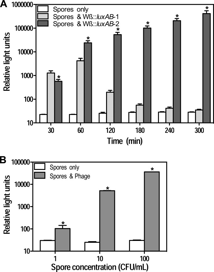
Signal kinetics between the Wβ::luxAB-1 and Wβ::luxAB-2 were compared by enriching B. anthracis spores (final concentration, 1 × 10⁶ CFU/ml) in tryptic soy broth (TSB) (containing 0.1 M l-alanine) for 3 h with shaking (250 rpm) at 35°C (n = 3). The culture was divided and infected with either Wβ::luxAB-1 or Wβ::luxAB-2 (final concentration, 1 × 10⁸ PFU/ml), incubated at designated time points (see Fig. 2A), and measured for bioluminescence. Detection limits of the Wβ::luxAB-2 reporter were determined by mixing B. anthracis spores (final concentration, 1 to 100 CFU/ml) with phage (final concentration, 3.4 × 10⁸ PFU/ml) in TSB containing 0.1 M l-alanine. Samples were incubated for 8 h with shaking (250 rpm) at 35°C before being assayed for bioluminescence.
FIG 2.
Reporter phage detection of B. anthracis Sterne 34F2. (A) Kinetics of light production by Wβ::luxAB-1 and Wβ::luxAB-2. Spores (1 × 10⁶ CFU/ml) were incubated in TSB (containing 0.1 M l-alanine) for 3 h at 35°C. Phage (final concentration, 1 × 10⁸ PFU/ml) were added, and bioluminescence was measured over time following the addition of n-decanal. (B) Detection sensitivity of Wβ::luxAB-2. Spores (1 to 100 CFU/ml) were mixed with reporter phage (final concentration, 3.4 × 10⁸ PFU/ml) and incubated in TSB (containing 0.1 M l-alanine) for 8 h at 35°C. Bioluminescence was measured following the addition of n-decanal. Values represent the means ± SD (n = 3). *, P < 0.05 (two-way ANOVA) for the signal response from Wβ::luxAB-1 compared to that of Wβ::luxAB-2 (A) or compared to that of spore-only controls (B).
Soil sample preparation and spore inoculation.
Mollisol HCB, a silty clay loam (USDA textural class), which is the predominant (21.5%) soil type in the United States (34), was purchased from Agvise Laboratories. A top layer (0 to 6 in.) of soil was passed through a 2-mm-pore-size sieve; samples (pH 7.8) contained 2.3% moisture and 7.5% organic matter. Where indicated (see Fig. 3 and 4), sterile soil was prepared by autoclaving (at 121°C for 60 min) soil samples three times in 50-ml Falcon tubes at 50% capacity. Sterility was assessed by the absence of growth following incubation on blood agar plates. For each experiment, the desired volume of soil (either 1.0 g or 0.1 g) was aliquoted into a 50-ml Falcon tube before 10 μl of an appropriate concentration of B. anthracis spores was inoculated into the center of each soil sample. Samples were maintained overnight (∼16 h) at 4°C before further use.
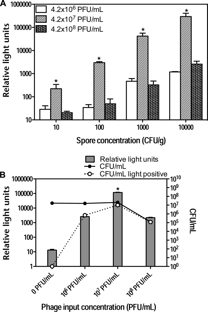
FIG 3.
Effect of varying the reporter phage concentration on B. anthracis detection. (A) Effect of varying the multiplicity of infection. Spores (10 to 10,000 CFU/g) were inoculated into sterile soil (1.0 g) and mixed with Wβ::luxAB-2 (final concentration, 4 × 10⁶ to 4 × 10⁸ PFU/ml) and TSB (containing 0.1 M l-alanine) at a 1:10 soil/medium ratio. Samples were then incubated for 12 h at 35°C and measured for bioluminescence following the addition of n-decanal. (B) Effect of the number of PFU/ml on bioluminescence and the number of CFU/ml. Spores (1 × 10⁴ CFU/g) were inoculated into soil (1.0 g) and mixed with a range of concentrations of Wβ::luxAB-2 (4 × 10⁶ to 4 × 10⁸ PFU/ml final) in TSB (containing 0.1 M l-alanine). After 12 h at 35°C, samples were measured for bioluminescence and plated for counts of CFU/ml. To determine the number of phage-infected colonies (light-positive lysogens) plates were exposed for 10-min to n-decanal and examined under dark-field illumination. Values represent the means ± SD (n = 3). *, P < 0.05 (two-way ANOVA) for comparisons of the results at the various phage concentrations used.
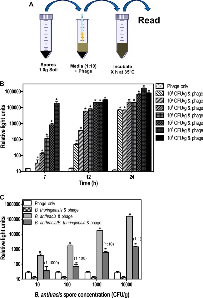
FIG 4.
Detection of B. anthracis spores from defined soil. (A) Assay procedure. Unless otherwise stated, spores at the desired concentration were inoculated into sterile soil (1.0 g) and maintained overnight (∼16 h) at 4°C before the addition of Wβ::luxAB-2 (final concentration, 4 × 10⁷ PFU/ml) and medium (containing 0.1 M l-alanine) at a 1:10 soil/medium ratio. Samples were then incubated for X h (where X is 7, 12, or 24 h) at 35°C and measured for bioluminescence following the addition of n-decanal. (B) Sensitivity limit of detection of B. anthracis spores in sterile soil. Spores (1.3 × 101 to 1.3 × 10⁷ CFU/g) were inoculated into soil and measured for bioluminescence after 7, 12, and 24 h of incubation. (C) Effect of mixed bacterial populations on detection in sterile soil. B. anthracis spores (10 to 1,000 CFU/g) were inoculated into soil in the presence or absence of an excess of spores (10,000 CFU/g) from the nonpermissive B. thuringiensis strain 4AG1. Bioluminescence was measured after 12 h of incubation. Values represent the means ± SD (n = 3). *, P < 0.05 (two-way ANOVA) for results compared to those with the phage-only controls. In panel C, values in parentheses indicate the initial ratio of B. anthracis spores to B. thuringiensis spores.
To assess the ability of Wβ::luxAB-2 to produce a bioluminescent signal response in the presence of other spore-forming bacteria, sterile soil was inoculated with B. anthracis spores at 1.1 × 101 to 1.1 × 10⁴ CFU/g and incubated in the presence of a fixed spore concentration (1.1 × 10⁴ CFU/g) of a nonpermissive B. thuringiensis strain (4AG1).
Phage-mediated detection of B. anthracis in soil.
TSB or beef infusion broth (containing 0.1 M l-alanine) was added to soil-spore mixtures at a 1:10 (for 1.0-g soil samples) or a 1:100 (for 0.1-g samples) ratio and vortexed vigorously before being mixed with Wβ::luxAB-2 at a final concentration of 4 × 10⁷ PFU/ml. Where indicated (see Fig. 6), spectinomycin (final concentration, 100 μg/ml) was added to the culture-soil mixes 4 h after phage infection. Samples were incubated at 35°C with shaking (250 rpm) before bioluminescence analyses at various times. Soil samples containing Wβ::luxAB-2 served as negative controls.
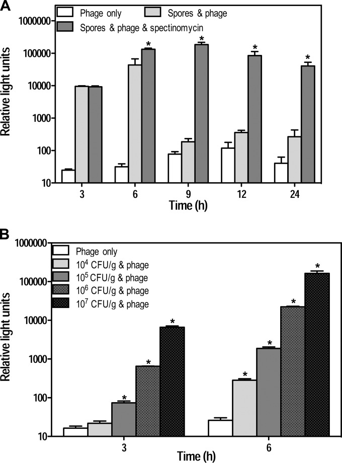
FIG 6.
Effect of spectinomycin on signal kinetics and limits of detection. (A) Detection of B. anthracis spores in soil in the presence of spectinomycin. Spores (1.3 × 10⁷ CFU/g) were inoculated into soil (0.1 g) and maintained overnight (∼16 h) at 4°C before being mixed with Wβ::luxAB-2 (final concentration, 4 × 10⁷ PFU/ml) and incubated in beef infusion broth (containing 0.1 M l-alanine) at 35°C. Spectinomycin (100 μg/ml) was added 4 h after phage infection, and bioluminescence was measured over time following the addition of n-decanal. (B) Limits of detection. B. anthracis spores (1.3 × 10⁴ to 1.3 × 10⁷ CFU/g) were inoculated into soil and incubated with the reporter phage and spectinomycin as described above. Samples were read for bioluminescence after 3 and 6 h. Numbers represent the means ± SD (n = 3). *, P < 0.05 (two-way ANOVA) for results with spectinomycin-treated samples compared to those with untreated samples (A) or for results compared with those of phage-only controls (B).
Bioluminescence assays and statistical analysis.
Samples (195 μl) were measured for bioluminescence at various times after phage infection using a Veritas Microplate Luminometer (Turner BioSystems). Samples were injected with n-decanal (67 μl of a 2% solution) and read for 10 s. Controls consisted of cells or phage alone. Bioluminescent signal intensities were reported as mean relative light units (RLU) from three experiments ± standard deviations (SD). Statistical analysis and graphing were performed with GraphPad Prism, version 5, software. Two-way analysis of variance (ANOVA) followed by post hoc Bonferroni multiple-comparisons tests were used to determine statistical significance (P < 0.05) between treatments.
RESULTS
Redesign of Wβ::luxAB-2 reporter phage and signal response.
Detection of B. anthracis spores from environmental samples poses significant challenges due to the complexity of the matrices and the requirement for low-level detection. To improve reporter phage sensitivity, we optimized the luxAB promoter to include all important conserved regions of Gram-positive bacterial promoters, in addition to the −35 and −10 hexamers (Fig. 1) (35–37). These include an A tract and T 5′ of the −35 hexamer, a TG dinucleotide 5′ of the −10 hexamer (extended −10), two A nucleotides 3′ of the −10 hexamer, and the optimal 17-bp spacer between the hexamers. These alterations generated significant changes in the characteristics of the reporter phage. B. anthracis lysogens harboring Wβ::luxAB-2 were brighter than lysogens harboring the Wβ::luxAB-1 reporter (data not shown). A more quantitative comparison of signal kinetics between Wβ::luxAB-1 and Wβ::luxAB-2 reporters indicated that the former elicits a peak signal 60 min after infection, followed by a general decline over time. In contrast, Wβ::luxAB-2 elicits a signal response that increases in strength over time (Fig. 2A). Wβ::luxAB is a temperate phage, and if the multiplicity of infection is high, the lysogenic pathway is typically favored over lytic growth (38). The reduction in luminescence after infection by Wβ::luxAB-1 suggests that luxAB expression decreases in the prophage state. The consensus luxAB promoter in Wβ::luxAB-2 therefore seems to be independent of other transcriptional regulatory processes acting on the phage genome. Nevertheless, the short-lived signal from Wβ::luxAB-1 necessitated that phage addition had to follow a period of incubation. However, Wβ::luxAB-2 can be added at the onset of germination. This allows a longer time frame for the reporter phage to interact with the cells and allows the spores to germinate and amplify during the course of the incubation time, a prerequisite for detection. As a result, the assay can readily detect a starting concentration of 1 CFU/ml of pure B. anthracis spores within 8 h (Fig. 2B). A similar level of sensitivity may be obtained from spores inoculated onto glass coupons, dried, and then immersed directly into outgrowth medium harboring the phage (data not shown).
Detection of B. anthracis in sterile soil.
Soil is a complex matrix containing a myriad of factors that interfere with biological assays (20, 39). Soil microbes, humic acids, heavy metals, and other factors can reduce spore germination and growth, prevent phage infection, and/or quench bioluminescence. To reduce sample complexity, we initially used sterile (autoclaved) soil and examined the effect of varying the multiplicity of infection on the ability to detect B. anthracis (Fig. 3A). Soil was inoculated with a range of spore concentrations (10 to 10,000 CFU/g) and incubated with reporter phage at concentrations varying from 106 to 108 PFU/ml. At each spore concentration, the highest signal response was obtained using 107 PFU/ml (P < 0.05), irrespective of the multiplicity of infection. To examine the relationship between the number of PFU/ml, B. anthracis growth, and the number of infected cells (lysogens), soil samples were inoculated with 104 CFU/g and incubated with a phage concentration ranging from 106 to 108 PFU/ml (Fig. 3B). After 12 h of incubation, the samples were measured for bioluminescence, the number of CFU/ml, and the number of light-positive CFU/ml (lysogens). In the absence of phage, B. anthracis amplified to 107 CFU/ml during the course of the experiment. At the highest phage concentrations analyzed (108 PFU/ml), B. anthracis growth was reduced 100-fold compared to growth of phage-free controls. However, a very high proportion of these colonies (96%) were light positive (lysogens). Samples incubated with the lowest concentration of phage (106 PFU/ml) contained similar counts of CFU/ml as phage-free controls, but <10% of these colonies were lysogens. B. anthracis samples incubated with 107 PFU/ml grew to similar CFU/ml levels as phage-free controls, harbored 37% light-positive colonies, and displayed the highest total bioluminescence (P < 0.05). Thus, a reporter phage concentration of 107 PFU/ml was used in the subsequent experiments.
The sensitivity limits of detection and signal response times were examined using sterile soil. Soil samples inoculated with spores were directly mixed with medium harboring reporter phage (Fig. 4A). Thus, spores were not extracted from the soil samples, and the reporter phage infected germinating cells directly in the soil-medium mix. A detection sensitivity of 100 CFU/g was achieved in 7 h but could be lowered to 10 CFU/g after an incubation time of 12 h (Fig. 4B). Signal responses were phage dose dependent at early times but saturated at 24 h after infection. These results indicate (i) that spore extraction from the (originally sterile) soil is unnecessary and (ii) that phage infection is not inhibited by the soil matrix.
Sterile soil lacks organisms that could compete with B. anthracis germination and growth. We therefore assayed for B. anthracis in the presence of B. thuringiensis 4AG1, which is nonpermissive to Wβ::luxAB. Sterile soil was inoculated with 10 to 10,000 B. anthracis spores in the presence of a fixed concentration (10,000 CFU/g) of B. thuringiensis spores and analyzed for bioluminescence after 12 h of incubation (Fig. 4C). The presence of B. thuringiensis reduced the signal elicited by phage-infected B. anthracis. Nevertheless, 100 CFU/g of B. anthracis bacteria is readily detected in the presence of 10,000 CFU/g of B. thuringiensis bacteria; similar data were recorded when a non-phage-susceptible B. cereus strain (ATCC 14579) was used (data not shown). Presumably, these closely related bacteria affected the luciferase assay by reducing B. anthracis germination or growth and/or by sequestering the phage.
Detection of B. anthracis in soil.
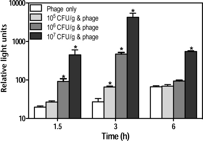
B. anthracis spores were tested in natural (nonsterile) soil samples. Soil was inoculated with 1.3 × 10⁵ to 1.3 × 10⁷ CFU/g and assayed for bioluminescence. Although 105 CFU/g could be detected within 3 h (Fig. 5), the signal strength was attenuated compared to that in sterile soil and then declined even further. The reduced signal may be due to (i) competitive exclusion of B. anthracis germination or growth, (ii) phage binding to nonspecific components in the soil, and/or (iii) vegetative cell death. In unsuccessful attempts to address the cause of the reduced signal, we changed the multiplicity of infection and incorporated a 1-h 70°C heat pretreatment prior to outgrowth in order to reduce the viability of vegetative cells and partially select for spores. Neither treatment increased signal intensity or prolonged signal kinetics (data not shown).
FIG 5.
Detection of B. anthracis spores in soil. B. anthracis spores (1.3 × 10⁵ to 1.3 × 10⁷ CFU/g) were inoculated into soil (1.0 g) and maintained overnight (∼16 h) at 4°C before being mixed with Wβ::luxAB-2 (final concentration, 4 × 10⁷ PFU/ml) and incubated in TSB (containing 0.1 M l-alanine) at 35°C. Bioluminescence was measured 1.5, 3, and 6 h following the addition of n-decanal. Values represent the means ± SD (n = 3). *, P < 0.05 (two-way ANOVA) for results compared to those with phage-only controls.
A spectinomycin resistance gene was used as a selection marker in the construction of Wβ::luxAB. Consequently, all phage-infected B. anthracis bacteria should have a competitive growth advantage in medium supplemented with spectinomycin. To test whether spectinomycin could improve signal responses, soil was inoculated with 1.3 × 10⁷ CFU/g and mixed with medium and phage. After 4 h of incubation, samples were supplemented with spectinomycin (100 μg/ml). In the absence of spectinomycin, peak signal intensity occurred at 6 h and then decreased (Fig. 6A). In contrast, the presence of spectinomycin allowed a significantly higher signal response by 6 h (P < 0.05), and the signal remained fairly stable up to 24 h (Fig. 6A). Spectinomycin also improved the assay sensitivity. Soil was inoculated with B. anthracis bacteria at 104 to 107 CFU/g and tested as described before; bacteria at 104 CFU/g were easily detected in 6 h, a 10-fold improvement over nonselective medium (Fig. 6B).
DISCUSSION
B. anthracis spores are the infectious agent of anthrax and may persist long-term in the environment if they are deliberately released (40–42). Viable B. anthracis spores were still detected 30 years after their release on Gruinard Island and have been revived from sediments ∼1,000 years old (42). For over a decade, scientists have been striving to develop rapid, sensitive, and specific methods for the detection of B. anthracis spores in environmental samples (8). Reporter phages constitute an alternative means that may fit this need. In contrast to conventional PCR methodologies, which are independent of host viability, reporter phages generate a signal only if metabolically active cells are present (43). This is a critical element required for environmental detection especially following remediation to ensure that viable and potentially infectious cells have been effectively decontaminated. However, while detection of viable cells may be considered an advantage, it introduces assay variability as individual cells in a population may not be at the same stages of growth (asynchronous) or may be compromised, leading to differences in signal strength. Moreover, detection requires spore germination, which in the presence of other microorganisms in soil samples may be subject to competitive exclusion of nutrients necessary for B. anthracis germination and growth. As assay success requires both germination and growth, we used a nonselective rich medium to achieve this objective. Moreover, in order to maximize signal expression, we initially engineered a second-generation reporter phage, Wβ::luxAB-2, which was able to detect 1 CFU/ml in 8 h and displayed improved sensitivity and kinetics. The improvements, in comparison to the performance of Wβ::luxAB-1, are likely due to (i) the presence of a Bacillus consensus promoter driving constitutive expression of the luxAB reporter genes, rendering it independent of a lytic or lysogenic response by the phage, and (ii) the addition of the reporter phage to the spores at the onset of the assay. Thus, there is a longer window for the phage to find and interact with the germinating cells and elicit a response. The improvement in assay sensitivity of nonsterile soil when spectinomycin was employed further suggests that use of the Wβ::luxAB reporter phage will be effective for the detection of moderately to grossly contaminated soil samples.
Other phage-based methodologies using the closely related γ phage are being explored for the detection of B. anthracis. Cox et al. utilized the fact that phage will amplify in the presence of their host as a means of detecting whether B. anthracis cells are present (44). When γ phage amplification was used in combination with an inexpensive lateral flow immunochromatographic device which harbors an antiphage antibody, 104 CFU/ml could be detected within 2 h. Alternatively, Schuch et al. used the phage lysin PlyG as a means of detecting B. anthracis (45). Germinating spores, when incubated in the presence of the B. anthracis-specific PlyG, rapidly lyse and release ATP. ATP is a cofactor for eukaryotic luciferase. Thus, when γ phage are mixed with luciferase/luciferin, light is emitted, indicating the presence of B. anthracis. Using this method, about 100 spores could be detected within 60 min. To the best of our knowledge, it is unknown if these methodologies have been assessed for the detection of B. anthracis from complex environmental samples. The transition from noncomplex to complex samples can be challenging. The differences in our results in terms of the efficiency of detection between pure cultures/cultures inoculated into sterile soil versus detection in “real” soil samples were significant. The limit of detection in pure cultures was 1 CFU/ml (in 8 h), but this decreased 1,000-fold to 104 CFU/g (in 6 h) in soil. The reasons for the drop in sensitivity in soil may be due to a multitude of factors, including a reduced ability of B. anthracis to grow during the course of incubation (C. Nguyen, N. J. Sharp, and D. A. Schofield, unpublished results). Future experiments may therefore require an initial spore extraction step to clean up and concentrate the spores and remove any soil-mitigating component prior to phage addition. It should be noted that for B. anthracis PCR detection methodologies, the use of commercial DNA extraction kits is dependent on the soil type, which can vary greatly and thus adds assay complexity. Moreover, input spore concentrations of ≥105 CFU/g are required (39). The reporter phage technology in its current configuration is therefore at least as sensitive as, or perhaps more sensitive than, currently available commercial kits. Importantly, spore extraction and extensive processing of soil samples are not necessary for the reporter phage assay. Therefore, it offers the potential for high throughput.
During the 2001 anthrax event, the Federal Bureau of Investigation opted for traditional culture methods as their simplicity permitted inoculation of culture medium in hot zones by nonspecialists (46). This eliminated the laboratory bottlenecks previously experienced with molecular techniques that necessitate multiple sample preparation and assay steps and require highly trained technicians. Reporter phage technology may provide a direct and more sensitive approach that is potentially compatible with large-scale, high-throughput sampling that can be processed by technicians with no prior experience with this phage assay.
ACKNOWLEDGMENT
We thank Tony Buhr at Naval Surface Warfare Center Dahlgren Division for kindly providing standardized spore preparations.
Funding Statement
Defense Threat Reduction Agency provided funding to the United States Army Engineer Research and Development Center under grant number BA11PHM148. United States Army Engineer Research and Development Center provided funding to David A. Schofield under grant number W9132T-12-C-0017. HHS | NIH | National Institute of Allergy and Infectious Diseases (NIAID) provided funding to David A. Schofield under grant number R01AI111535.
REFERENCES
- 1.Dixon TC, Meselson M, Guillemin J, Hanna PC. 1999. Anthrax. N Engl J Med 341:815–826. doi: 10.1056/NEJM199909093411107. [DOI] [PubMed] [Google Scholar]
- 2.Inglesby TV, O'Toole T, Henderson DA, Bartlett JG, Ascher MS, Eitzen E, Friedlander AM, Gerberding J, Hauer J, Hughes J, McDade J, Osterholm MT, Parker G, Perl TM, Russell PK, Tonat K. 2002. Anthrax as a biological weapon, 2002: updated recommendations for management. JAMA 287:2236–2252. doi: 10.1001/jama.287.17.2236. [DOI] [PubMed] [Google Scholar]
- 3.Sweeney DA, Hicks CW, Cui X, Li Y, Eichacker PQ. 2011. Anthrax infection. Am J Respir Crit Care Med 184:1333–1341. doi: 10.1164/rccm.201102-0209CI. [DOI] [PMC free article] [PubMed] [Google Scholar]
- 4.Spencer RC. 2003. Bacillus anthracis. J Clin Pathol 56:182–187. doi: 10.1136/jcp.56.3.182. [DOI] [PMC free article] [PubMed] [Google Scholar]
- 5.Schmitt K, Zacchia NA. 2012. Total decontamination cost of the anthrax letter attacks. Biosecur Bioterror 10:98–107. doi: 10.1089/bsp.2010.0053. [DOI] [PubMed] [Google Scholar]
- 6.Manchee RJ, Broster MG, Stagg AJ, Hibbs SE. 1994. Formaldehyde solution effectively inactivates spores of Bacillus anthracis on the Scottish island of Gruinard. Appl Environ Microbiol 60:4167–4171. [DOI] [PMC free article] [PubMed] [Google Scholar]
- 7.Whitman WB, Coleman DC, Wiebe WJ. 1998. Prokaryotes: the unseen majority. Proc Natl Acad Sci U S A 95:6578–6583. doi: 10.1073/pnas.95.12.6578. [DOI] [PMC free article] [PubMed] [Google Scholar]
- 8.Irenge LM, Gala JL. 2012. Rapid detection methods for Bacillus anthracis in environmental samples: a review. Appl Microbiol Biotechnol 93:1411–1422. doi: 10.1007/s00253-011-3845-7. [DOI] [PubMed] [Google Scholar]
- 9.Beesley CA, Vanner CL, Helsel LO, Gee JE, Hoffmaster AR. 2010. Identification and characterization of clinical Bacillus spp. isolates phenotypically similar to Bacillus anthracis. FEMS Microbiol Lett 313:47–53. doi: 10.1111/j.1574-6968.2010.02120.x. [DOI] [PubMed] [Google Scholar]
- 10.Knisely RF. 1966. Selective medium for Bacillus anthracis. J Bacteriol 92:784–786. [DOI] [PMC free article] [PubMed] [Google Scholar]
- 11.Luna VA, Gulledge J, Cannons AC, Amuso PT. 2009. Improvement of a selective media for the isolation of B. anthracis from soils. J Microbiol Methods 79:301–306. doi: 10.1016/j.mimet.2009.09.023. [DOI] [PubMed] [Google Scholar]
- 12.Marston CK, Beesley C, Helsel L, Hoffmaster AR. 2008. Evaluation of two selective media for the isolation of Bacillus anthracis. Lett Appl Microbiol 47:25–30. doi: 10.1111/j.1472-765X.2008.02375.x. [DOI] [PubMed] [Google Scholar]
- 13.Perlman D, Mahler I. March 2004. Selective growth medium for Bacillus anthracis and methods of use. US patent 20040058428.
- 14.Be NA, Thissen JB, Gardner SN, McLoughlin KS, Fofanov VY, Koshinsky H, Ellingson SR, Brettin TS, Jackson PJ, Jaing CJ. 2013. Detection of Bacillus anthracis DNA in complex soil and air samples using next-generation sequencing. PLoS One 8:e73455. doi: 10.1371/journal.pone.0073455. [DOI] [PMC free article] [PubMed] [Google Scholar]
- 15.Zasada AA, Gierczynski R, Raddadi N, Daffonchio D, Jagielski M. 2006. Some Bacillus thuringiensis strains share rpoB nucleotide polymorphisms also present in Bacillus anthracis. J Clin Microbiol 44:1606–1607. doi: 10.1128/JCM.44.4.1606-1607.2006. [DOI] [PMC free article] [PubMed] [Google Scholar]
- 16.Rasko DA, Worsham PL, Abshire TG, Stanley ST, Bannan JD, Wilson MR, Langham RJ, Decker RS, Jiang L, Read TD, Phillippy AM, Salzberg SL, Pop M, Van Ert MN, Kenefic LJ, Keim PS, Fraser-Liggett CM, Ravel J. 2011. Bacillus anthracis comparative genome analysis in support of the Amerithrax investigation. Proc Natl Acad Sci U S A 108:5027–5032. doi: 10.1073/pnas.1016657108. [DOI] [PMC free article] [PubMed] [Google Scholar]
- 17.Chen PE, Willner KM, Butani A, Dorsey S, George M, Stewart A, Lentz SM, Cook CE, Akmal A, Price LB, Keim PS, Mateczun A, Brahmbhatt TN, Bishop-Lilly KA, Zwick ME, Read TD, Sozhamannan S. 2010. Rapid identification of genetic modifications in Bacillus anthracis using whole genome draft sequences generated by 454 pyrosequencing. PLoS One 5:e12397. doi: 10.1371/journal.pone.0012397. [DOI] [PMC free article] [PubMed] [Google Scholar]
- 18.Beyer W, Pocivalsek S, Bohm R. 1999. Polymerase chain reaction-ELISA to detect Bacillus anthracis from soil samples—limitations of present published primers. J Appl Microbiol 87:229–236. doi: 10.1046/j.1365-2672.1999.00875.x. [DOI] [PubMed] [Google Scholar]
- 19.Cheun HI, Makino SI, Watarai M, Erdenebaatar J, Kawamoto K, Uchida I. 2003. Rapid and effective detection of anthrax spores in soil by PCR. J Appl Microbiol 95:728–733. doi: 10.1046/j.1365-2672.2003.02038.x. [DOI] [PubMed] [Google Scholar]
- 20.Dineen SM, Aranda R IV, Anders DL, Robertson JM. 2010. An evaluation of commercial DNA extraction kits for the isolation of bacterial spore DNA from soil. J Appl Microbiol 109:1886–1896. doi: 10.1111/j.1365-2672.2010.04816.x. [DOI] [PubMed] [Google Scholar]
- 21.Silvestri EE, Perkins SD, Feldhake D, Nichols T, Schaefer FW III. 2015. Recent literature review of soil processing methods for recovery of Bacillus anthracis. Ann Microbiol 65:1215–1226. doi: 10.1007/s13213-014-0932-x. [DOI] [Google Scholar]
- 22.Letant SE, Kane SR, Murphy GA, Alfaro TM, Hodges LR, Rose LJ, Raber E. 2010. Most-probable-number rapid viability PCR method to detect viable spores of Bacillus anthracis in swab samples. J Microbiol Methods 81:200–202. doi: 10.1016/j.mimet.2010.02.011. [DOI] [PubMed] [Google Scholar]
- 23.Kane S, Shah S, Letant S, Murphy G, Alfaro T, Avila J, Salazer E, Mullins M, Nichols T. 2013. Operational evaluation of the rapid viability PCR method for post-decontamination clearance sampling. J Bioterr Biodef S3:016. [Google Scholar]
- 24.EPA. 2011. Development and verification of rapid viability polymerase chain reaction (RV-PCR) protocols for Bacillus anthracis—for application to air filters, water and surface samples. Document EPA/600/R-10/156. US Environmental Protection Agency, Washington, DC. [Google Scholar]
- 25.Singh A, Poshtiban S, Evoy S. 2013. Recent advances in bacteriophage based biosensors for food-borne pathogen detection. Sensors (Basel) 13:1763–1786. doi: 10.3390/s130201763. [DOI] [PMC free article] [PubMed] [Google Scholar]
- 26.Smartt AE, Ripp S. 2011. Bacteriophage reporter technology for sensing and detecting microbial targets. Anal Bioanal Chem 400:991–1007. doi: 10.1007/s00216-010-4561-3. [DOI] [PubMed] [Google Scholar]
- 27.Brown ER, Cherry WB. 1955. Specific identification of Bacillus anthracis by means of a variant bacteriophage. J Infect Dis 96:34–39. doi: 10.1093/infdis/96.1.34. [DOI] [PubMed] [Google Scholar]
- 28.McCloy EW. 1951. Studies on a lysogenic Bacillus strain. I. A bacteriophage specific for Bacillus anthracis. J Hyg (Lond) 49:114–125. [DOI] [PMC free article] [PubMed] [Google Scholar]
- 29.Schuch R, Fischetti VA. 2006. Detailed genomic analysis of the Wβ and γ phages infecting Bacillus anthracis: implications for evolution of environmental fitness and antibiotic resistance. J Bacteriol 188:3037–3051. doi: 10.1128/JB.188.8.3037-3051.2006. [DOI] [PMC free article] [PubMed] [Google Scholar]
- 30.Schofield DA, Westwater C. 2009. Phage-mediated bioluminescent detection of Bacillus anthracis. J Appl Microbiol 107:1468–1478. doi: 10.1111/j.1365-2672.2009.04332.x. [DOI] [PubMed] [Google Scholar]
- 31.Schofield DA, Sharp NJ, Vandamm J, Molineux IJ, Spreng KA, Rajanna C, Westwater C, Stewart GC. 2013. Bacillus anthracis diagnostic detection and rapid antibiotic susceptibility determination using “bioluminescent” reporter phage. J Microbiol Methods 95:156–161. doi: 10.1016/j.mimet.2013.08.013. [DOI] [PubMed] [Google Scholar]
- 32.Carlson K. 2005. Working with bacteriophages: common techniques and methodological approaches, p 437–494. In Kutter E, Sulakvelidze A (ed), Bacteriophages: biology and applications. CRC Press, Boca Raton, FL. [Google Scholar]
- 33.Buhr TL, Young AA, Minter ZA, Wells CM, Shegogue DA. 2011. Decontamination of a hard surface contaminated with Bacillus anthracis ΔSterne and B. anthracis Ames spores using electrochemically generated liquid-phase chlorine dioxide (eClO2). J Appl Microbiol 111:1057–1064. doi: 10.1111/j.1365-2672.2011.05122.x. [DOI] [PubMed] [Google Scholar]
- 34.USDA Natural Resources Conservation Service. 1999. Soil taxonomy: a basic system of soil classification for making and interpreting soil surveys, 2nd ed US Department of Agriculture, Washington, DC. [Google Scholar]
- 35.Helmann JD. 1995. Compilation and analysis of Bacillus subtilis σA-dependent promoter sequences: evidence for extended contact between RNA polymerase and upstream promoter DNA. Nucleic Acids Res 23:2351–2360. doi: 10.1093/nar/23.13.2351. [DOI] [PMC free article] [PubMed] [Google Scholar]
- 36.Voskuil MI, Chambliss GH. 1998. The −16 region of Bacillus subtilis and other gram-positive bacterial promoters. Nucleic Acids Res 26:3584–3590. doi: 10.1093/nar/26.15.3584. [DOI] [PMC free article] [PubMed] [Google Scholar]
- 37.Voskuil MI, Voepel K, Chambliss GH. 1995. The −16 region, a vital sequence for the utilization of a promoter in Bacillus subtilis and Escherichia coli. Mol Microbiol 17:271–279. doi: 10.1111/j.1365-2958.1995.mmi_17020271.x. [DOI] [PubMed] [Google Scholar]
- 38.Kourilsky P. 1973. Lysogenization by bacteriophage lambda. I. Multiple infection and the lysogenic response. Mol Gen Genet 122:183–195. [DOI] [PubMed] [Google Scholar]
- 39.Gulledge JS, Luna VA, Luna AJ, Zartman R, Cannons AC. 2010. Detection of low numbers of Bacillus anthracis spores in three soils using five commercial DNA extraction methods with and without an enrichment step. J Appl Microbiol 109:1509–1520. doi: 10.1111/j.1365-2672.2010.04774.x. [DOI] [PubMed] [Google Scholar]
- 40.Kim HS, Sherman D, Johnson F, Aronson AI. 2004. Characterization of a major Bacillus anthracis spore coat protein and its role in spore inactivation. J Bacteriol 186:2413–2417. doi: 10.1128/JB.186.8.2413-2417.2004. [DOI] [PMC free article] [PubMed] [Google Scholar]
- 41.Sharp RJ, Roberts AG. 2006. Anthrax: the challenges for decontamination. J Chem Technol Biotechnol 81:1612–1625. doi: 10.1002/jctb.1591. [DOI] [Google Scholar]
- 42.Sinclair R, Boone SA, Greenberg D, Keim P, Gerba CP. 2008. Persistence of category A select agents in the environment. Appl Environ Microbiol 74:555–563. doi: 10.1128/AEM.02167-07. [DOI] [PMC free article] [PubMed] [Google Scholar]
- 43.Schofield DA, Sharp NJ, Westwater C. 2012. Phage-based platforms for the clinical detection of human bacterial pathogens. Bacteriophage 2:105–283. doi: 10.4161/bact.19274. [DOI] [PMC free article] [PubMed] [Google Scholar]
- 44.Cox CR, Jensen KR, Mondesire RR, Voorhees KJ. 2015. Rapid detection of Bacillus anthracis by gamma phage amplification and lateral flow immunochromatography. J Microbiol Methods 118:51–56. doi: 10.1016/j.mimet.2015.08.011. [DOI] [PubMed] [Google Scholar]
- 45.Schuch R, Nelson D, Fischetti VA. 2002. A bacteriolytic agent that detects and kills Bacillus anthracis. Nature 418:884–889. doi: 10.1038/nature01026. [DOI] [PubMed] [Google Scholar]
- 46.Beecher DJ. 2006. Forensic application of microbiological culture analysis to identify mail intentionally contaminated with Bacillus anthracis spores. Appl Environ Microbiol 72:5304–5310. doi: 10.1128/AEM.00940-06. [DOI] [PMC free article] [PubMed] [Google Scholar]


