

Abstract
Background
Internal fixation with plates is a reliable fixation technique for the treatment of distal radius fractures. An ongoing discussion exists whether volar or dorsal plating is the appropriate technique. In clinical practice, volar plate fixation is usually preferred because of the assumed lower complication frequency. However, recent studies with the newer generation low-profile dorsal plates reported lower complication rates.
Purpose
The aim of our study was to evaluate the differences in complication rates between volar and dorsal plate for the treatment of distal radius fractures in adult patients.
Patients and Methods
A total of 214 patients with acute distal radius fractures were included in this retrospective study with a minimum 2 years of follow-up. In total, 123 patients were treated with dorsal plate fixation and 91 patients with volar plate fixation. Our primary study outcome was complication rate.
Results
The overall risk for complications was 15.4% in the dorsal group and 14.3% in the volar group (p = 0.81). A total of 19 patients had implant removal due to complications: 11 patients in the dorsal group and 8 patients in the volar group (p = 0.97).
Conclusion
There is no preferred plate fixation technique based on these study results. In our opinion, decision for type of plate fixation should be based on fracture type and surgeon's experience with the specific approach and plate types.
Level of Evidence
Therapeutic level III.
Keywords: distal radius fractures, internal fixation, dorsal plates, volar plates, complications
Distal radius fractures represent 17.5% of all fractures treated by orthopedic trauma surgeons.1 In the last two decades, open reduction and internal fixation with dorsal or volar plates has gained widespread popularity.2 It is now the most commonly used surgical technique for displaced distal radius fractures in young active patients.3 Decision between volar and dorsal plate fixation of the acute distal radius fracture is based on the direction of fragment displacement and the surgeon's experience and preference.4 5 A major advantage of dorsal plating in dorsal displacement is direct visualization of the fracture fragments. Furthermore, the plate provides a buttress against dorsal collapse.6 Despite these advantages, several studies reported a higher incidence of complications associated with dorsal plate fixation, especially when using Pi-plates.4 6 7 The newer generation lower profile dorsal plates appear to have a lower complication rate.8 9
Volar plate fixation was originally reserved for volarly displaced fractures, but it has become the standard approach for dorsally displaced fractures in clinical practice.10 11 Close contact between volar plate and flexor tendons is avoided because of the longer distance between flexor tendons and volar cortex, and the pronator quadratus muscle covers the plate.12 In addition, dorsal collapse of distal fragments can be prevented by inserting distal locking screws in the volar plate.10
Until now, there is no consensus in literature about which approach is the most appropriate approach for internal fixation of distal radius fractures when using the newer generation plates. Studies showed contradictory results regarding the complication rates.13 14 15 16 17 Therefore, the aim of this study was to evaluate differences in complication rates between dorsal and volar plate fixation for the treatment of acute distal radius fractures in adult patients. This study tested the null hypothesis that both approaches show comparable complication rates after at least 2 years of follow-up.
Methods
Study Design
This study is a retrospective cohort study comparing dorsal with volar plate fixation in adult patients with acute distal radius fractures. The study was conducted at the Department of Trauma Surgery, Maastricht University Medical Centre in Maastricht (MUMC Þ), the Netherlands. Institutional approval was obtained from the local medical research ethics committee.
Inclusion and Exclusion Criteria
From January 2003 to December 2013, 301 adult patients were treated with dorsal or volar plate fixation in our hospital. Indications for operative treatment were unstable distal radius fractures. Definitions for unstable distal radius fractures were: loss of angulation > 15 degrees, radial shortening of at least 5 mm, comminution, intra-articular gap > 2 mm, and loss of reduction > 15 degrees after closed reduction and during follow-up.18 19 Exclusion criteria were: bilateral fractures, previous fractures of the wrist at the ipsilateral or contralateral arm, other fractures at the ipsilateral arm (except for distal ulnar fractures), and fractures associated with neurovascular injury. Patients who had local disorders (e.g., tumors, Paget disease) and motor function disorders (e.g., central motor disorder, myasthenia gravis) were also excluded. Another exclusion criterion was a follow-up period of less than 2 years. After applying exclusion criteria, 214 patients were eligible for the study. A total of 111 eligible patients were contacted by phone to be invited for measurement of functional and radiographic outcomes. Forty-one patients refused to participate for several reasons such as no interest, too anxious to come to the hospital, too busy, cognitive impairment, and personal circumstances. Four patients could not be reached by phone because of unknown phone number.
Study Population
Demographic and baseline characteristics of all 214 eligible patients are summarized in Table 1. A total of 123 patients (57.5%) with a distal radius fracture were treated with dorsal plate fixation and 91patients (42.5%) were treated with volar plate fixation. The mean age of patients with dorsal plate fixation was 58.2 years (range: 18–83 years). Patients with volar plate fixation had a mean age of 57.2 years (range: 18–83 years). More women were included in this study than men: 71.5% in dorsal group and 82.4% in volar group. In most cases, trauma mechanism was falling from standing height (70.7% in dorsal group and 67.0% in volar group). Three patients had crush injuries without neurovascular injury. One of these injuries was classified as an open fracture grade 1 according to Gustilo et al.20 Most patients sustained AO type C fractures: 63.4% in the dorsal group and 61.5% in the volar group. Twenty patients had concomitant fractures; nine of these fractures involved the lower limb, three involved the thorax, four involved the contralateral arm (except the wrist), and four involved the skull without severe brain damage. Median follow-up was 47 months in patients with dorsal plate fixation and 50 months in patients with volar plate fixation.
Table 1. Demographic and baseline characteristics.
| Demographic and baseline characteristics | Dorsal | Volar | p-Value |
|---|---|---|---|
| Number of patients | 123 | 91 | |
| Mean age (y) | 58.2 (range: 18–83) | 57.2 (range: 18–83) | 0.95 |
| Gender | |||
| Female | 88 | 75 | 0.07 |
| Male | 35 | 16 | |
| Hand dominancea | |||
| Right | 44 | 38 | 0.70 |
| Left | 10 | 7 | |
| Injured side | |||
| Right | 51 | 49 | 0.40 |
| Left | 72 | 42 | |
| AO classification | |||
| A | 38 | 21 | 0.06 |
| B | 7 | 13 | |
| C | 78 | 57 | |
| Mechanism of injurya | |||
| Fall from ground level | 87 | 61 | 0.50 |
| Fall from height | 14 | 6 | |
| High-energy trauma | 7 | 7 | |
| Crush injury | 1 | 2 | |
| Multiple injuries (except for ulna fracture) | |||
| Yes | 12 | 8 | 0.81 |
| No | 111 | 83 | |
| Median time of follow-up (mo) | 47 (range: 24–99) | 50 (range: 24–108) | 0.53 |
Note: An overview of demographic and baseline characteristics of all 214 eligible patients.
Numbers do not sum to n = 214 because of missing values.
Surgical Technique
All surgical procedures were performed by one of the fellowship-trained institutional trauma surgeons who had experience with both approaches and all plate materials. Decision for plate type was based on fracture type and biomechanical considerations, that is, dorsal or volar dislocation, shortening, and intra-articular gap. In general, dorsal plates were used in cases of dorsal angulation and volar plates in cases of volar angulation.
Under general or brachial block anesthesia, patients were operated in supine position with the forearm in abduction on a radiolucent hand table. A nonsterile tourniquet was used at the discretion of the operating surgeon. In the volar approach, an incision was made between the flexor carpi radialis tendon and the radial artery to expose the distal part of the radius. This approach is also known as the modified Henry approach. Then, the pronator quadratus muscle was released from the radial shaft. In the dorsal approach, an incision was performed immediately ulnar to the Lister tubercle. The radial column was approached with subcutaneous dissection toward the radial side, which opened between the first and second extensor compartment. The intermediate column was approached with a straight incision in the extensor retinaculum to open the third extensor compartment. After mobilization of extensor pollicis longus tendon and subperiosteal elevation of the fourth extensor compartment, the intermediate column was exposed. After reduction, internal fixation was performed with fragment-specific locking plates in the dorsal approach. Two dorsal plates were used in most cases (96.7%). Three patients had one dorsal plate and one patient had three dorsal plates. In the volar approach, T-plates (N = 52), double-column plate (N = 31), or fragment-specific plates (N = 8) were used for internal fixation. Six of the T-plates were nonlocking plates. See Table 2 for the use of different volar plates divided into the three main AO fracture types. All plates were provided by Synthes, Oberdorf, Switzerland. Examples of plate fixation are shown in Figs. 1 and 2. After surgery, the wrist was immobilized in a below-the-elbow splint for pain reduction for 10 days. After removal of the splint, all patients started with active and passive exercises.
Table 2. Overview of the different volar plates divided into the three main AO fracture types.
| Fracture type | No. of patients | T-plate | Double-column plate | Fragment-specific plate | Nonlocking plates | Locking plates |
|---|---|---|---|---|---|---|
| A | 21 | 12 (57.2%) | 8 (38.0%) | 1 (4.8%) | 1 (4.8%) | 20 (95.2%) |
| B | 13 | 9 (69.2%) | 3 (23.1%) | 1 (7.7%) | 1 (7.7%) | 12 (92.3%) |
| C | 57 | 31 (54.4%) | 20 (35.1%) | 6 (10.5%) | 4 (7.0%) | 53 (93%) |
Note: An overview of all used volar plates divided into the three main AO fracture types. The percentages are reported in parentheses.
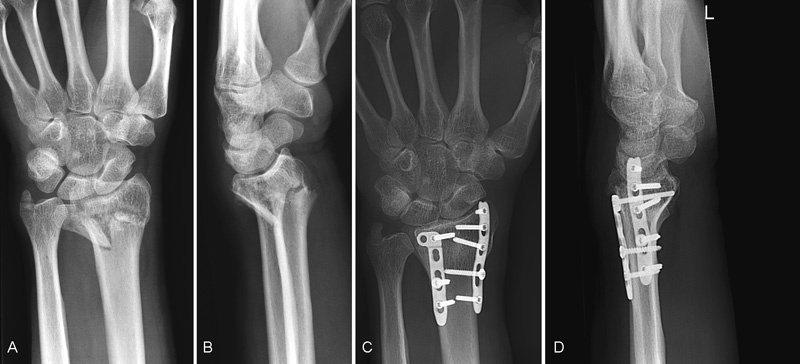
Fig. 1.

A 42-year-old woman is treated with volar plate fixation for AO type C fracture. (A, B) Preoperative anteroposterior and lateral radiographs. (C, D) Postoperative anteroposterior and lateral radiographs after 36 months of follow-up.
Fig. 2.
An AO type C fracture in a 58-year-old man who is treated with dorsal plate fixation. (A, B) Preoperative anteroposterior and lateral radiographs. (C, D) Postoperative anteroposterior and lateral radiographs after 38 months of follow-up.
Complication Assessment
The complication rate was our primary study outcome. All complications were obtained from medical records of all 214 patients. Medical information was retrieved of patients who had their follow-up in another hospital. Complications were divided in three groups: soft tissue/wounds, implant/surgery, and bone/fractures.14
Functional Assessment
Our secondary study outcomes were the functional and radiographic outcome measurements. Subjective functional outcome was measured with the Disabilities of the Arm, Shoulder and Hand (DASH) questionnaire and the Patient-Rated Wrist/Hand Evaluation (PRWHE) questionnaire. The DASH questionnaire is a self-reporting questionnaire with 30 items. These items evaluate symptoms and physical function of the whole upper extremity. The final score ranges from 0 to 100 points, in which lower numbers indicate a lower level of disability.21
The PRWHE is a 15-item self-reporting questionnaire with the focus on wrist pain and disability in daily activities.22 The PRWHE score ranges from 0 to 100, in which 0 means no disability and 100 the worst disability.23
After completing the questionnaires, the patients were clinically examined by the same researcher. Both wrists were tested, in which values for the injured side were compared with those for the contralateral side. Wrist and forearm range of motion were measured with a computerized goniometer (MicroFET 6 Dual Inclinometer, Hoggan Scientific, Salt Lake City, UT). Movements were performed according to the American Medical Association (AMA) guidelines.24 Grip strength was measured with Jamar Hand dynamometer (Sammons Preston Rolyan, Bolingbrook, IL) and pinch strength was measured with hand-held dynamometer (MircoFET 2, Biometrics, Hoggan Scientific, Salt Lake City, UT). For each measurement, mean of three repetitions was recorded because of highest test–retest reliability.25
Radiographic Assessment
Standard posteroanterior and lateral radiographs of both wrists were made at the end of follow-up. Two authors independently evaluated these radiographs. The uninjured side was used as a template to assess whether radiographic parameters of the injured side had the original values as before trauma.26 The following radiographic parameters were evaluated on posteroanterior view: radial inclination, ulnar variance, and radial length. Palmar tilt was assessed on lateral view. To determine fracture type, initial radiographs immediately after trauma of the injured wrist were evaluated. Fractures were classified in three main fracture types according to AO classification system. This leads to a substantial level of interobserver reliability and intraobserver reproducibility.27 28
Statistical Analysis
Baseline demographic variables and relevant clinical variables are summarized descriptively to characterize study population. Any patients requiring additional intervention were identified and their treatments were documented. Data are presented as mean scores with 95% confidence intervals. Results of both treatment groups were compared with the use of Pearson chi-square test for categorical variables and unpaired t-test for continuous variables. The objective functional and radiographic outcomes were compared between the injured side and contralateral side with paired t-test where the values were normally distributed and with Wilcoxon test in cases of nonnormal distributed data. A p-value of < 0.05 was considered as significant. All computations were performed by using SPSS Statistics Software Version 22 (SPSS Inc., Chicago, IL). Sample size was calculated with two averages (α = 0.05 and 1 − β = 0.9), and it was estimated to include at least 60 patients in each study arm, based on the complication rates of prospective clinical study.14 Assuming a 20% dropout in every group, a minimum of 72 patients should be included in every study arm.
Results
Complications
A total of 22 complications were reported in the dorsal group and 14 complications in the volar group (Table 3). Tendon rupture occurred in three patients who were treated with dorsal plate fixation. Two of these patients had tendon rupture of the extensor pollicis longus and one patient had extensor tendon rupture of the fourth finger. All patients underwent implant removal and tendon transfer. One patient with volar plate fixation had a rupture of the extensor polices longus tendon as a result of the fracture itself. After primary tendon repair, a re-rupture occurred due to a prominent screw of the volar implant. The implant was removed and followed by tendon transfer. Six patients had tendinitis due to the implant and screws, of which five patients were treated with dorsal plates. Implant removal was performed in all patients. Three patients with dorsal plate fixation and one patient with volar plate fixation had findings suggestive of complex regional pain syndrome. All patients had implant removal. Most neurologic complications were noted in the volar group. Three patients were diagnosed with carpal tunnel syndrome (CTS) and one patient had neuropraxia of the median nerve. One patient had complete neurologic loss of the median nerve, which required implant removal. After the implant removal, the patient developed a wound infection. In the dorsal group, one patient had CTS and one other patient had neuropraxia of the superficial branch of the radial nerve. Skin infection was noted in one patient with dorsal plate fixation who was treated with antibiotics. Another patient had tendon adhesions of the extensors after dorsal plate fixation. One patient with volar plate fixation developed synostosis after 1 year postoperatively.
Table 3. Reported complications.
| Complications | Dorsal | Volar | p-Value |
|---|---|---|---|
| Soft tissue/wound | 15 (12.2%) | 10 (11.0%) | 0.95 |
| CRPS/dystrophy | 3 (2.4%) | 1 (1.1%) | |
| Skin infection | 1 (0.8%) | 1 (1.1%) | |
| Tendinitis | 5 (4.1%) | 1 (1.1%) | |
| Tendon (re)rupture | 3 (2.4%) | 1 (1.1%) | |
| Neurologic problems | 2 (1.6%) | 5 (5.5%) | |
| Other soft tissue problems | 1 (0.8%) | 1 (1.1%) | |
| Implant/surgery | 4 (3.3%) | 1 (1.1%) | 0.30 |
| Plate/screw pullout | 3 (2.4%) | 1 (1.1%) | |
| Broken plate/screw | 1 (0.8%) | 0 (0%) | |
| Bone/fracture | 3 (2.4%) | 3 (3.3%) | 0.71 |
| Healing problems | 2 (1.6%) | 2 (2.2%) | |
| Loss of reduction | 1 (0.8%) | 0 (0%) | |
| Malunion | 0 (0%) | 1 (1.1%) | |
| Complication risk (%)a | 15.4 | 14.3 | 0.81 |
Abbreviation: CRPS, complex regional pain syndrome.
Note: An overview of all complications of the 214 eligible patients. The percentages are reported in parentheses.
The estimation of the complication risk was based on experiencing at least one complication. Sixteen patients who were treated with dorsal plate fixation had one complication. Three patients experienced two complications. Twelve patients in the volar group experienced one complication and one patient had two complications.
One patient with dorsal plate fixation experienced pullout of the implant twice, which required multiple additional surgical interventions. In another patient, the implant was broken after dorsal plate fixation. Afterwards, the patient underwent volar plate fixation. Three patients had a prominent screw which required implant removal. Two of these patients were treated with dorsal plate fixation. Two patients had a delayed union, of which one patient was treated with volar plate fixation. Eventually, all fractures healed within 12 months. Two patients of both groups had a nonunion. Both fractures healed after reintervention. One patient with dorsal plate fixation had loss of reduction without functional impairment in daily activities. Another patient with volar plate fixation had a malunion which required corrective osteotomy with plate fixation.
Table 4 summarizes the complication rates between the three main AO fracture types. Most complications occurred in patients with AO type C fractures (52.8%), followed by AO type A fractures (33.3%). However, the differences were not significant. In addition, Table 5 summarizes the complication rates between the different volar plates used. Although volar T-plates showed a higher incidence in complications, the differences were not significant (p = 0.23).
Table 4. An overview of complications rates between the three main AO fracture types.
| Complications | Dorsal | Volar | ||||
|---|---|---|---|---|---|---|
| A | B | C | A | B | C | |
| Soft tissue/wound | 9 (40.1%) | 1 (4.6%) | 5 (22.7%) | 1 (7.1%) | 2 (14.3%) | 7 (50%) |
| Implant/surgery | 0 (0%) | 0 (0%) | 3 (13.6%) | 1 (7.1%) | 1 (7.1%) | 1 (7.1%) |
| Bone/fracture | 1 (4.5%) | 0 (0%) | 3 (13.6%) | 0 (0%) | 1 (7.1%) | 0 (0%) |
| Overall complications | 10 (45.5%) | 1 (4.5%) | 11 (50%) | 2 (14.3%) | 4 (30.8%) | 8 (57.1%) |
Note: An overview of all complications between the three main AO fracture types. In the majority of the cases, the complications occurred in AO type C fractures (52.8%), followed by AO type A fractures (33.3%). The volar group showed a stronger tendency than the dorsal group. The differences were not significant.
Table 5. An overview of complication rates between the different volar plate types.
| Complications | T-plate | Double-column plate | Fragment-specific plate | Nonlocking plates | Locking plates |
|---|---|---|---|---|---|
| Soft tissue/wound | 9 (64.3%) | 1 (7.1%) | 0 (0%) | 0 (0%) | 10 (71.4%) |
| Implant/surgery | 1 (7.1%) | 0 (0%) | 0 (0%) | 0 (0%) | 1 (7.1%) |
| Bone/fracture | 1 (7.1%) | 2 (14.3%) | 0 (0%) | 0 (0%) | 3 (21.4%) |
| Overall | 11 (78.6%) | 3 (21.4%) | 0 (0%) | 0 (0%) | 14 (100%) |
Note: An overview of all complications between the different types of volar plates. The percentages are reported in parentheses. Overall, the complication rate was higher in patients who were treated with volar T-plates (p > 0.05).
A total of 19 patients had implant removal due to complications, of which 11 patients were treated with dorsal plate fixation and 8 patients with volar plate fixation (p = 0.97).
Functional Outcome
The functional outcomes are summarized in Tables 6 and 7. In both groups, significant differences were observed in grip strength, palmar flexion, and dorsal extension when the values were compared with the contralateral side. The dorsal group also showed significant differences in supination, ulnar, and radial deviation. The range of motion was 84.4% in dorsal group and 94.0% in the volar group compared with the contralateral side.
Table 6. Subjective functional outcomes.
| Functional scores | Dorsal (N = 31) | Volar (N = 29) | p-Value |
|---|---|---|---|
| DASH score | 19.3 ± 21.3 | 13.3 ± 15.0 | 0.21 |
| PRWHE pain score | 22.0 ± 27.2 | 17.6 ± 19.3 | 0.47 |
| PRWHE function score | 21.4 ± 28.2 | 15.5 ± 17.3 | 0.33 |
| PRWHE total score | 21.7 ± 27.0 | 16.5 ± 17.1 | 0.38 |
Abbreviations: DASH, Disabilities of the Arm, Shoulder and Hand; PRWHE, Patient-Rated Wrist/Hand Evaluation.
Note: All values are given as mean and standard deviation.
Table 7. Objective functional outcomes.
| Functional outcomes | Dorsal (N = 31) | p-Value | Volar (N = 29) | p-Value |
|---|---|---|---|---|
| Grip strength (kg) | 28 ± 13 (31 ± 13) | 0.01 | 29 ± 13 (31 ± 13) | 0.01 |
| Pinch strength (N) | 51 ± 21 (53 ± 18) | 0.19 | 47 ± 19 (51 ± 23) | 0.27 |
| Palmar flexion (degree) | 35 ± 11 (50 ± 17) | 0.00 | 52 ± 13 (59 ± 14) | 0.03 |
| Dorsal extension (degree) | 41 ± 17 (51 ± 13) | 0.00 | 53 ± 13 (60 ± 12) | 0.01 |
| Ulnar deviation (degree) | 32 ± 9 (38 ± 8) | 0.00 | 36 ± 6 (38 ± 7) | 0.07 |
| Radial deviation (degree) | 21 ± 6 (24 ± 6) | 0.01 | 24 ± 6 (26 ± 6) | 0.12 |
| Pronation (degree) | 76 ± 12 (80 ± 8) | 0.14 | 77 ± 9 (75 ± 13) | 0.27 |
| Supination (degree) | 70 ± 17 (79 ± 12) | 0.01 | 78 ± 10 (79 ± 11) | 0.37 |
Note: All values are given as mean and standard deviation. Values of the contralateral healthy side are given in parentheses. Comparison was made between injured side and contralateral side.
Radiographic Outcome
Both groups showed significant differences in comparison with the contralateral side in terms of radial inclination, ulnar variance, and palmar tilt (Table 8).
Table 8. Radiographic outcomes.
| Radiographic parameters | Dorsal (N = 27) | p-Value | Volar (N = 25) | p-Value |
|---|---|---|---|---|
| Radial inclination (degree) | 18 ± 6 (22 ± 4) | 0.00 | 19 ± 6 (21 ± 3) | 0.04 |
| Radial height (mm) | 12 ± 3 (13 ± 2) | 0.09 | 12 ± 4 (13 ± 2) | 0.15 |
| Ulnar variance (mm) | 0.7 ± 3 (−0.5 ± 2) | 0.02 | 0.4 ± 2 (−0.4 ± 1) | 0.03 |
| Palmar tilt (degree) | 8 ± 6 (12 ± 3) | 0.00 | 7 ± 6 (13 ± 3) | 0.01 |
Note: All values are given as mean and standard deviation. Values of the contralateral side are given in parentheses. Comparison was made between injured side and contralateral side.
Discussion
Throughout the years, an ongoing discussion exists whether volar or dorsal plating is the appropriate technique for internal fixation of distal radius fractures. Studies showed contradictory results regarding the complication rates. Our study showed no significant differences in complication rates between the volar and dorsal approach. The estimated risk of experiencing at least one local complication was 15.4% in the dorsal group and 14.3% in the volar group (p = 0.81). A tendency of more cases of tendinitis was observed in patients with dorsal plate fixation compared to patients with volar plate fixation: 4.1 versus 1.1% (p = 0.19), respectively. However, patients with volar plate fixation had higher risk of developing neurologic problems: 5.5% in the volar group versus 1.6% in the dorsal group (p = 0.12). These observations are consistent with a recently published meta-analysis.3 This meta-analysis evaluated complications between dorsal and volar approach with different types of plates.3 The overall complication rate of all included studies was 23.9% in the volar approach and 24.2% in the dorsal approach (p > 0.05). Subgroup analyses showed a significant higher incidence of neuropathy and CTS in the volar group. A significant higher risk of tendon irritation was noted in patients who were treated by dorsal plate fixation.3 This suggests that the development of complications is mainly caused by the different anatomical structures which are involved in dorsal or volar plate fixation.3 The study by Ruch and Papadonikolakis reported a statistically significant association of volar collapse of the distal fragment when the distal screws were pointing proximally in patients with dorsal plate fixation.5 Through this loss of reduction, the plate becomes more prominent in its relation to the overlying extensor tendons and may cause tendon problems.5 We evaluated this finding in our patients who were treated with dorsal plate fixation and had tendon or implant problems (Table 9). More negative screw angles were observed in patients with tendinitis. However, these patients had no loss of reduction during follow-up. Still, it is possible that the plate was prominent in relation to the overlying extensor tendons.
Table 9. Screw angle of patients with dorsal plate fixation and tendon or implant problems.
| Patient | Complication | No. of plates | Plate position | Screw angle (degree) |
|---|---|---|---|---|
| 1 | Rupture | 2 | Radial and dorsoulnar | +1 |
| 2 | Rupture | 2 | Radial and dorsoulnar | +4 |
| 3 | Rupture | 2 | Radial and dorsoradial | −13 |
| 4 | Tendinitis | 2 | Radial and dorsoulnar | −21 |
| 5 | Tendinitis | 2 | Radial and dorsoradial | −30 |
| 6 | Tendinitis | 2 | Dorsoradial and dorsoulnar | −7 |
| 7 | Tendinitis | 2 | Radial and dorsoulnar | 0 |
| 8 | Tendinitis | 2 | Dorsoradial and dorsoulnar | +11 |
| 9 | Tendon adhesions | 2 | Dorsoradial and dorsoulnar | +1 |
| 10 | Pullout of implant | 2 | Dorsoradial and dorsoulnar | +11 |
Note: We summarized the amount of used plates, the plate position, and the screw angle in patients who were treated with dorsal plate fixation and had tendon or implant problems. In the patient with the broken plates, no postoperative radiographs were available with intact plates. Therefore, the screw angle could not be measured in this patient.
At present, only a few studies have been published in which a comparison is made between volar plates and low-profile dorsal plates for internal fixation of the distal radius (Table 10). Most studies showed lower complication rate after dorsal plate fixation.13 14 15 Meanwhile, another study reported lower complication rate after volar plate fixation.16 Like our study, the study of Zettl et al showed no significant difference in complication rates between dorsal and volar plate fixation.17 Several factors such as fracture type, type of plate, and the surgeon's experience are possible causes for the contradictory results. In addition, different definitions for complications were used in the several studies. For example, study of Yu et al defined complications as any adverse outcome in which additional surgical interventions was required.13 Other studies also reported complications in which no additional surgical interventions were performed.14 15 16 17 Based on the heterogeneity of all studies, we argued that surgical decision for surgical approach should be based on fracture type and surgeon's experience with the specific approach and plate types.
Table 10. Overview of studies comparing volar and dorsal plate fixation.
| Author | Year of publication | Number of patients | Study design | Time of follow-up (mo) | Overall complication rate (%) | Number of surgical reinterventions due to complications (%) |
|---|---|---|---|---|---|---|
| Chou et al | 2011 | 19/22 | RCS | 24–53 | 21.1/4.5 | None |
| Matschke et al | 2011 | 266/39 | PCS | 24 | 15/5 | 3.8/2.6 |
| Wichlas et al | 2014 | 225/60 | RCS | 33a | 3.6/16.7 | 2.2/13.3 |
| Yu et al | 2011 | 47/57 | RCS | 12–80 | 14.9/10.5 | 21.3/14.0 |
| Zettl et al | 2009 | 60/60 | RCT | 12 | 33.3/28.3 | 11.7/6.7 |
Abbreviations: PCS, prospective cohort study; RCS, retrospective cohort study; RCT, randomized controlled trial.
Note: An overview of all studies which compares volar plates and low-profile dorsal plates for internal fixation in patients with distal radius fractures.13 14 15 16 17 The values before slash (/) belongs to the volar group and the values after the slash are from the dorsal group.
No range was reported.
Our study has some limitations. A major limitation is the retrospective study design. The surgeon himself decided which plate had to be used based on fracture type and mechanical considerations. Although demographic and baseline characteristics were similar, both groups can differ due to unknown factors for which no correction is possible because of the nonrandomized retrospective nature of the study. Therefore, the risk of confounding is not completely excluded. Although we performed a sample size calculation based on the study results of Matschke et al,14 the difference in complication rates between the two groups is more likely to be smaller than 10%. The study results of the meta-analysis of Wei et al14 already showed a few percent difference between the complication rates. Therefore, a much larger sample size is needed to provide 90% power. Another limitation is the small number of participants in which the functional and radiographic outcomes were assessed. Therefore, no firm conclusions can be drawn regarding the significant results in functional and radiographic outcomes.
In conclusion, there is no preferred plate fixation technique based on these study results. Other factors such as type of fracture and type of plate can also affect the complication rates in dorsal or volar plate fixation of distal radius fractures.
Footnotes
Conflict of Interest None.
References
- 1.Court-Brown C M, Caesar B. Epidemiology of adult fractures: a review. Injury. 2006;37(8):691–697. doi: 10.1016/j.injury.2006.04.130. [DOI] [PubMed] [Google Scholar]
- 2.Handoll H H, Madhok R. WITHDRAWN: Surgical interventions for treating distal radial fractures in adults. Cochrane Database Syst Rev. 2009;(3):CD003209. doi: 10.1002/14651858.CD003209.pub2. [DOI] [PMC free article] [PubMed] [Google Scholar]
- 3.Wei J, Yang T B, Luo W, Qin J B, Kong F J. Complications following dorsal versus volar plate fixation of distal radius fracture: a meta-analysis. J Int Med Res. 2013;41(2):265–275. doi: 10.1177/0300060513476438. [DOI] [PubMed] [Google Scholar]
- 4.Martineau P A Berry G K Harvey E J Plating for distal radius fractures Orthop Clin North Am 2007382193–201., vi [DOI] [PubMed] [Google Scholar]
- 5.Ruch D S, Papadonikolakis A. Volar versus dorsal plating in the management of intra-articular distal radius fractures. J Hand Surg Am. 2006;31(1):9–16. doi: 10.1016/j.jhsa.2005.09.011. [DOI] [PubMed] [Google Scholar]
- 6.Tavakolian J D, Jupiter J B. Dorsal plating for distal radius fractures. Hand Clin. 2005;21(3):341–346. doi: 10.1016/j.hcl.2005.02.001. [DOI] [PubMed] [Google Scholar]
- 7.Jakubietz M G, Gruenert J G, Jakubietz R G. Palmar and dorsal fixed-angle plates in AO C-type fractures of the distal radius: is there an advantage of palmar plates in the long term? J Orthop Surg. 2012;7(1):8. doi: 10.1186/1749-799X-7-8. [DOI] [PMC free article] [PubMed] [Google Scholar]
- 8.Day C S, Franko O I. Low-profile dorsal plating for dorsally angulated distal radius fractures. Tech Hand Up Extrem Surg. 2007;11(2):142–148. doi: 10.1097/01.bth.0000248361.91577.fc. [DOI] [PubMed] [Google Scholar]
- 9.Kamath A F, Zurakowski D, Day C S. Low-profile dorsal plating for dorsally angulated distal radius fractures: an outcomes study. J Hand Surg Am. 2006;31(7):1061–1067. doi: 10.1016/j.jhsa.2006.05.008. [DOI] [PubMed] [Google Scholar]
- 10.Orbay J L, Fernandez D L. Volar fixation for dorsally displaced fractures of the distal radius: a preliminary report. J Hand Surg Am. 2002;27(2):205–215. doi: 10.1053/jhsu.2002.32081. [DOI] [PubMed] [Google Scholar]
- 11.Kamano M, Honda Y, Kazuki K, Yasuda M. Palmar plating for dorsally displaced fractures of the distal radius. Clin Orthop Relat Res. 2002;(397):403–408. doi: 10.1097/00003086-200204000-00047. [DOI] [PubMed] [Google Scholar]
- 12.Vasenius J Operative treatment of distal radius fractures Scand J Surg 2008974290–296., discussion 296–297 [DOI] [PubMed] [Google Scholar]
- 13.Yu Y R, Makhni M C, Tabrizi S, Rozental T D, Mundanthanam G, Day C S. Complications of low-profile dorsal versus volar locking plates in the distal radius: a comparative study. J Hand Surg Am. 2011;36(7):1135–1141. doi: 10.1016/j.jhsa.2011.04.004. [DOI] [PubMed] [Google Scholar]
- 14.Matschke S, Wentzensen A, Ring D, Marent-Huber M, Audigé L, Jupiter J B. Comparison of angle stable plate fixation approaches for distal radius fractures. Injury. 2011;42(4):385–392. doi: 10.1016/j.injury.2010.10.010. [DOI] [PubMed] [Google Scholar]
- 15.Chou Y C, Chen A C, Chen C Y, Hsu Y H, Wu C C. Dorsal and volar 2.4-mm titanium locking plate fixation for AO type C3 dorsally comminuted distal radius fractures. J Hand Surg Am. 2011;36(6):974–981. doi: 10.1016/j.jhsa.2011.02.024. [DOI] [PubMed] [Google Scholar]
- 16.Wichlas F, Haas N P, Disch A, Machó D, Tsitsilonis S. Complication rates and reduction potential of palmar versus dorsal locking plate osteosynthesis for the treatment of distal radius fractures. J Orthop Traumatol. 2014;15(4):259–264. doi: 10.1007/s10195-014-0306-y. [DOI] [PMC free article] [PubMed] [Google Scholar]
- 17.Zettl R P, Clauberg E, Nast-Kolb D, Ruchholtz S, Kühne C A. Volar locking compression plating versus dorsal plating for fractures of the distal radius: a prospective, randomized study [in German] Unfallchirurg. 2009;112(8):712–718. doi: 10.1007/s00113-008-1526-5. [DOI] [PubMed] [Google Scholar]
- 18.Lafontaine M, Hardy D, Delince P. Stability assessment of distal radius fractures. Injury. 1989;20(4):208–210. doi: 10.1016/0020-1383(89)90113-7. [DOI] [PubMed] [Google Scholar]
- 19.Altissimi M, Mancini G B, Azzarà A, Ciaffoloni E. Early and late displacement of fractures of the distal radius. The prediction of instability. Int Orthop. 1994;18(2):61–65. doi: 10.1007/BF02484412. [DOI] [PubMed] [Google Scholar]
- 20.Gustilo R B, Mendoza R M, Williams D N. Problems in the management of type III (severe) open fractures: a new classification of type III open fractures. J Trauma. 1984;24(8):742–746. doi: 10.1097/00005373-198408000-00009. [DOI] [PubMed] [Google Scholar]
- 21.Hudak P L Amadio P C Bombardier C; The Upper Extremity Collaborative Group (UECG). Development of an upper extremity outcome measure: the DASH (disabilities of the arm, shoulder and hand) [corrected] Am J Ind Med 1996296602–608. [DOI] [PubMed] [Google Scholar]
- 22.MacDermid J C, Turgeon T, Richards R S, Beadle M, Roth J H. Patient rating of wrist pain and disability: a reliable and valid measurement tool. J Orthop Trauma. 1998;12(8):577–586. doi: 10.1097/00005131-199811000-00009. [DOI] [PubMed] [Google Scholar]
- 23.Changulani M, Okonkwo U, Keswani T, Kalairajah Y. Outcome evaluation measures for wrist and hand: which one to choose? Int Orthop. 2008;32(1):1–6. doi: 10.1007/s00264-007-0368-z. [DOI] [PMC free article] [PubMed] [Google Scholar]
- 24.Rondinelli R D, Katz R T. Chicago, IL: American Medical Association; 2007. Guides to the Evaluation of Permanent Impairment. 5th ed. [Google Scholar]
- 25.Mathiowetz V, Weber K, Volland G, Kashman N. Reliability and validity of grip and pinch strength evaluations. J Hand Surg Am. 1984;9(2):222–226. doi: 10.1016/s0363-5023(84)80146-x. [DOI] [PubMed] [Google Scholar]
- 26.van Eerten P V, Lindeboom R, Oosterkamp A E, Goslings J C. An X-ray template assessment for distal radial fractures. Arch Orthop Trauma Surg. 2008;128(2):217–221. doi: 10.1007/s00402-007-0391-y. [DOI] [PMC free article] [PubMed] [Google Scholar]
- 27.Müller M. New York, NY: Springer-Verlag; 1991. The principle of the classification; p. 118. [Google Scholar]
- 28.Andersen D J, Blair W F, Steyers C M Jr, Adams B D, el-Khouri G Y, Brandser E A. Classification of distal radius fractures: an analysis of interobserver reliability and intraobserver reproducibility. J Hand Surg Am. 1996;21(4):574–582. doi: 10.1016/s0363-5023(96)80006-2. [DOI] [PubMed] [Google Scholar]